

The DOI matches its Crossref record; 37 of the 37 references it cites were found in a registry or an index.
TP53 copy number and protein expression inform mutation status across risk categories in acute myeloid leukemia
Open accessAbstract
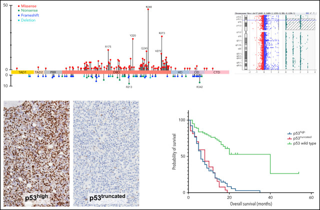
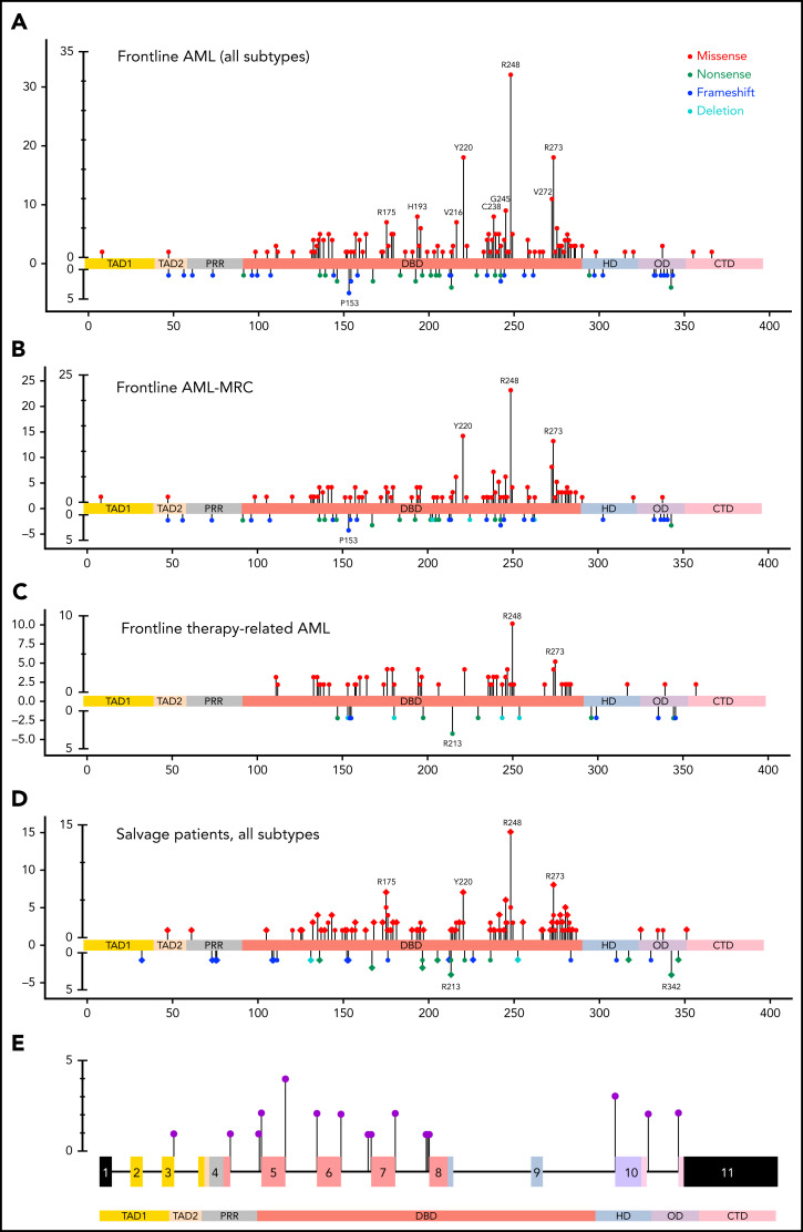
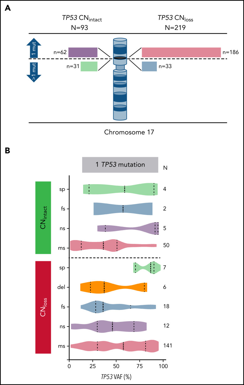
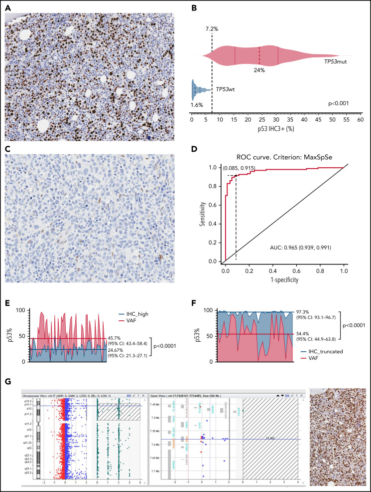
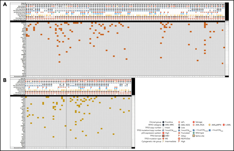
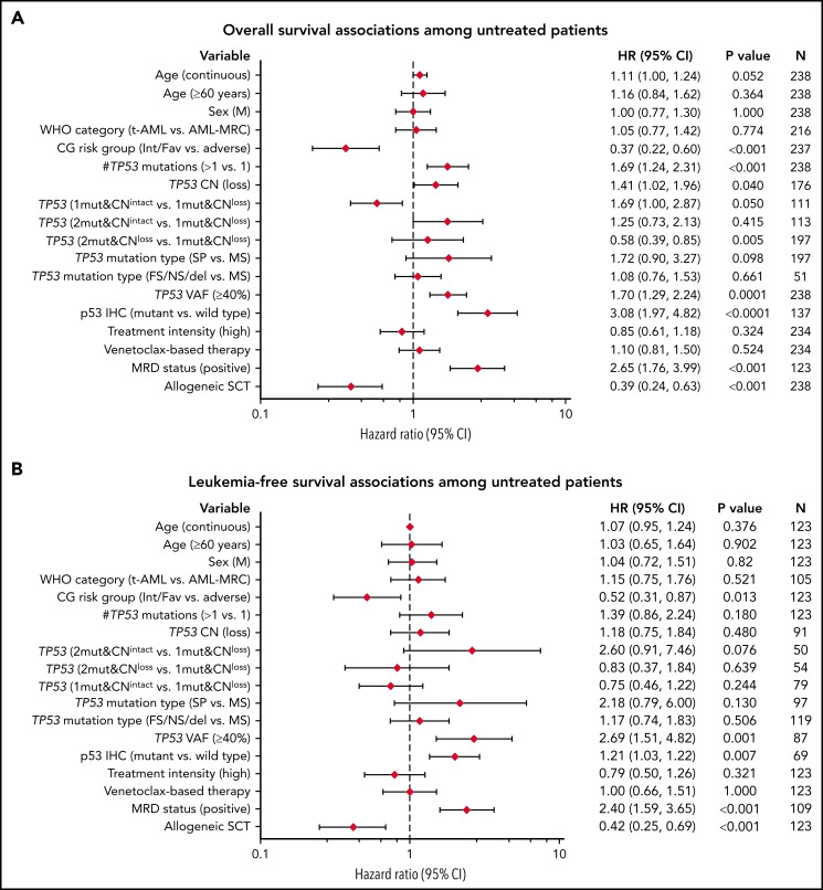
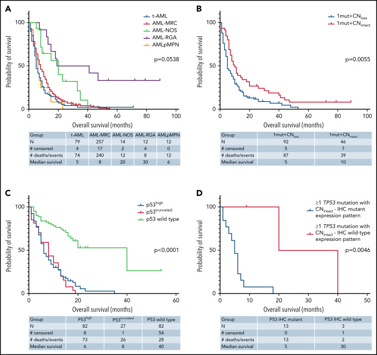
Mutant TP53 is an adverse risk factor in acute myeloid leukemia (AML), but large-scale integrated genomic-proteomic analyses of TP53 alterations in patients with AML remain limited. We analyzed TP53 mutational status, copy number (CN), and protein expression data in AML (N = 528) and provide a compilation of mutation sites and types across disease subgroups among treated and untreated patients. Our analysis shows differential hotspots in subsets of AML and uncovers novel pathogenic variants involving TP53 splice sites. In addition, we identified TP53 CN loss in 70.2% of TP53-mutated AML cases, which have more deleterious TP53 mutations, as well as copy neutral loss of heterozygosity in 5/32 (15.6%) AML patients who had intact TP53 CN. Importantly, we demonstrate that mutant p53 protein expression patterns by immunohistochemistry evaluated using digital image-assisted analysis provide a robust readout that integrates TP53 mutation and allelic states in patients with AML. Expression of p53 by immunohistochemistry informed mutation status irrespective of TP53 CN status. Genomic analysis of comutations in TP53-mutant AML shows a muted landscape encompassing primarily mutations in genes involved in epigenetic regulation (DNMT3A and TET2), RAS/MAPK signaling (NF1, KRAS/NRAS, PTPN11), and RNA splicing (SRSF2). In summary, our data provide a rationale to refine risk stratification of patients with AML on the basis of integrated molecular and protein-level TP53 analyses.
Overview
The claims and statistics of this paper haven’t been checked yet.
Run the check — free, no account needed. It reads the open-access text and checks the sources it cites.
Integrity signals
Registry
Cites
Statistics
Figures
Verification
Claims the paper makes
The claims and statistics of this paper haven’t been checked yet.
Run the check — free, no account needed. It reads the open-access text and checks the sources it cites.
Claims other papers make about this one
Each is checked against this paper’s own text: does it say what the citing paper says it does?
The source backs part of the claim, not all of it — or backs it for a different population, condition or comparison.
In the citing paper
“there is a very strong interplay between VAF, allelic state, and cytogenetic complexity strongly predicting outcomes in this molecular subset”
From Long‐term follow‐up and combined Phase 2 results of eprenetapopt and azacitidine in patients with TP53 mutant MDS/AML (2025) · cites this paper as [10]
Experimental — accuracy not yet measuredThe source backs part of the claim, not all of it — or backs it for a different population, condition or comparison.
In the citing paper
“with a recent study identifying 92% of TP53 mutant MDS/AML patients to represent a poor risk population.”
From Long‐term follow‐up and combined Phase 2 results of eprenetapopt and azacitidine in patients with TP53 mutant MDS/AML (2025) · cites this paper as [10]
Experimental — accuracy not yet measured
Self-check
Percentages, sums and reported differences re-computed from the paper’s own numbers.
The self-check needs the paper’s full text.
Citations
Cites
Döhner, H., Estey, E. H., Grimwade, D. J., Amadori, S., Appelbaum, F. R., Büchner, T., Dombret, H., Ebert, B. L., Fenaux, P., Larson, R. A., et al. (2016) · Blood · doi:10.1182/blood-2016-08-733196 · Cited by 5,938
Genomic classification and prognosis in acute myeloid leukemia
Papaemmanuil, E., Gerstung, M., Bullinger, L., Gaidzik, V. I., Paschka, P., Roberts, N. D., Potter, N. E., Heuser, M., Thol, F., Bolli, N., et al. (2016) · New England Journal of Medicine · doi:10.1056/nejmoa1516192 · Cited by 4,350
Kastenhuber, E. R., & Lowe, S. W. (2017) · Cell · doi:10.1016/j.cell.2017.08.028 · Cited by 1,906
p53: 800 million years of evolution and 40 years of discovery
Levine, A. J. (2020) · Nature reviews. Cancer · doi:10.1038/s41568-020-0262-1 · Cited by 876
Prognostic Mutations in Myelodysplastic Syndrome after Stem-Cell Transplantation
Lindsley, R. C., Saber, W., Mar, B. G., Redd, R., Wang, T., Haagenson, M. D., Grauman, P. V., Hu, Z.-H., Spellman, S. R., Lee, S. J., et al. (2017) · New England Journal of Medicine · doi:10.1056/nejmoa1611604 · Cited by 776
Bouaoun, L., Sonkin, D., Ardin, M., Hollstein, M., Byrnes, G., Zavadil, J., & Olivier, M. (2016) · Human Mutation · doi:10.1002/humu.23035 · Cited by 730
Rücker, F. G., Schlenk, R. F., Bullinger, L., Käyser, S., Teleanu, V., Kett, H., Habdank, M., Kugler, C.-M., Holzmann, K., Gaidzik, V. I., et al. (2011) · Blood · doi:10.1182/blood-2011-08-375758 · Cited by 694
Bernard, E., Nannya, Y., Hasserjian, R. P., Devlin, S. M., Tuechler, H., Medina-Martinez, J. S., Yoshizato, T., Shiozawa, Y., Saiki, R., Malcovati, L., et al. (2020) · Nature Medicine · doi:10.1038/s41591-020-1008-z · Cited by 648
Eprenetapopt (APR-246) and Azacitidine in TP53 -Mutant Myelodysplastic Syndromes
Sallman, D. A., DeZern, A. E., Garcia-Manero, G., Steensma, D. P., Roboz, G. J., Sekeres, M. A., Cluzeau, T., Sweet, K. L., McLemore, A., McGraw, K. L., et al. (2021) · Journal of Clinical Oncology · doi:10.1200/jco.20.02341 · Cited by 465
Self-citationA dominant-negative effect drives selection of TP53 missense mutations in myeloid malignancies
Boettcher, S., Miller, P. G., Sharma, R., McConkey, M., Leventhal, M., Krivtsov, A. V., Giacomelli, A. O., Wong, W., Kim, J., Chao, S., et al. (2019) · Science · doi:10.1126/science.aax3649 · Cited by 441
Cited by
Sorted by citations
Items 101–112 of 112
Zhang, R., Kou, W., Wu, T., & Zhou, R. (2026) · Frontiers in Medicine · doi:10.3389/fmed.2025.1712877
Wang, X., Huang, Y., Sun, M., Cai, S., Hu, X., Zheng, Y., Fang, S., Li, S., Tu, Y., & Tang, H. (2026) · Translational Oncology · doi:10.1016/j.tranon.2026.102705
Francesco, M. A. D., Ribolla, R., Sottini, A., Bertoli, D., Giupponi, C., Bonuccelli, O., Bresciani, R., Tucci, A., & Biasiotto, G. (2026) · Blood Science · doi:10.1097/bs9.0000000000000282
Ji, M., Hao, Y., Li, W., Zhou, Y., Jia, R., Li, W., Jin, S., Dai, M., Chang, M., Feng, L., et al. (2026) · The Oncologist · doi:10.1093/oncolo/oyag088
Wu, W., Chen, T., Tao, N., Li, P. M., Gao, M. M., Wang, M., Zhang, X. G., Tang, L., Liu, M., & Liu, J. (2026) · Research Square · doi:10.21203/rs.3.rs-8955297/v1
Acute Myeloid Leukemia with Mutated TP53
Sameeta, F., & Wang, W. J. (2026) · Cambridge University Press eBooks · doi:10.1017/9781009397797.032
Niche-level immune evasion in TP53 mutant AML residual disease revealed by spatial proteomicsPreprint
Hideaki, M., Nishida, Y., Bedoy, A. D., Ayoub, E., Lee, Y., Basi, A., Sasaki, K., Garcia‐Manero, G., Burks, J., Kanagal‐Shamanna, R., et al. (2026) · bioRxiv (Cold Spring Harbor Laboratory) · doi:10.64898/2026.05.15.725421
Self-citationContemporary approach to diagnosis and classification of acute myeloid leukemia
Loneman, D. M., Nardi, V., & Hasserjian, R. P. (2026) · Human Pathology · doi:10.1016/j.humpath.2026.106197 · Cited by 1
Fang, H., Khoury, J. D., Tang, Z., Wang, S., Dinardo, C. D., Loghavi, S., Wang, W. J., Dimopoulos, Y. P., Hu, S., Yin, C. C., et al. (2026) · American Journal of Hematology · doi:10.1002/ajh.70426
Self-citationInterpretation of p53 immunohistochemical stain of TP53-mutated myeloid neoplasms
Barwari, N., Morrow, W. P., Evans, J. K., Rosenthal, N., Beaty, M. W., Hsi, E. D., Insuasti‐Beltran, G., & Maracaja, D. L. V. (2026) · Frontiers in Oncology · doi:10.3389/fonc.2026.1916425
Fitzpatrick, M. J., Yuan, J., Akuma, D., Allbee, A. W., Bledsoe, J. R., Capa, I., Chen, E., Chen, M., Chen, P.-H., Chen, X., et al. (2026) · Modern Pathology · doi:10.1016/j.modpat.2026.101107
Self-citationWannaphut, C., Chiraphapphaiboon, W., Sachs, Z., Gooptu, M., & Sumransub, N. (2026) · Blood Reviews · doi:10.1016/j.blre.2026.101438
Metadata
- Authors
- Tashakori, M., Kadia, T., Loghavi, S., Daver, N., Kanagal-Shamanna, R., Pierce, S., Sui, D., Wei, P., Khodakarami, F., Tang, Z., Routbort, M., Bivins, C. A., Jabbour, E. J., Medeiros, L. J., Bhalla, K., Kantarjian, H. M., Ravandi, F., & Khoury, J. D.
- Affiliations
- University of Minnesota (US)
- 3Department of Leukemia, and
- The University of Texas MD Anderson Cancer Center (US)
- 1Department of Hematopathology, The University of Texas MD Anderson Cancer Center, Houston, TX;
- 5Cyclica Inc, Toronto, ON, Canada
- Corresponding author
- Khoury, Joseph D.
- ORCID
- Venue
- Blood
- Publisher
- American Society of Hematology
- Published
- 2022-07-07
- Volume
- 140
- Issue
- 1
- Pages
- 58-72
- ISSN
- 0006-4971 (ISSN-L)
- 1528-0020
- PMC
- PMC9346958
- OpenAlex
- W4226526247
- Cited by
- 111
- References
- 36
- Citations by year
- 2026: 17 · 2025: 24 · 2024: 31 · 2023: 27 · 2022: 12
- FWCI
- 17.28
- Type
- journal article
- Language
- en
- Open access
- hybrid
- doi.org
- Licence
- Locations
- Blood (publishedVersion, cc-by-nc-nd)
- PubMed Central (publishedVersion, cc-by-nc-nd)
- PubMed (publishedVersion)
- Digital Commons-TMC (Texas Medical Center) (submittedVersion)
- Topics
- Health Sciences › Medicine › Hematology › Acute Myeloid Leukemia Research
- Life Sciences › Biochemistry, Genetics and Molecular Biology › Cancer Research › Cancer Genomics and Diagnostics
- Health Sciences › Medicine › Oncology › Cancer-related Molecular Pathways
- Keywords
- acute myeloid leukemia, TP53 mutation, p53 expression, immunohistochemistry, copy-neutral loss of heterozygosity, co-occurring mutations, DNMT3A, TET2, RAS/MAPK pathway, SRSF2, risk stratification
- MeSH terms
- Humans, Mutation, Prognosis, Leukemia, Myeloid, Acute, Tumor Suppressor Protein p53, Proteomics, Epigenesis, Genetic, DNA Copy Number Variations
About these checks
Registry, retraction, Cites and statistics results are computed, not judged, with one exception: a printed reference that no index lists is read once by a model before it counts as a citation error. Claims are extracted by a model and kept only when their quote appears verbatim in the text; each cited source is then checked, with a model’s help, for whether it says what the claim relies on. Those verdicts can be wrong — read the claim’s quote and the cited source before relying on one.
Statistics and arithmetic are recomputed, not judged.
Confidence bands are shown only for fields calibrated on our annotated test set. None is calibrated yet, so no band is shown.
What counts as a claim ›How we measure confidence ›How we check this ›
Are you an author, or do you see a problem with this page? Request a review
