

The DOI matches its Crossref record; 37 of the 38 references it cites were found in a registry or an index; 29 of the 50 sources its claims cite were checked for support.
Computer-aided detection thresholds for digital chest radiography interpretation in tuberculosis diagnostic algorithms.
Open accessAbstract
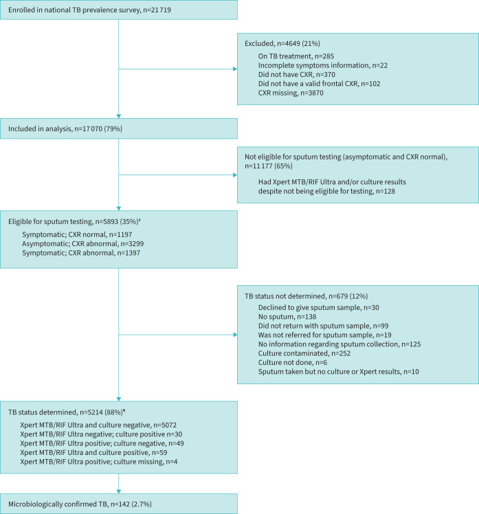
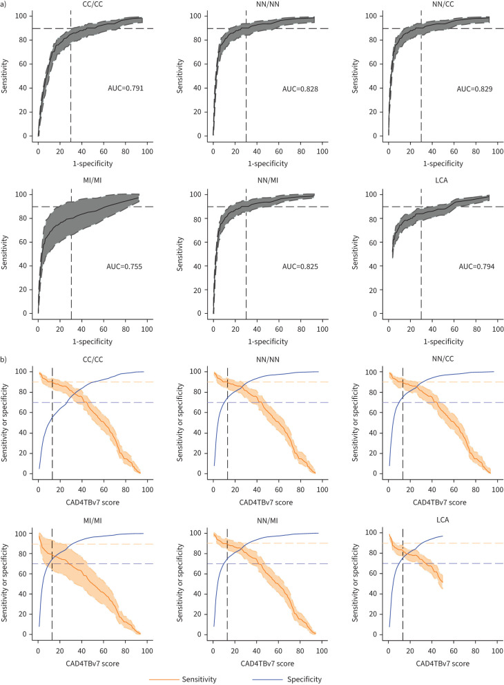
Objectives: Use of computer-aided detection (CAD) software is recommended to improve tuberculosis screening and triage, but threshold determination is challenging if reference testing has not been performed in all individuals. We aimed to determine such thresholds through secondary analysis of the 2019 Lesotho national tuberculosis prevalence survey. Methods: Symptom screening and chest radiographs were performed in participants aged ≥15 years; those symptomatic or with abnormal chest radiographs provided samples for Xpert MTB/RIF and culture testing. Chest radiographs were processed using CAD4TB version 7. We used six methodological approaches to deal with participants who did not have bacteriological test results to estimate pulmonary tuberculosis prevalence and assess diagnostic accuracy. Results: Among 17 070 participants, 5214 (31%) had their tuberculosis status determined; 142 had tuberculosis. Prevalence estimates varied between methodological approaches (0.83-2.72%). Using multiple imputation to estimate tuberculosis status for those eligible but not tested, and assuming those not eligible for testing were negative, a CAD4TBv7 threshold of 13 had a sensitivity of 89.7% (95% CI 84.6-94.8) and a specificity of 74.2% (73.6-74.9), close to World Health Organization (WHO) target product profile criteria. Assuming all those not tested were negative produced similar results. Conclusions: This is the first study to evaluate CAD4TB in a community screening context employing a range of approaches to account for unknown tuberculosis status. The assumption that those not tested are negative - regardless of testing eligibility status - was robust. As threshold determination must be context specific, our analytically straightforward approach should be adopted to leverage prevalence surveys for CAD threshold determination in other settings with a comparable proportion of eligible but not tested participants.
Overview
CoChat Summary
## TL;DR
Using secondary analysis of the 2019 Lesotho national tuberculosis prevalence survey, the paper estimated a CAD4TBv7 threshold of 13 for chest radiograph screening that achieved about 90% sensitivity and 74% specificity under its preferred assumptions. The main message is that threshold calibration is context-specific, and that treating untested participants as tuberculosis-negative gave robust results in this setting.
## Key findings
- Among 17,070 participants analyzed, 5,214 (31%) had tuberculosis status determined, and 142 had tuberculosis.
- Estimated tuberculosis prevalence varied by method from 0.83% to 2.72%; the authors’ “best” estimate was 0.97%.
- Under the NN/NN approach, the CAD4TBv7 threshold of 13 gave sensitivity of 90.1% (95% CI 84.0–94.1) and specificity of 74.2% (95% CI 73.6–74.9); under NN/MI, sensitivity was 89.7% (95% CI 84.6–94.8) with the same specificity.
- Subgroup analysis found lower specificity among men, symptomatic participants, and those with a history of tuberculosis; sensitivity was worse for women than men, with differences of around 30% at some thresholds.
## Methods
- Secondary analysis of the 2019 Lesotho national tuberculosis prevalence survey; adults aged ≥15 years were screened with symptoms and chest radiography, and symptomatic or radiographically abnormal participants provided sputum for Xpert MTB/RIF Ultra and culture.
- Chest radiographs were processed with CAD4TB versions 6 and 7, and the study focused on CAD4TBv7 threshold determination.
- Six approaches handled missing bacteriological status, including complete case analysis, treating untested as negative, multiple imputation, and Bayesian latent class analysis; sensitivity, specificity, ROC curves, and AUC were estimated across thresholds.
## Limitations
- The paper states that its tuberculosis case definition differed from the national survey’s definition, which risked including false positives; it also notes that Ultra and culture are imperfect reference standards, especially in community screening.
- Diagnostic accuracy for chest radiography comparisons is said to be cautious to interpret because field readers were instructed to over-read, and abnormal chest radiographs were an inclusion criterion for bacteriological testing, creating selection bias.
- The text makes evident that many participants had missing chest radiographs and that 19% of invited individuals refused participation; the authors note these issues could affect representativeness, though they expect little impact on threshold determination.
Model-written summary. Check it against the paper before relying on it.
Integrity signals
Registry
Cites
Statistics
Figures

Verification
Claims the paper makes
16 Valid · 0 Not valid · 5 Not checked · 61 Own results and interpretations
The cited sources support only part of this claim — read each source’s verdict below.
In the paper
“There remains a paucity of guidance for threshold determination when the majority of individuals did not undergo microbiological testing [30].”
§ Computer-aided detection thresholds for digital chest radiography interpretation in tuberculosis diagnostic algorithms > Introduction
What the sources say
- Partly supportedcites [30] Geric, 2023, The rise of artificial intelligence reading of chest X-rays for enhanced TB diagnosis and elimination, Int J Tuberc Lung Dis sourceExperimental — accuracy not yet measured
The Diagnostic Performance of CAD for TB section says threshold selection is challenging, requires local calibration, and can be prone to error. It also warns that testing only participants with abnormal chest X-rays can bias accuracy estimates. It does not specifically say that guidance is scarce when most individuals have not undergone microbiological testing; no page number was available in our copy. (LLM, conf=0.82)
plain- Asserted: passes — The authors put it forward as true.
- Checkable: passes — It can be checked against evidence.
- Load-bearing: passes — The paper’s conclusions depend on it.
- Based: passes — The paper gives a reason why it holds.
- Partly supported
The cited sources support only part of this claim — read each source’s verdict below.
In the paper
“yet context-specific determination is often not done due to logistical and financial constraints [8, 14, 15].”
§ Computer-aided detection thresholds for digital chest radiography interpretation in tuberculosis diagnostic algorithms > Introduction
What the sources say
- Partly supportedcites [8] Tavaziva, 2021, Chest X-ray analysis with deep learning-based software as a triage test for pulmonary tuberculosis: an individual patient data meta-analysis of diagnostic accuracy, Clin Infect Dis sourceExperimental — accuracy not yet measured
The Discussion says setting-specific threshold selection requires local patient data, resources, protocols, and tools, and calls this an important implementation challenge. It does not say that such determination is often omitted or identify financial constraints as a reason. (LLM, conf=0.83)
- Not checked — no full text of the sourcecites [14] source
- Partly supportedcites [15] Odume, 2022, Portable digital X-ray for TB pre-diagnosis screening in rural communities in Nigeria, Public Health Action sourceExperimental — accuracy not yet measured
The METHODS section of Odume et al. (2022) confirms only the factual core for this one project: 'The CAD4TB score threshold was based on the manufacturer's specification, without further calibration in the study population.' Nothing in our copy gives a reason for that omission — no passage attributes it to logistical or financial constraints (searched: logistical/financial constraints, cost, local calibration of the cut-off, resource-limited settings); the only cost remark in the DISCUSSION concerns Xpert savings from the low NNT, not threshold calibration. Nor does the paper make the frequency claim that context-specific determination is 'often' not done; it reports a single Nigerian pilot, so the generalisation and the causal explanation rest on the other citations, not this source. (LLM, conf=0.72)
plainstates a cause- Asserted: passes — The authors put it forward as true.
- Checkable: passes — It can be checked against evidence.
- Load-bearing: passes — The paper’s conclusions depend on it.
- Based: passes — The paper gives a reason why it holds.
- Partly supported
The cited sources support only part of this claim — read each source’s verdict below.
In the paper
“The Lesotho Ministry of Health conducted a nationwide tuberculosis prevalence survey in 2019 [34].”
§ Computer-aided detection thresholds for digital chest radiography interpretation in tuberculosis diagnostic algorithms > Material and methods > Participants and study design
What the sources say
- Partly supportedcites [34] Matji, 2023, Policy and programmatic directions for the Lesotho tuberculosis programme: findings of the national tuberculosis prevalence survey, 2019, PLoS ONE sourceExperimental — accuracy not yet measured
The Methods (Survey design and population; page unavailable) state that a national TB prevalence survey was conducted from March through November 2019 and that no areas of Lesotho were excluded from the sampling frame. The Introduction identifies the National TB Programme as a beneficiary of the survey, but the passages do not establish that the Ministry of Health conducted it. (LLM, conf=0.83)
plain- Asserted: passes — The authors put it forward as true.
- Checkable: passes — It can be checked against evidence.
- Load-bearing: unclear — It is unclear whether the paper’s conclusions depend on it.
- Based: passes — The paper gives a reason why it holds.
- Partly supported
The sources with a verdict support this claim; the rest weren’t checked yet, have no full text we could read, or their verdicts aren’t rated yet.
In the paper
“Assuming that those who did not undergo testing did not have tuberculosis is commonly done [12, 28, 29], but this will misclassify some individuals who have subclinical tuberculosis with normal chest radiograph, and addressing subclinical tuberculosis is essential if WHO incidence targets are to be met [40].”
§ Computer-aided detection thresholds for digital chest radiography interpretation in tuberculosis diagnostic algorithms > Discussion
What the sources say
- Supportedcites [12] Fehr, 2021, Computer-aided interpretation of chest radiography reveals the spectrum of tuberculosis in rural South Africa, NPJ Digital Med source
The Performance of CAD4TB section reports 19 microbiologically confirmed TB cases with radiologist-assessed normal lung fields; 15 were asymptomatic and would have gone undetected under radiologist-based triage. The Discussion notes that the TB status of participants who did not provide sputum is unknown, so treating all untested participants as TB-negative could misclassify cases of this kind. Page numbers were not available in our copy. (LLM, conf=0.87)
- Not checked — no full text of the sourcecites [28] Koesoemadinata, 2018, Computer-assisted chest radiography reading for tuberculosis screening in people living with diabetes mellitus, Int J Tuberc Lung Dis source
- Not checked — no full text of the sourcecites [29] source
- Supportedcites [40] source
The source's 'The Large Burden of Subclinical TB' section states that most prevalence surveys only submit sputum for bacteriologic testing from those screening positive on symptoms or chest radiography, so 'the TB status of those with normal chest X-ray findings and no reported symptoms is unknown' (Figure 2 legend), and that if chest X-ray sensitivity is no better for subclinical than symptomatic TB (likely worse), 'the burden of subclinical TB will be underestimated in prevalence surveys by at least 10%' — i.e., treating the untested as TB-negative misclassifies some individuals with subclinical TB who have a normal chest radiograph. The quantified inference (at least 81 culture-positive cases missed per 100,000 in the 2011 Cambodia survey extrapolation, and substantially more after accounting for symptom/X-ray correlation) directly supports the paraphrase. Only the 'commonly done' clause of the citing sentence is carried by the other citations [12, 28, 29], not this source. (LLM, conf=0.86)
plainstates a cause- Asserted: passes — The authors put it forward as true.
- Checkable: passes — It can be checked against evidence.
- Load-bearing: passes — The paper’s conclusions depend on it.
- Based: passes — The paper gives a reason why it holds.
- Supported
The cited sources support only part of this claim — read each source’s verdict below.
In the paper
“To date, only one study has attempted to address this problem in a principled way, using latent class analyses (LCA) to account for the missingness in tuberculosis status [11].”
§ Computer-aided detection thresholds for digital chest radiography interpretation in tuberculosis diagnostic algorithms > Introduction
What the sources say
- Partly supportedcites [11] Mungai, 2022, Accuracy of computer-aided chest X-ray in community-based tuberculosis screening: Lessons from the 2016 Kenya National Tuberculosis Prevalence Survey, PLOS Glob Public Health sourceExperimental — accuracy not yet measured
The Methods (Statistical analysis; page not provided) say the study used Bayesian latent class modelling to infer TB status among participants without sputum testing and estimate CAD4TBv6 accuracy across thresholds. The Discussion (page not provided) explains that excluding untested participants could bias accuracy estimates. Our copy does not establish the claim that this was the only study to use this approach. (LLM, conf=0.86)
plain- Asserted: passes — The authors put it forward as true.
- Checkable: passes — It can be checked against evidence.
- Load-bearing: passes — The paper’s conclusions depend on it.
- Based: passes — The paper gives a reason why it holds.
- Partly supported
33 more claims · 5 with their cited sources checked
Create a free account to see all 82 claims and to verify the 9 references still open.
- Probably not a substantial claim. 19
- It may state two separate claims as one. 25
- The quote only partly states this claim. 3
Citations
Cites
Multiple imputation using chained equations: Issues and guidance for practice
White, I. R., Royston, P., & Wood, A. M. (2011) · Statistics in Medicine · doi:10.1002/sim.4067 · Cited by 9,792 · cited 3×
“In the fourth approach, we used multiple imputation (MI) to impute unknown tuberculosis statuses under a missing at random assumption (MI/MI) [36].”
BackgroundMethodInfluentialHow to improve access to medical imaging in low- and middle-income countries
Frija, G., Blažić, I., Frush, D. P., Hierath, M., Kawooya, M., Donoso-Bach, L., & Brkljačić, B. (2021) · eClinicalMedicine · doi:10.1016/j.eclinm.2021.101034 · Cited by 310 · cited 1×
“The World Health Organization (WHO) recommends incorporating chest radiography in screening and triaging algorithms but this relies on expert interpretation thus limiting its reach [3, 4].”
BackgroundQin, Z. Z., Ahmed, S., Sarker, M. S., Paul, K., Adel, A. S. S., Naheyan, T., Barrett, R., Banu, S., & Creswell, J. (2021) · The Lancet Digital Health · doi:10.1016/s2589-7500(21)00116-3 · Cited by 290 · cited 2×
“In the first, a large study in nearly 24 000 participants, a much higher threshold of 50 was identified (for 90% sensitivity with 73% specificity) [23].”
BackgroundResultQin, Z. Z., Sander, M. S., Rai, B., Titahong, C. N., Sudrungrot, S., Laah, S. N., Adhikari, L. M., Carter, E. J., Puri, L., Codlin, A. J., et al. (2019) · Scientific Reports · doi:10.1038/s41598-019-51503-3 · Cited by 289
The Epidemiological Importance of Subclinical Tuberculosis. A Critical Reappraisal
Kendall, E. A., Shrestha, S., & Dowdy, D. W. (2021) · American Journal of Respiratory and Critical Care Medicine · doi:10.1164/rccm.202006-2394pp · Cited by 185
Melendez, J., Sánchez, C. I., Philipsen, R. H. H. M., Maduskar, P., Dawson, R., Theron, G., Dheda, K., & van Ginneken, B. (2016) · Scientific Reports · doi:10.1038/srep25265 · Cited by 152
Khan, F. A., Majidulla, A., Tavaziva, G., Nazish, A., Abidi, S. K., Benedetti, A., Menzies, D., Johnston, J. C., Khan, A. J., & Saeed, S. (2020) · The Lancet Digital Health · doi:10.1016/s2589-7500(20)30221-1 · Cited by 134
Computer aided detection of tuberculosis on chest radiographs: An evaluation of the CAD4TB v6 system
Murphy, K., Habib, S. S., Zaidi, S. M. A., Khowaja, S., Khan, A. J., Melendez, J., Scholten, E. T., Amad, F., Schalekamp, S., Verhagen, M., et al. (2020) · eCommons - AKU (Aga Khan University) · Cited by 125
Self-citationComputer aided detection of tuberculosis on chest radiographs: An evaluation of the CAD4TB v6 system
Murphy, K., Habib, S. S., Zaidi, S. M. A., Khowaja, S., Khan, A., Melendez, J., Scholten, E. T., Amad, F., Schalekamp, S., Verhagen, M., et al. (2020) · Scientific Reports · doi:10.1038/s41598-020-62148-y · Cited by 122
Self-citationIndependent evaluation of 12 artificial intelligence solutions for the detection of tuberculosis
Codlin, A. J., Dao, T. P., Vo, L. N. Q., Forse, R. J., Van Truong, V., Dang, H. M., Nguyen, L. H., Nguyen, H. B., Nguyen, N. V., Sidney-Annerstedt, K., et al. (2021) · Scientific Reports · doi:10.1038/s41598-021-03265-0 · Cited by 106 · cited 1×
“Lastly, specificity was poorer in those with history of tuberculosis, as expected and corresponding to findings in other settings [11, 17, 23, 43].”
Result
Cited by
Scott, A. J., Perumal, T., Pooran, A., Oelofse, S., Jaumdally, S. Z., Swanepoel, J., Gina, P., Mthiyane, T., Qin, Z. Z., Fehr, J., et al. (2025) · The Lancet Global Health · doi:10.1016/s2214-109x(24)00516-3 · Cited by 19
Signorell, A., van Heerden, A. C., Ayakaka, I., Jacobs, B. K., Antillón, M., Tediosi, F., Verjans, A., Brugger, C., Harkare, H. V., Labhardt, N. D., et al. (2025) · BMJ Open · doi:10.1136/bmjopen-2024-093989 · Cited by 13
Self-citationKaewwilai, L., Yoshioka, H., Choppin, A., Prueksaritanond, T., Ayuthaya, T. P. N., Brukesawan, C., Matupumanon, S., Kawabe, S., Shimahara, Y., Phosri, A., et al. (2025) · Global Transitions · doi:10.1016/j.glt.2025.02.005 · Cited by 14
Bosman, S., Ayakaka, I., Muhairwe, J. A., Kamele, M., van Heerden, A. C., Madonsela, T., Labhardt, N. D., Sommer, G., Bremerich, J., Zöller, T., et al. (2024) · Clinical Infectious Diseases · doi:10.1093/cid/ciae378 · Cited by 19
Self-citationNzimande, N., Murphy, K., Reither, K., Bosman, S., Ayakaka, I., Glass, T. R., Vanobberghen, F. M., Jacobs, B. K. M., Signorell, A., & Ncayiyana, J. R. (2025) · Journal of Clinical Tuberculosis and Other Mycobacterial Diseases · doi:10.1016/j.jctube.2025.100540 · Cited by 15
“[36] Operational research studies are not always viable in resource-constrained settings like SA and Lesotho; in that case, these various other approaches should be considered.”
InfluentialSelf-citation
Metadata
- Authors
- Vanobberghen, F., Keter, A. K., Jacobs, B. K. M., Glass, T. R., Lynen, L., Law, I., Murphy, K., van Ginneken, B., Ayakaka, I., van Heerden, A., Maama, L., & Reither, K.
- Affiliations
- Swiss Tropical and Public Health Institute (CH)
- Human Sciences Research Council (ZA)
- Instituut voor Tropische Geneeskunde (BE)
- World Health Organization (CH)
- Radboud University Medical Center (NL)
- SolidarMed (CH)
- Ministry of Health (LS)
- Corresponding author
- Vanobberghen, Fiona
- ORCID
- Venue
- ERJ Open Research
- Publisher
- European Respiratory Society (ERS)
- Published
- 2023-12-07
- Volume
- 10
- Issue
- 1
- Pages
- 00508-2023
- ISSN
- 2312-0541 (ISSN-L)
- PMC
- PMC10772898
- OpenAlex
- W4389431098
- Cited by
- 13
- References
- 38
- Citations by year
- 2026: 3 · 2025: 8 · 2024: 2
- FWCI
- 2.68
- Type
- journal article
- Language
- en
- Open access
- gold
- doi.org
- Licence
- Locations
- ERJ Open Research (publishedVersion, cc-by-nc)
- PubMed Central (publishedVersion, cc-by-nc)
- PubMed (publishedVersion)
- Radboud Repository (Radboud University) (submittedVersion)
- DOAJ (DOAJ: Directory of Open Access Journals) (submittedVersion, cc-by-sa)
- Funders
- European and Developing Countries Clinical Trials Partnership (RIA2018D-2498)
- Topics
- Health Sciences › Medicine › Infectious Diseases › Tuberculosis Research and Epidemiology
- Physical Sciences › Computer Science › Artificial Intelligence › AI in cancer detection
- Health Sciences › Medicine › Pulmonary and Respiratory Medicine › Lung Cancer Diagnosis and Treatment
- Keywords
- computer-aided detection, CAD4TB, tuberculosis screening, chest radiography, diagnostic thresholds, diagnostic accuracy, multiple imputation, Xpert MTB/RIF, culture testing, Lesotho
About these checks
Registry, retraction, Cites and statistics results are computed, not judged, with one exception: a printed reference that no index lists is read once by a model before it counts as a citation error. Claims are extracted by a model and kept only when their quote appears verbatim in the text; each cited source is then checked, with a model’s help, for whether it says what the claim relies on. Those verdicts can be wrong — read the claim’s quote and the cited source before relying on one.
The claim verdicts this report counts are experimental: their accuracy is not yet measured.
A language model drew the claims from the paper on 2026-10-10, under version 3 of the claim definition (model deepseek-v4.1-flash, prompt extract-v3).
A second model labelled each claim on 2026-10-10 (model jev-1.13-20260917, definition revision gold-guideline-v0.2).
Cited sources are checked with a language model’s help (gpt-6-sol and qwen3.8-omni-flash).
Statistics and arithmetic are recomputed, not judged.
Confidence bands are shown only for fields calibrated on our annotated test set. None is calibrated yet, so no band is shown.
What counts as a claim ›How we measure confidence ›How we check this ›
Are you an author, or do you see a problem with this page? Request a review
How to cite
Cite CoChat for the checks on this page, and the paper for its own findings.
- This page
- CoChat. (2026). Citation check: Computer-aided detection thresholds for digital chest radiography interpretation in tuberculosis diagnostic algorithms. https://app.cochat.ai/papers/10.1183/23120541.00508-2023
- The paper
- Vanobberghen, F., Keter, A. K., Jacobs, B. K. M., Glass, T. R., Lynen, L., Law, I., Murphy, K., van Ginneken, B., Ayakaka, I., van Heerden, A., Maama, L., & Reither, K. (2024). Computer-aided detection thresholds for digital chest radiography interpretation in tuberculosis diagnostic algorithms. ERJ Open Research. https://doi.org/10.1183/23120541.00508-2023
Data sources
Bibliographic data: Semantic Scholar, OpenAlex, Crossref.
Contains information from the Semantic Scholar Academic Graph Datasets, made available under the ODC Attribution License (ODC-BY 1.0).
Semantic Scholar’s data is described in: Kinney, R., et al. (2023). The Semantic Scholar Open Data Platform. arXiv:2301.10140.
