

The DOI matches its Crossref record; 49 of the 49 references it cites were found in a registry or an index.
Immunohistological detection of Chlamydia pneumoniae in the Alzheimer's disease brain
Open accessAbstract
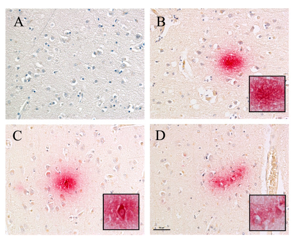
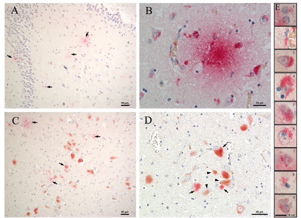
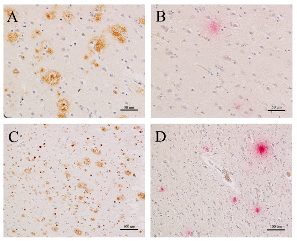
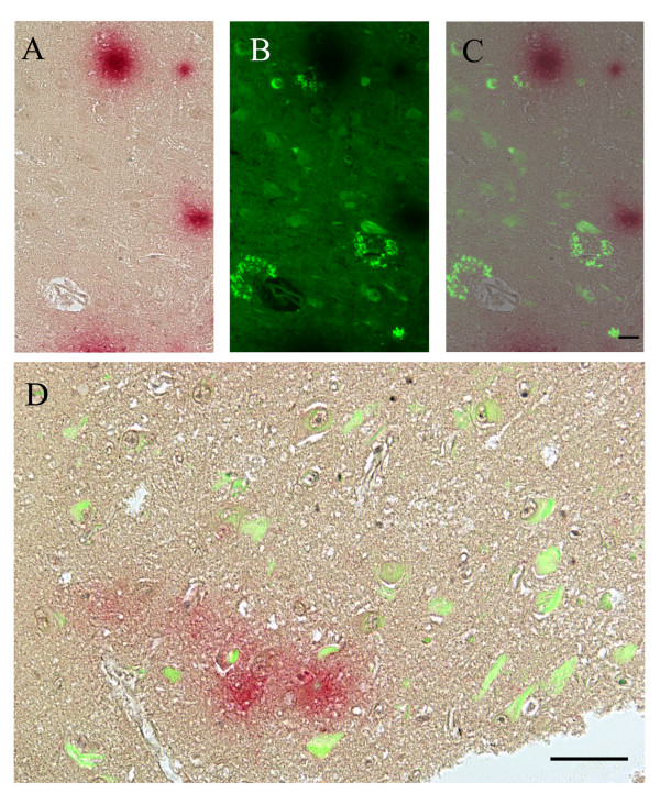
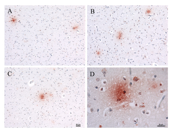
Background: Sporadic late-onset Alzheimer's disease (AD) appears to evolve from an interplay between genetic and environmental factors. One environmental factor that continues to be of great interest is that of Chlamydia pneumoniae infection and its association with late-onset disease. Detection of this organism in clinical and autopsy samples has proved challenging using a variety of molecular and histological techniques. Our current investigation utilized immunohistochemistry with a battery of commercially available anti-C. pneumoniae antibodies to determine whether C. pneumoniae was present in areas typically associated with AD neuropathology from 5 AD and 5 non-AD control brains. Results: Immunoreactivity for C. pneumoniae antigens was observed both intracellularly in neurons, neuroglia, endothelial cells, and peri-endothelial cells, and extracellularly in the frontal and temporal cortices of the AD brain with multiple C. pneumoniae-specific antibodies. This immunoreactivity was seen in regions of amyloid deposition as revealed by immunolabeling with two different anti-beta amyloid antibodies. Thioflavin S staining, overlaid with C. pneumoniae immunolabeling, demonstrated no direct co-localization of the organism and amyloid plaques. Further, the specificity of C. pneumoniae labeling of AD brain sections was demonstrated using C. pneumoniae antibodies pre-absorbed against amyloid β 1-40 and 1-42 peptides. Conclusions: Anti-C. pneumoniae antibodies, obtained commercially, identified both typical intracellular and atypical extracellular C. pneumoniae antigens in frontal and temporal cortices of the AD brain. C. pneumoniae, amyloid deposits, and neurofibrillary tangles were present in the same regions of the brain in apposition to one another. Although additional studies are required to conclusively characterize the nature of Chlamydial immunoreactivity in the AD brain, these results further implicate C. pneumoniae infection with the pathogenesis of Alzheimer's disease.
Overview
The claims and statistics of this paper haven’t been checked yet.
Run the check — free, no account needed. It reads the open-access text and checks the sources it cites.
Integrity signals
Registry
Cites
Statistics
Figures
Verification
Claims the paper makes
The claims and statistics of this paper haven’t been checked yet.
Run the check — free, no account needed. It reads the open-access text and checks the sources it cites.
Claims other papers make about this one
Each is checked against this paper’s own text: does it say what the citing paper says it does?
The source backs part of the claim, not all of it — or backs it for a different population, condition or comparison.
In the citing paper
“C. pneumoniae antigens have been detected inside both astrocytes and microglia in post-mortem human brains9,11,67,68.”
From Chlamydia pneumoniae can infect the central nervous system via the olfactory and trigeminal nerves and contributes to Alzheimer’s disease risk (2022) · cites this paper as [67]
Experimental — accuracy not yet measured
Self-check
Percentages, sums and reported differences re-computed from the paper’s own numbers.
The self-check needs the paper’s full text.
Citations
Cites
Mirra, S. S., Heyman, A., McKeel, D., Sumi, S. M., Crain, B. J., Brownlee, L. M., Vogel, F. S., Hughes, J. P., Belle, G. V., Berg, L., et al. (1991) · Neurology · doi:10.1212/wnl.41.4.479 · Cited by 4,961
Diffusible, nonfibrillar ligands derived from Aβ 1–42 are potent central nervous system neurotoxins
Lambert, M. P., Barlow, A. K., Chromy, B. A., Edwards, C., Freed, R., Liosatos, M., Morgan, T. E., Rozovsky, I., Trommer, B., Viola, K. L., et al. (1998) · Proceedings of the National Academy of Sciences · doi:10.1073/pnas.95.11.6448 · Cited by 3,506
Consortium to Establish a Registry for Alzheimer's Disease (CERAD)
Fillenbaum, G., & Heyman, A. (2002) · doi:10.1002/0470846410.ch42(iii · Cited by 2,859
The Alzheimer's Disease-Associated Amyloid β-Protein Is an Antimicrobial Peptide
Soscia, S. J., Kirby, J. E., Washicosky, K. J., Tucker, S. M., Ingelsson, M., Hyman, B., Burton, M. A., Goldstein, L. E., Duong, S., Tanzi, R. E., et al. (2010) · PLoS ONE · doi:10.1371/journal.pone.0009505 · Cited by 1,060
Classification and basic pathology of Alzheimer disease
Duyckaerts, C., Delatour, B., & Potier, M.-C. (2009) · Acta Neuropathologica · doi:10.1007/s00401-009-0532-1 · Cited by 1,031
Age, Neuropathology, and Dementia
Savva, G. M., Wharton, S. B., Ince, P. G., Forster, G., Matthews, F. E., & Brayne, C. (2009) · New England Journal of Medicine · doi:10.1056/nejmoa0806142 · Cited by 900
Epidemiology and Clinical Characteristics of Community-Acquired Pneumonia in Hospitalized Children
Michelow, I. C., Olsen, K. D., Lozano, J., Rollins, N. K., Duffy, L. B., Ziegler, T. T., Kauppila, J., Leinonen, M., & McCracken, G. H. (2004) · PEDIATRICS · doi:10.1542/peds.113.4.701 · Cited by 709
Takahashi, R. H., Milner, T. A., Li, F., Nam, E. E., Edgar, M. A., Yamaguchi, H., Beal, M. F., Xu, H., Greengard, P., & Gouras, G. K. (2002) · The American Journal of Pathology · doi:10.1016/s0002-9440(10)64463-x · Cited by 706
Herpes simplex virus type 1 in brain and risk of Alzheimer's disease
Itzhaki, R. F., Lin, W., Shang, D., Wilcock, G. K., Faragher, B. E., & Jamieson, G. A. (1997) · The Lancet · doi:10.1016/s0140-6736(96)10149-5 · Cited by 671
Rasmussen, S. J., Eckmann, L., Quayle, A. J., Shen, L., Zhang, Y., Anderson, D. J., Fierer, J., Stephens, R. S., & Kagnoff, M. F. (1997) · Journal of Clinical Investigation · doi:10.1172/jci119136 · Cited by 514
Cited by
Sorted by citations
Interactions between the microbiota, immune and nervous systems in health and disease
Fung, T. C., Olson, C. A., & Hsiao, E. Y. (2017) · Nature Neuroscience · doi:10.1038/nn.4476 · Cited by 1,900
Association between periodontal pathogens and systemic disease
Bui, F. Q., Almeida-da-Silva, C. L. C., Huynh, B. C., Trinh, A., Liu, J., Woodward, J., Asadi, H., & Ojcius, D. M. (2019) · Biomedical Journal · doi:10.1016/j.bj.2018.12.001 · Cited by 882
Brain-Gut-Microbiota Axis in Alzheimer’s Disease
Kowalski, K., & Mulak, A. (2019) · Journal of Neurogastroenterology and Motility · doi:10.5056/jnm18087 · Cited by 826
Poole, S., Singhrao, S. K., Kesavalu, L. N., Curtis, M. A., & Crean, S. (2013) · Journal of Alzheimer s Disease · doi:10.3233/jad-121918 · Cited by 610
Role of gut-brain axis, gut microbial composition, and probiotic intervention in Alzheimer's disease
Kesika, P., Suganthy, N., Sivamaruthi, B. S., & Chaiyasut, C. (2021) · Life Sciences · doi:10.1016/j.lfs.2020.118627 · Cited by 479
Metadata
- Authors
- Hammond, C. J., Hallock, L. R., Howanski, R. J., Appelt, D. M., Little, C. S., & Balin, B. J.
- Affiliations
- Philadelphia College of Osteopathic Medicine (US)
- Corresponding author
- Balin, Brian J
- Venue
- BMC Neuroscience
- Published
- 2010-09-23
- Volume
- 11
- Issue
- 1
- Pages
- 121-121
- ISSN
- 1471-2202 (ISSN-L)
- PMC
- PMC2949767
- OpenAlex
- W2056188402
- Cited by
- 156
- References
- 49
- Citations by year
- 2026: 5 · 2025: 11 · 2024: 6 · 2023: 4 · 2022: 21 · 2021: 12 · 2020: 17 · 2019: 11 · 2018: 9 · 2017: 8 · 2016: 12 · 2015: 7 · 2014: 11 · 2013: 11 · 2012: 4
- FWCI
- 4.76
- Type
- journal article
- Language
- en
- Open access
- gold
- doi.org
- Licence
- cc-by
- Locations
- BMC Neuroscience (publishedVersion, cc-by)
- PubMed Central (publishedVersion, cc-by)
- PubMed (publishedVersion)
- Digital Commons - PCOM (Philadelphia College of Osteopathic Medicine) (submittedVersion, cc-by)
- DOAJ (DOAJ: Directory of Open Access Journals) (submittedVersion, cc-by-sa)
- Funders
- Drexel University
- Topics
- Life Sciences › Neuroscience › Cellular and Molecular Neuroscience › Alzheimer's disease research and treatments
- Life Sciences › Neuroscience › Neurology › Neuroinflammation and Neurodegeneration Mechanisms
- Health Sciences › Medicine › Psychiatry and Mental health › Dementia and Cognitive Impairment Research
- Keywords
- Chlamydia pneumoniae, Alzheimer's disease, late-onset Alzheimer's disease, chlamydia, amyloid plaques, amyloid-beta, neurofibrillary tangles, frontal and temporal cortex, antibody specificity, neuroinflammation, Alzheimer's disease pathogenesis
- MeSH terms
- Aged, Alzheimer Disease, Brain, Brain Chemistry, Cerebral Cortex, Chlamydia Infections, Coloring Agents, Female, Humans, Immunohistochemistry, Male, Thiazoles, Tissue Banks, Reproducibility of Results, Amyloid beta-Peptides, Neurofibrillary Tangles, Chlamydophila pneumoniae, Benzothiazoles, Plaque, Amyloid
About these checks
Registry, retraction, Cites and statistics results are computed, not judged, with one exception: a printed reference that no index lists is read once by a model before it counts as a citation error. Claims are extracted by a model and kept only when their quote appears verbatim in the text; each cited source is then checked, with a model’s help, for whether it says what the claim relies on. Those verdicts can be wrong — read the claim’s quote and the cited source before relying on one.
Statistics and arithmetic are recomputed, not judged.
Confidence bands are shown only for fields calibrated on our annotated test set. None is calibrated yet, so no band is shown.
What counts as a claim ›How we measure confidence ›How we check this ›
Are you an author, or do you see a problem with this page? Request a review
