

The DOI matches its Crossref record; 43 of the 43 references it cites were found in a registry or an index.
Identification of the collagen type 1 alpha 1 gene (COL1A1) as a candidate survival-related factor associated with hepatocellular carcinoma
Open accessAbstract
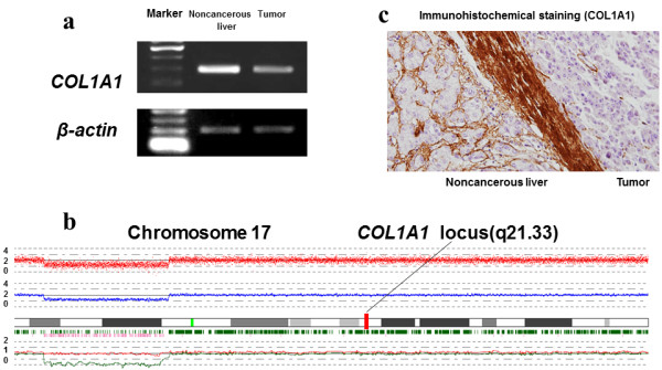
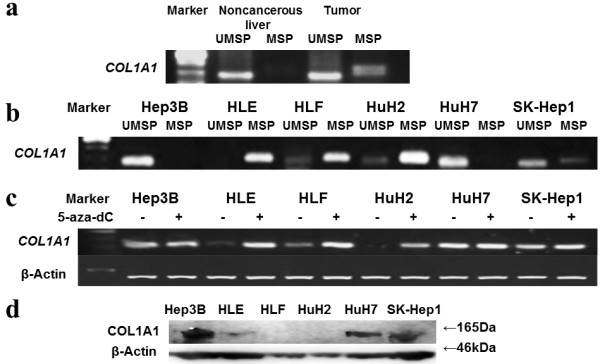
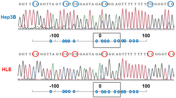
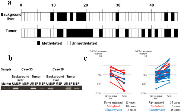
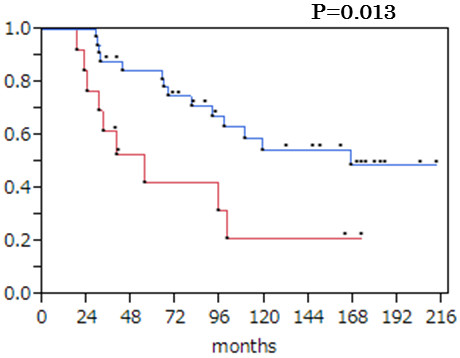
BACKGROUND: Hepatocellular carcinoma (HCC) is one of the major causes of cancer-related death especially among Asian and African populations. It is urgent that we identify carcinogenesis-related genes to establish an innovative treatment strategy for this disease. METHODS: Triple-combination array analysis was performed using one pair each of HCC and noncancerous liver samples from a 68-year-old woman. This analysis consists of expression array, single nucleotide polymorphism array and methylation array. The gene encoding collagen type 1 alpha 1 (COL1A1) was identified and verified using HCC cell lines and 48 tissues from patients with primary HCC. RESULTS: Expression array revealed that COL1A1 gene expression was markedly decreased in tumor tissues (log2 ratio -1.1). The single nucleotide polymorphism array showed no chromosomal deletion in the locus of COL1A1. Importantly, the methylation value in the tumor tissue was higher (0.557) than that of the adjacent liver tissue (0.008). We verified that expression of this gene was suppressed by promoter methylation. Reactivation of COL1A1 expression by 5-aza-2'-deoxycytidine treatment was seen in HCC cell lines, and sequence analysis identified methylated CpG sites in the COL1A1 promoter region. Among 48 pairs of surgical specimens, 13 (27.1%) showed decreased COL1A1 mRNA expression in tumor sites. Among these 13 cases, 10 had promoter methylation at the tumor site. The log-rank test indicated that mRNA down-regulated tumors were significantly correlated with a poor overall survival rate (P = 0.013). CONCLUSIONS: Triple-combination array analysis successfully identified COL1A1 as a candidate survival-related gene in HCCs. Epigenetic down-regulation of COL1A1 mRNA expression might have a role as a prognostic biomarker of HCC.
Overview
The claims and statistics of this paper haven’t been checked yet.
Run the check — free, no account needed. It reads the open-access text and checks the sources it cites.
Integrity signals
Registry
Cites
Statistics
Figures
Verification
Claims the paper makes
The claims and statistics of this paper haven’t been checked yet.
Run the check — free, no account needed. It reads the open-access text and checks the sources it cites.
Claims other papers make about this one
Each is checked against this paper’s own text: does it say what the citing paper says it does?
In the citing paper
“In three breast cancer tissues, H&E regions corresponding to tumor tissue showed high similarity with the bulk RNA-seq data from tumor biopsies, which include tumor-related markers such as COL1A1 (ref. 38) and ACTB39 (Fig. 3b and Supplementary Fig. 5).”
From A visual–omics foundation model to bridge histopathology with spatial transcriptomics (2025) · cites this paper as [38]
Self-check
Percentages, sums and reported differences re-computed from the paper’s own numbers.
The self-check needs the paper’s full text.
Citations
Cites
Jemal, A., Bray, F., Ferlay, J., Ward, E., & Forman, D. (2011) · CA A Cancer Journal for Clinicians · doi:10.3322/caac.20107 · Cited by 55,077
El-Serag, H. B. (2011) · New England Journal of Medicine · doi:10.1056/nejmra1001683 · Cited by 3,529
Decitabine improves patient outcomes in myelodysplastic syndromes
Kantarjian, H. M., Issa, J. J., Rosenfeld, C. S., Bennett, J. M., Albitar, M. X., DiPersio, J. F., Klimek, V. M., Slack, J. L., de Castro, C., Ravandi, F., et al. (2006) · Cancer · doi:10.1002/cncr.21792 · Cited by 1,539
Hotary, K. B., Allen, E. D., Brooks, P. C., Datta, N. S., Long, M. W., & Weiss, S. J. (2003) · Cell · doi:10.1016/s0092-8674(03)00513-0 · Cited by 674
Over- and under-expressed microRNAs in human colorectal cancer
Mori (2009) · International Journal of Oncology · doi:10.3892/ijo_00000233 · Cited by 349
Huang, J., Wang, Y., Guo, Y., & Sun, S. (2010) · Hepatology · doi:10.1002/hep.23660 · Cited by 345
Zeller, C., Dai, W., Steele, N. L., Siddiq, A., Walley, A. J., Wilhelm-Benartzi, C. S., Rizzo, S., van der Zee, A. G. J., Plumb, J. A., & Brown, R. E. (2012) · Oncogene · doi:10.1038/onc.2011.611 · Cited by 286
Genome-wide screen of promoter methylation identifies novel markers in melanomaPreprint
Koga, Y., Pelizzola, M., Cheng, E., Krauthammer, M. O., Sznol, M., Ariyan, S., Narayan, D., Molinaro, A. M., Halaban, R., & Weissman, S. M. (2009) · Genome Research · doi:10.1101/gr.091447.109 · Cited by 197
Ishigami, K., Yoshimitsu, K., Nishihara, Y., Irié, H., Asayama, Y., Tajima, T., Nishie, A., Hirakawa, M., Ushijima, Y., Okamoto, D., et al. (2008) · Radiology · doi:10.1148/radiol.2501071702 · Cited by 188
Identification of Novel Target Genes by an Epigenetic Reactivation Screen of Renal Cancer
de Cáceres, I. I., Dulaimi, E., Hoffman, A. M., Al‐Saleem, T., Uzzo, R. G., & Cairns, P. (2006) · Cancer Research · doi:10.1158/0008-5472.can-05-3365 · Cited by 154
Research works printed but not linked: 1
- Saito Y, Hibino S, Saito H: Alterations of epigenetics and microRNA in hepatocellular carcinoma. Hepatol Res. in press
Cited by
Sorted by citations
Ma, H., Chang, H., Bamodu, O. A., Yadav, V. K., Huang, T., Wu, A. T. H., Yeh, C., Tsai, S. H., & Lee, W. (2019) · Cancers · doi:10.3390/cancers11060786 · Cited by 212
Identification of COL1A1 and COL1A2 as candidate prognostic factors in gastric cancer
Li, J., Ding, Y., & Li, A. (2016) · World Journal of Surgical Oncology · doi:10.1186/s12957-016-1056-5 · Cited by 199
MiR-129-5p suppresses gastric cancer cell invasion and proliferation by inhibiting COL1A1
Wang, Q., & Yu, J. (2017) · Biochemistry and Cell Biology · doi:10.1139/bcb-2016-0254 · Cited by 127
A visual–omics foundation model to bridge histopathology with spatial transcriptomics
Chen, W., Zhang, P., Tran, T. N., Xiao, Y., Li, S., Shah, V. V., Cheng, H., Brannan, K. W., Youker, K., Lai, L., et al. (2025) · Nature Methods · doi:10.1038/s41592-025-02707-1 · Cited by 118
“38) and ACTB 39 (Fig.”
He, B., Lin, X., Tian, F., Yu, W., & Qiao, B. (2017) · Journal of Cellular Biochemistry · doi:10.1002/jcb.26182 · Cited by 116
Hua, X., Wang, Y., Jia, P., Xiong, Q., Hu, Y., Chang, Y., Lai, S., Xu, Y. J., Zhao, Z., & Song, J. (2020) · BMC Medicine · doi:10.1186/s12916-019-1469-4 · Cited by 114
Aspects of the Epigenetic Regulation of EMT Related to Cancer Metastasis
Nowak, E., & Bednarek, I. A. (2021) · Cells · doi:10.3390/cells10123435 · Cited by 110
Angel, P. M., Comte‐Walters, S., Ball, L. E., Talbot, K. M., Mehta, A. S., Brockbank, K. G. M., & Drake, R. R. (2017) · Journal of Proteome Research · doi:10.1021/acs.jproteome.7b00713 · Cited by 104
Jiang, K., Liu, H., Xie, D., & Xiao, Q. (2019) · Oncology Letters · doi:10.3892/ol.2019.9952 · Cited by 87
Thangavelu, P. U., Krenács, T., Dray, E. V., & Duijf, P. H. G. (2016) · Clinical Epigenetics · doi:10.1186/s13148-016-0290-6 · Cited by 80
Zhang, R., Ma, M., Lin, X.-H., Liu, H.-H., Chen, J., Chen, J., Gao, D.-M., Cui, J.-F., Ren, Z.-G., & Chen, R.-X. (2018) · BMC Cancer · doi:10.1186/s12885-018-4820-9 · Cited by 68
Regulatory effects of COL1A1 on apoptosis induced by radiation in cervical cancer cells
Liu, S., Liao, G., & Li, G. (2017) · Cancer Cell International · doi:10.1186/s12935-017-0443-5 · Cited by 68
Chen, C., Chung, T., Wu, C., Ng, K., Yu, J., Tsai, C.-H., Chang, Y., Liang, Y. J., Tsui, K., & Chen, Y. (2015) · Molecular & Cellular Proteomics · doi:10.1074/mcp.m115.051524 · Cited by 64
Filliol, A. A., & Schwabe, R. F. (2019) · Seminars in Liver Disease · doi:10.1055/s-0039-1685539 · Cited by 60
Yin, Y., Du, L., Li, X., Zhang, X., & Gao, Y. (2018) · Journal of Cellular Physiology · doi:10.1002/jcp.27896 · Cited by 56
Manjubaashini, N., Purushothaman, B., & Balakumar, S. (2024) · Journal of Photochemistry and Photobiology A Chemistry · doi:10.1016/j.jphotochem.2024.115702 · Cited by 52
Mann, S. N., Hadad, N., Holte, M. N., Rothman, A. R., Sathiaseelan, R., Mondal, S. A., Agbaga, M., Unnikrishnan, A., Subramaniam, M., Hawse, J. R., et al. (2020) · eLife · doi:10.7554/elife.59616 · Cited by 52
Benlebna, M., Balas, L., Bonafos, B., Pessemesse, L., Vigor, C., Grober, J., Bernex, F., Fouret, G., Paluchová, V., Gaillet, S., et al. (2020) · The Journal of Nutritional Biochemistry · doi:10.1016/j.jnutbio.2020.108361 · Cited by 49
Mitochondrial DNA plasticity is an essential inducer of tumorigenesis
Lee, W., Cain, J. E., Cuddihy, A. R., Johnson, J., Dickinson, A., Yeung, K.-Y., Kumar, B., Johns, T. G., Watkins, D. N., Spencer, A. G., et al. (2016) · Cell Death Discovery · doi:10.1038/cddiscovery.2016.16 · Cited by 40
Identification of hub genes and outcome in colon cancer based on bioinformatics analysis
Yang, W., Ma, J., Zhou, W. J., Li, Z., Zhou, X., Cao, B., Zhang, Y., Liu, J., Yang, Z., Zhang, H., et al. (2018) · Cancer Management and Research · doi:10.2147/cmar.s173240 · Cited by 38
Atia, M. M., Abdel-Tawab, H. S., Mostafa, A., & Mobarak, S. A. (2022) · Scientific Reports · doi:10.1038/s41598-022-12406-y · Cited by 37
miR-133b Suppresses Invasion and Migration of Gastric Cancer Cells via the COL1A1/TGF-β Axis
Guo, Y., Lu, G., Mao, H., Zhou, S., Tong, X., Wu, J., Sun, Q., Xu, H., & Fang, F. (2020) · OncoTargets and Therapy · doi:10.2147/ott.s249667 · Cited by 36
Wu, W., Zhu, Y., Long, F., Luo, L., Deng, Q., Wu, J.-L., Ouyang, S., & Tang, D. (2019) · Bioorganic Chemistry · doi:10.1016/j.bioorg.2019.103457 · Cited by 34
Lv, J., Guo, L., Wang, J.-H., Yan, Y., Zhang, J., Wang, Y., Yu, Y. P., Huang, Y., & Zhao, H. (2019) · World Journal of Gastroenterology · doi:10.3748/wjg.v25.i2.233 · Cited by 33
High expression of collagen 1A2 promotes the proliferation and metastasis of esophageal cancer cells
Li, G., Jiang, W., Kang, Y., Yu, X., Zhang, C., & Yu, F. (2020) · Annals of Translational Medicine · doi:10.21037/atm-20-7867 · Cited by 33
Min, S., Lee, Y., Hong, J., Park, T. J., Woo, H. G., Kwon, S. M., & Yoon, G. (2021) · Cell Death and Disease · doi:10.1038/s41419-021-04370-8 · Cited by 34
Yang, W., Zhao, X., Han, Y., Duan, L., Lü, X., Wang, X., Zhang, Y., Zhou, W. J., Liu, J., Zhang, H., et al. (2019) · Cancer Cell International · doi:10.1186/s12935-019-0854-6 · Cited by 31
Bibaki, E., Tsitoura, E., Vasarmidi, E., Margaritopoulos, G., Trachalaki, A., Koutoulaki, C., Georgopoulou, T., Spandidos, D., Tzanakis, N., & Antoniou, K. (2018) · Molecular Medicine Reports · doi:10.3892/mmr.2018.8778 · Cited by 29
Zarzour, R. A., Alshawsh, M. A., Asif, M., Al‐Mansoub, M. A., Mohamed, Z., Ahmad, M., Majid, A. M. S. A., Asmawi, M. Z., Kaur, G., Al-dualimi, D. W., et al. (2018) · Nutrients · doi:10.3390/nu10081057 · Cited by 28
Influence of surface topography on PCL electrospun scaffolds for liver tissue engineering
Gao, Y., & Callanan, A. (2021) · Journal of Materials Chemistry B · doi:10.1039/d1tb00789k · Cited by 29
Shen, L., Xiao, Y., Wu, Q., Liu, L., Zhang, C., & Pan, X. (2019) · Europe PMC (PubMed Central) · Cited by 25
Song, Y., Wang, X., Wang, F., Peng, X., Li, P., Liu, S., & Zhang, D. (2021) · Cancer Cell International · doi:10.1186/s12935-021-01814-1 · Cited by 23
Nondegradable Collagen Increases Liver Fibrosis but Not Hepatocellular Carcinoma in Mice
Baglieri, J., Zhang, C., Liang, S., Liu, X., Nishio, T., Rosenthal, S. B., Dhar, D., Su, H., Cong, M., Jia, J., et al. (2021) · American Journal Of Pathology · doi:10.1016/j.ajpath.2021.05.019 · Cited by 23
Identity of MMP1 and its effects on tumor progression in head and neck squamous cell carcinoma
Zhang, G., Li, T., Tan, G., Song, Y., Liu, Q., Wang, K., Ai, J., Zhou, Z. Y., & Li, W. (2022) · Cancer Medicine · doi:10.1002/cam4.4623 · Cited by 23
Zhao, W. G., Jiang, X., & Yang, S. (2020) · Cancer Management and Research · doi:10.2147/cmar.s256624 · Cited by 23
Hepatotoxicity assessment of Rhizoma Paridis in adult zebrafish through proteomes and metabolome
Jia, Z., Zhao, C., Wang, M., Zhao, X., Zhang, W., Han, T., Xia, Q., Han, Z., Lin, R., & Li, X.-R. (2019) · Biomedicine & Pharmacotherapy · doi:10.1016/j.biopha.2019.109558 · Cited by 21
Identification of COL1A1 associated with immune infiltration in brain lower grade glioma
Ren, J., Da, J., & Hu, N. (2022) · PLoS ONE · doi:10.1371/journal.pone.0269533 · Cited by 17
MALDI-MSI: A Powerful Approach to Understand Primary Pancreatic Ductal Adenocarcinoma and Metastases
Gonçalves, J. P. L., Bollwein, C., Schlitter, A. M., Kriegsmann, M., Jacob, A., Weichert, W., & Schwamborn, K. (2022) · Molecules · doi:10.3390/molecules27154811 · Cited by 18
Inokawa, Y., Inaoka, K., Sonohara, F., Hayashi, M., Kanda, M., & Nomoto, S. (2016) · Oncology Letters · doi:10.3892/ol.2016.5141 · Cited by 16
Self-citationIdentification of six candidate genes for endometrial carcinoma by bioinformatics analysis
Zhu, Y., Shi, L., Chen, P., Zhang, Y., & Zhu, T. (2020) · World Journal of Surgical Oncology · doi:10.1186/s12957-020-01920-w · Cited by 15
Panneerselvam, J., Mohamed, J. M. M., Annamalai, D., Al‐Serwi, R. H., Ibrahim, A. M., El‐Sherbiny, M., Rajam, A. M., Eldesoqui, M. B., & Mansour, N. (2023) · International Journal of Biological Macromolecules · doi:10.1016/j.ijbiomac.2023.125948 · Cited by 15
Igreja, I. C., Tripská, K., Hroch, M., Hyšpler, R., Tichá, A., Laštůvková, H., Schreiberová, J., Doleželová, E., Eissazadeh, S., Vitverová, B., et al. (2020) · International Journal of Molecular Sciences · doi:10.3390/ijms21239021 · Cited by 14
Search for useful biomarkers in hepatocellular carcinoma, tumor factors and background liver factors
Shimizu, D., Inokawa, Y., Sonohara, F., Inaoka, K., & Nomoto, S. (2017) · Oncology Reports · doi:10.3892/or.2017.5541 · Cited by 13
Self-citationHansdah, K., Singh, N., Bouzid, A., Priyadarshi, S., Ray, C. S., Desai, A., Panda, K. C., Choudhury, J. C., Biswal, N. C., Tekari, A., et al. (2020) · Genetic Testing and Molecular Biomarkers · doi:10.1089/gtmb.2019.0235 · Cited by 12
Zhang, R., Zhang, L., Yang, M., Huang, L., Chen, G., & Feng, Z. (2017) · Molecular Medicine Reports · doi:10.3892/mmr.2017.8167 · Cited by 12
Patil, A., Salvatori, R., Smith, L., Jenkins, S. M., Cannon, A. C., Hartley, C., Graham, R. P., & Moreira, R. K. (2023) · Histopathology · doi:10.1111/his.15001 · Cited by 12
Ding, B., Ye, Z., Yin, H., Hong, X., Feng, S., Xu, J., & Shen, Y. (2023) · Genomics · doi:10.1016/j.ygeno.2023.110703 · Cited by 11
Genomic-Wide Analysis with Microarrays in Human Oncology
Inaoka, K., Inokawa, Y., & Nomoto, S. (2015) · Microarrays · doi:10.3390/microarrays4040454 · Cited by 11
Self-citationSun, W., Zhang, Y., Liu, B., Duan, Y., Li, W., & Chen, J. (2021) · Canadian Journal of Gastroenterology and Hepatology · doi:10.1155/2021/9990305 · Cited by 12
Hishida, M., Inokawa, Y., Takano, N., Nishikawa, Y., Iwata, N., Kanda, M., Tanaka, C., Kobayashi, D., Yamada, S., Nakayama, G., et al. (2015) · Journal of Surgical Research · doi:10.1016/j.jss.2014.12.045 · Cited by 11
Self-citationIdentifying hepatocellular carcinoma-related genes and pathways by system biology analysis
Wang, P., Ouyang, L., Zheng, L., & Wang, Z. (2014) · Irish Journal of Medical Science (1971 -) · doi:10.1007/s11845-014-1119-y · Cited by 10
Jiang, A., Liu, J.-T., Zhao, K., Wu, H., Wang, S., Yan, K., & Yang, W. (2020) · Frontiers in Oncology · doi:10.3389/fonc.2020.561805 · Cited by 10
Xu, J., Ma, J., Shi, Y., Yin, D., Zhang, Y., Dai, P., Zhao, W., & Zhang, T. (2020) · PROTEOMICS - CLINICAL APPLICATIONS · doi:10.1002/prca.201900112 · Cited by 10
Tandem mass tag‐based quantitative proteomic analysis of cervical cancer
Aljawad, M. F., Faisal, A. H. M. A., Alqanbar, M. F., Wilmarth, P. A., & Hassan, B. Q. (2022) · PROTEOMICS - CLINICAL APPLICATIONS · doi:10.1002/prca.202100105 · Cited by 10
Proteomic Signatures of Thymomas
Wang, L., Branson, O. E., Shilo, K., Hitchcock, C. L., & Freitas, M. A. (2016) · PLoS ONE · doi:10.1371/journal.pone.0166494 · Cited by 12
Propolis compound inhibits profibrotic TGF-β1/SMAD signalling in human fibroblasts
Randall, L. J., Bajan, S., Tran, T. D., Harvey, R. J., & Russell, F. D. (2025) · Scientific Reports · doi:10.1038/s41598-025-07918-2 · Cited by 9
Macdonald, J. K., Taylor, H. B., Wang, M., DelaCourt, A. T., Edge, C., Lewin, D., Kubota, N., Fujiwara, N., Rasha, F. A., Marquez, C. A., et al. (2024) · Journal of Proteome Research · doi:10.1021/acs.jproteome.4c00099 · Cited by 9
Zhu, M., Gong, J., Zhan, M., Xi, C., Shen, G., & Shen, H. (2021) · Comparative Biochemistry and Physiology Part D Genomics and Proteomics · doi:10.1016/j.cbd.2021.100916 · Cited by 9
Castro‐Gil, M. P., Torres‐Mena, J. E., Salgado, R. M., Muñoz-Montero, S. A., Martínez-Garcés, J. M., López‐Torres, C. D., Mendoza-Vargas, A., Gabiño‐López, N. B., Villa‐Treviño, S., del Pozo‐Yauner, L., et al. (2021) · Genomics · doi:10.1016/j.ygeno.2021.11.035 · Cited by 9
Exploring the key genes and pathways in enchondromas using a gene expression microarray
Shi, Z., Zhou, H., Pan, B., Lu, L., Kang, Y., Liu, L., Wei, Z., & Feng, S. (2017) · Oncotarget · doi:10.18632/oncotarget.16700 · Cited by 9
Bioinformatic and experimental analyses of key biomarkers in pancreatic cancer
Ren, T., Xue, X., Wang, X., Zhou, X., & Dang, S.-C. (2021) · Experimental and Therapeutic Medicine · doi:10.3892/etm.2021.10794 · Cited by 8
Quah, Y., Chan, J. Y.-L., Park, N., Lee, Y. Y., Lee, E., Jang, S.-H., Kim, M.-J., Rhee, M. H., Lee, S., & Park, S.-C. (2022) · Chinese Medicine · doi:10.1186/s13020-022-00622-7 · Cited by 8
Integrated miRNA-mRNA analysis of Epstein-Barr virus-positive nasopharyngeal carcinoma
Zhu, L., Miao, X. T., & Wang, N. (2015) · Genetics and Molecular Research · doi:10.4238/2015.june.1.20 · Cited by 8
Identification of a Combined RNA Prognostic Signature in Adenocarcinoma of the Lung
He, S.-Y., Xi, W., Wang, X., Xu, C., Cheng, L., Liu, S.-Y., Meng, Q., Li, B., Wang, Y., Shi, H., et al. (2019) · Medical Science Monitor · doi:10.12659/msm.913727 · Cited by 7
Yang, W., Pan, Y., Guan, P., Li, X., & You, C. (2019) · Europe PMC (PubMed Central) · doi:10.12122/j.issn.1673-4254.2019.05.07 · Cited by 7
ZIC5 promotes human hepatocellular carcinoma cell proliferation through upregulating COL1A1
Song, W., Yu, W., Ding, L., Cheng, C., Wu, X., Chen, J., & Zhang, W. (2022) · Journal of Gastrointestinal Oncology · doi:10.21037/jgo-22-335 · Cited by 7
A prognostic model based on the COL1A1-network in gastric cancer.
Liu, S., Chen, L., Zeng, J., & Chen, Y. (2023) · PubMed Central · Cited by 7
Hansen, R. L., & Lange, J. (2013) · PubMed · Cited by 5
Mao, M., Li, Z., Zheng, W., Xia, H., Fei, Z., Yao, Q., & Zheng, R. (2025) · Journal of Neuro-Oncology · doi:10.1007/s11060-025-05132-8 · Cited by 4
Guo, Y., Wang, X., Ning, W., Zhang, H., & Yu, C. (2020) · Molecular Biology Reports · doi:10.1007/s11033-020-05804-w · Cited by 4
Hu, Y., Hu, Z., Ding, H., Li, Y., Zhao, X., Shao, T. M., & Pan, Y. (2020) · Translational Cancer Research · doi:10.21037/tcr-20-926 · Cited by 4
Wihastyoko, H. Y. L., Soeharto, S., Widjajanto, E., Handono, K., & Pardjianto, B. (2021) · Research Journal of Pharmacy and Technology · doi:10.52711/0974-360x.2021.00862 · Cited by 4
Mohamed, A. A., Abo-Amer, Y. E.-E., Aalkhalegy, A., Fathalla, L. A., Elmaghraby, M. B., Elhoseeny, M. M., Mostafa, S. M., El-Abgeegy, M., Khattab, R. A., El-damasy, D. A., et al. (2021) · The Open Biomarkers Journal · doi:10.2174/1875318302111010108 · Cited by 3
Sztolsztener, K., Konstantynowicz-Nowicka, K., Pędzińska‐Betiuk, A., & Chabowski, A. (2023) · Cells · doi:10.3390/cells12182243 · Cited by 3
Alsahli, T. G., Khalid, M., Malik, M. N. H., & Alshammari, S. O. (2025) · Medicina · doi:10.3390/medicina61061048 · Cited by 2
Molecular Profiling of Human Hepatocellular Carcinoma
Budhu, A., & Wang, X.-W. (2016) · Springer eBooks · doi:10.1007/978-3-319-34214-6_5 · Cited by 1
Yang, Y., Dai, E., Wang, S., & Bai, Y. (2020) · DNA and Cell Biology · doi:10.1089/dna.2019.5320 · Cited by 2
The P2X7 Receptor Promotes Intestinal Fibrosis by Modulating the Gut Microbiota and the Inflammasome
Ribeiro, B. E., de Lima, I. S., Souza, K. C. D. S. E., Rosas, S. L. B., Santana, P. T., Amaral, G. R. S., Machado, J. C., de Oliveira, R. P., Leal, C. V., Thompson, C. C., et al. (2025) · Cellular and Molecular Gastroenterology and Hepatology · doi:10.1016/j.jcmgh.2025.101718 · Cited by 1
Li, X., Wang, Z., Xie, J., & Zhu, N. (2021) · Research Square · doi:10.21203/rs.3.rs-386986/v1
Wagle, M. M., Rao, A. K., Kabekkodu, S. P., & Mallya, S. P. (2021) · Research Square · doi:10.21203/rs.3.rs-879048/v1
Wagle, M. M., Rao, A. K., Kabekkodu, S. P., & Mallya, S. P. (2022) · Research Square · doi:10.21203/rs.3.rs-879048/v2
Causin, R. L., da Silva, L. S., Leal, L. F., Resende, J. C. P., Evangelista, A. F., Matsushita, G. M., Scapulatempo‐Neto, C., Fregnani, J. H. T. G., de Oliveira, M. A., Musselwhite, L. W., et al. (2023) · Cancer Cytopathology · doi:10.1002/cncy.22704
Wagle, M. M., Rao, A. K., Kabekkodu, S. P., & Mallya, S. P. (2023) · Asian Pacific Journal of Cancer Prevention · doi:10.31557/apjcp.2023.24.5.1601
Ghosh, S., Datta, R., & Thakur, S. (2024) · Human Gene · doi:10.1016/j.humgen.2024.201357
Effectiveness of a Novel CTGF LNA GapmeR Sequence in Gastric Cancer Cells
Bilecen, D. S. (2024) · Süleyman Demirel Üniversitesi Fen Edebiyat Fakültesi Fen Dergisi · doi:10.29233/sdufeffd.1395816
Boonto, T., & Ariyachet, C. (2025) · World Journal of Hepatology · doi:10.4254/wjh.v17.i7.106795
Song, Y., Wang, X., Wang, F., Peng, X., Li, P., Liu, S., & Zhang, D. (2021) · Figshare · doi:10.6084/m9.figshare.14059482
Song, Y., Wang, X., Wang, F., Peng, X., Li, P., Liu, S., & Zhang, D. (2021) · Figshare · doi:10.6084/m9.figshare.14059476
Quah, Y., Chan, J. Y.-L., Park, N.-H., Lee, Y. Y., Lee, E.-B., Jang, S.-H., Kim, M.-J., Rhee, M. H., Lee, S.-J., & Park, S.-C. (2022) · Figshare · doi:10.6084/m9.figshare.20070891
Quah, Y., Chan, J. Y.-L., Park, N.-H., Lee, Y. Y., Lee, E.-B., Jang, S.-H., Kim, M.-J., Rhee, M. H., Lee, S.-J., & Park, S.-C. (2022) · Figshare · doi:10.6084/m9.figshare.20070903
Quah, Y., Chan, J. Y.-L., Park, N.-H., Lee, Y. Y., Lee, E.-B., Jang, S.-H., Kim, M.-J., Rhee, M. H., Lee, S.-J., & Park, S.-C. (2022) · Figshare · doi:10.6084/m9.figshare.20070894
Quah, Y., Chan, J. Y.-L., Park, N.-H., Lee, Y. Y., Lee, E.-B., Jang, S.-H., Kim, M.-J., Rhee, M. H., Lee, S.-J., & Park, S.-C. (2022) · Figshare · doi:10.6084/m9.figshare.20070876
Quah, Y., Chan, J. Y.-L., Park, N.-H., Lee, Y. Y., Lee, E.-B., Jang, S.-H., Kim, M.-J., Rhee, M. H., Lee, S.-J., & Park, S.-C. (2022) · Figshare · doi:10.6084/m9.figshare.20070888
Quah, Y., Chan, J. Y.-L., Park, N.-H., Lee, Y. Y., Lee, E.-B., Jang, S.-H., Kim, M.-J., Rhee, M. H., Lee, S.-J., & Park, S.-C. (2022) · Figshare · doi:10.6084/m9.figshare.20070885
Quah, Y., Chan, J. Y.-L., Park, N.-H., Lee, Y. Y., Lee, E.-B., Jang, S.-H., Kim, M.-J., Rhee, M. H., Lee, S.-J., & Park, S.-C. (2022) · Figshare · doi:10.6084/m9.figshare.20070882
Quah, Y., Chan, J. Y.-L., Park, N.-H., Lee, Y. Y., Lee, E.-B., Jang, S.-H., Kim, M.-J., Rhee, M. H., Lee, S.-J., & Park, S.-C. (2022) · Figshare · doi:10.6084/m9.figshare.20070867
Song, Y., Wang, X., Wang, F., Peng, X., Li, P., Liu, S., & Zhang, D. (2021) · Figshare · doi:10.6084/m9.figshare.14059479
Quah, Y., Chan, J. Y.-L., Park, N.-H., Lee, Y. Y., Lee, E.-B., Jang, S.-H., Kim, M.-J., Rhee, M. H., Lee, S.-J., & Park, S.-C. (2022) · Figshare · doi:10.6084/m9.figshare.20070870
EVALUATION OF THE EFFECTS OF SULFAMETHOXAZOLE ON ENDOTHELIAL AND FIBROBLAST CELLS
Şen, M. B., Kaçaroğlu, D., & Erikçi, A. (2026) · Ankara Universitesi Eczacilik Fakultesi Dergisi · doi:10.33483/jfpau.1661024
Metadata
- Authors
- Hayashi, M., Nomoto, S., Hishida, M., Inokawa, Y., Kanda, M., Okamura, Y., Nishikawa, Y., Tanaka, C., Kobayashi, D., Yamada, S., Nakayama, G., Fujii, T., Sugimoto, H., Koike, M., Fujiwara, M., Takeda, S., & Kodera, Y.
- Affiliations
- Nagoya University (JP)
- Corresponding author
- Nomoto, Shuji
- ORCID
- Venue
- BMC Cancer
- Published
- 2014-02-19
- Volume
- 14
- Issue
- 1
- Pages
- 108-108
- ISSN
- 1471-2407 (ISSN-L)
- PMC
- PMC4015503
- OpenAlex
- W2152909439
- Cited by
- 101
- References
- 43
- Citations by year
- 2026: 2 · 2025: 6 · 2024: 4 · 2023: 7 · 2022: 17 · 2021: 16 · 2020: 14 · 2019: 10 · 2018: 5 · 2017: 7 · 2016: 7 · 2015: 4 · 2014: 1
- FWCI
- 2.60
- Type
- journal article
- Language
- en
- Open access
- gold
- doi.org
- Licence
- Locations
- BMC Cancer (publishedVersion, cc-by)
- Europe PMC (PubMed Central) (publishedVersion, cc-by)
- PubMed Central (publishedVersion, cc-by)
- PubMed (publishedVersion)
- Funders
- Japan Society for the Promotion of Science (22591427)
- Topics
- Life Sciences › Biochemistry, Genetics and Molecular Biology › Molecular Biology › Epigenetics and DNA Methylation
- Health Sciences › Medicine › Hepatology › Hepatocellular Carcinoma Treatment and Prognosis
- Life Sciences › Biochemistry, Genetics and Molecular Biology › Cancer Research › Cancer-related molecular mechanisms research
- Keywords
- hepatocellular carcinoma, COL1A1, promoter methylation, epigenetic silencing, gene downregulation, prognostic factors, overall survival, decitabine, single nucleotide polymorphism array, DNA methylation arrays
- MeSH terms
- Collagen Type I, alpha 1 Chain, Aged, Base Sequence, Female, Carcinoma, Hepatocellular, Humans, Liver Neoplasms, Molecular Sequence Data, Survival Rate, Collagen Type I, Cell Line, Tumor, Genetic Association Studies
About these checks
Registry, retraction, Cites and statistics results are computed, not judged, with one exception: a printed reference that no index lists is read once by a model before it counts as a citation error. Claims are extracted by a model and kept only when their quote appears verbatim in the text; each cited source is then checked, with a model’s help, for whether it says what the claim relies on. Those verdicts can be wrong — read the claim’s quote and the cited source before relying on one.
Statistics and arithmetic are recomputed, not judged.
Confidence bands are shown only for fields calibrated on our annotated test set. None is calibrated yet, so no band is shown.
What counts as a claim ›How we measure confidence ›How we check this ›
Are you an author, or do you see a problem with this page? Request a review
