

The DOI matches its Crossref record.
Antioxidant activity of Coriandrum sativum and protection against DNA damage and cancer cell migration
Abstract
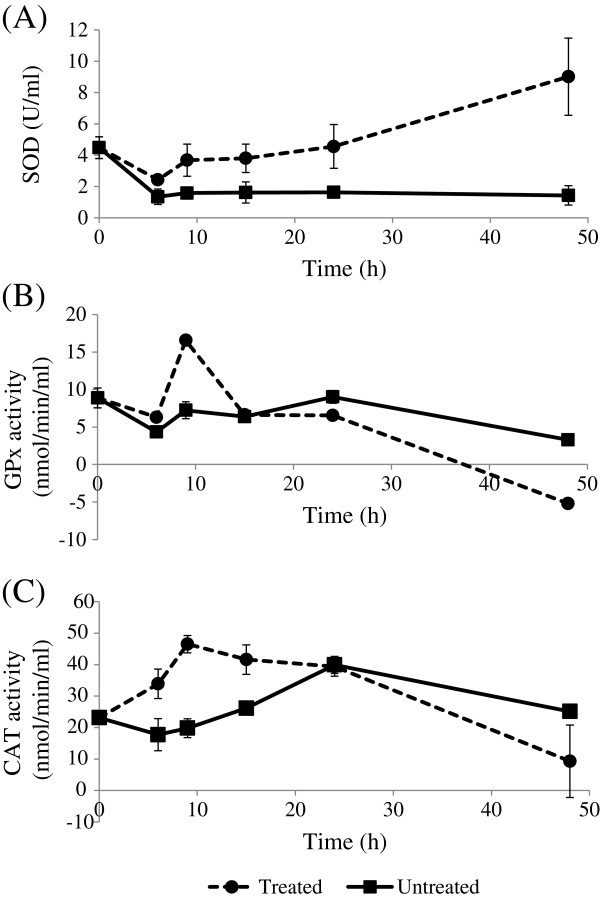
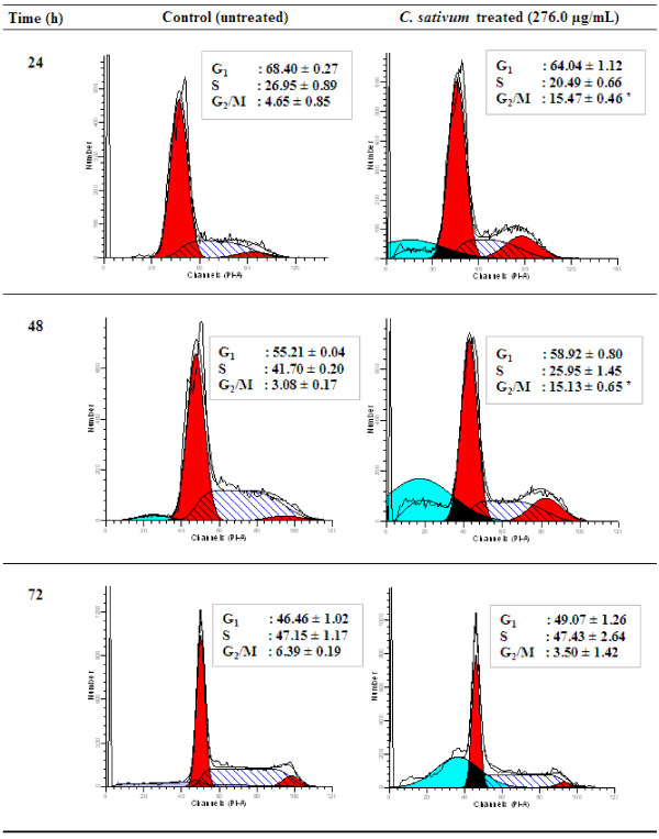
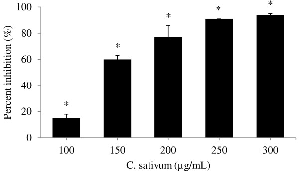
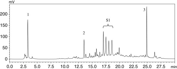
Background: Coriandrum sativum is a popular culinary and medicinal herb of the Apiaceae family. Health promoting properties of this herb have been reported in pharmacognostical, phytochemical and pharmacological studies.However, studies on C. sativum have always focused on the aerial parts of the herb and scientific investigation on the root is limited. The aim of this research was to investigate the antioxidant and anticancer activities of C. sativum root, leaf and stem, including its effect on cancer cell migration, and its protection against DNA damage, with special focus on the roots. Methods: Powdered roots, leaves and stems of C. sativum were extracted through sequential extraction using hexane, dichloromethane, ethyl acetate, methanol and water. Total phenolic content, FRAP and DPPH radical scavenging activities were measured. Anti-proliferative activitiy on the breast cancer cell line, MCF-7, was assayed using the MTT assay. Activities of the antioxidant enzymes, catalase, superoxide dismutase, glutathione peroxidase, and of the caspases-3, -8 and -9 were assayed on treatment with the extract. Cell cycle progression was analysed using flow cytometry. The scratch motility assay was used to assess inhibition of MCF-7 cell migration. DNA damage in 3 T3-L1 fibroblasts was evaluated by the comet assay. The components in the extract were identified by HPLC and GC-MS. Results: The ethyl acetate extract of C. sativum roots showed the highest antiproliferative activity on MCF-7 cells (IC 50 = 200.0 ± 2.6 μg/mL) and had the highest phenolic content, FRAP and DPPH scavenging activities among the extracts.C. sativum root inhibited DNA damage and prevented MCF-7 cell migration induced by H 2 O 2, suggesting its potential in cancer prevention and inhibition of metastasis. The extract exhibited anticancer activity in MCF-7 cells by affecting antioxidant enzymes possibly leading to H 2 O 2 accumulation, cell cycle arrest at the G 2/M phase and apoptotic cell death by the death receptor and mitochondrial apoptotic pathways. Conclusions: This study is the first report on the antioxidant and anticancer properties of C. sativum root. The herb shows potential in preventing oxidative stress-related diseases and would be useful as supplements used in combination with conventional drugs to enhance the treatment of diseases such as cancer.
Overview
The claims and statistics of this paper haven’t been checked yet.
Run the check — free, no account needed. It reads the open-access text and checks the sources it cites.
Integrity signals
Registry
Cites
Statistics
Figures
Verification
Claims the paper makes
The claims and statistics of this paper haven’t been checked yet.
Run the check — free, no account needed. It reads the open-access text and checks the sources it cites.
Claims other papers make about this one
Each is checked against this paper’s own text: does it say what the citing paper says it does?
In the citing paper
“managing gastric ulcers.”
From Unlocking the Apiaceae: comprehensive advances in agronomy, biotechnology, phytochemistry, and health applications of major seed spices (2025) · cites this paper as Tang et al. 2013
In the citing paper
“hepatoprotection”
From Unlocking the Apiaceae: comprehensive advances in agronomy, biotechnology, phytochemistry, and health applications of major seed spices (2025) · cites this paper as Tang et al. 2013
In the citing paper
“studies support its role in breast”
From Unlocking the Apiaceae: comprehensive advances in agronomy, biotechnology, phytochemistry, and health applications of major seed spices (2025) · cites this paper as Tang et al. 2013
Self-check
Percentages, sums and reported differences re-computed from the paper’s own numbers.
The self-check needs the paper’s full text.
Citations
Cites
No references could be resolved yet.
Research works printed but not linked: 57
- DeSylvia D, Stuber M, Fung CC, Bazargan-Hejazi S, Cooper E: The knowledge, attitudes and usage of complementary and alternative medicine of medical students. Evid-Based Compl Alt. 2011, 10.1093/ecam/nen075 · DOI
- Benz CC, Yau C: Ageing, oxidative stress and cancer: paradigms in parallax. Nat Rev Cancer. 2008, 8: 875-879. 10.1038/nrc2522. · DOI
- Jiménez-Estrada M, Velázquez-Contreras C, Garibay-Escobar A, Sierras-Canchola D, Lapizco-Vázquez R, Ortiz-Sandoval C, Burgos-Hernández A, Robles-Zepeda RE: In vitro antioxidant and antiproliferative activities of plants of the ethnopharmacopeia from northwest of Mexico. BMC Complem Altern M. 2013, 13: 12-10.1186/1472-6882-13-12. · DOI
- Imlay JA, Linn S: DNA damage and oxygen radical toxicity. Science. 1988, 240: 1302-10.1126/science.3287616. · DOI · Paper page
- Nelson KK, Ranganathan AC, Mansouri J, Rodriguez AM, Providence KM, Rutter JL, Pumiglia K, Bennett JA, Melendez JA: Elevated sod2 activity augments matrix metalloproteinase expression evidence for the involvement of endogenous hydrogen peroxide in regulating metastasis. Clin Cancer Res. 2003, 9: 424-432.
- López-Lázaro M: Dual role of hydrogen peroxide in cancer: possible relevance to cancer chemoprevention and therapy. Cancer Lett. 2007, 252: 1-8. 10.1016/j.canlet.2006.10.029. · DOI
- Taraphdar AK, Roy M, Bhattacharya R: Natural products as inducers of apoptosis: implication for cancer therapy and prevention. Curr Sci India. 2001, 80: 1387-1396.
- Sreelatha S, Padma P, Umadevi M: Protective effects of Coriandrum sativum extracts on carbon tetrachloride-induced hepatotoxicity in rats. Food Chem Toxicol. 2009, 47: 702-708. 10.1016/j.fct.2008.12.022. · DOI
- Padmaa M: Coriandrum sativum linn: a review. Pharmacol online Newsl. 2009, 3: 561-573.
- Asgarpanah J, Kazemivash N: Phytochemistry, pharmacology and medicinal properties of Coriandrum sativum L. Afr J Pharm Pharmaco. 2012, 6: 2340-2345.
- Oganesyan E, Nersesyan Z, Parkhomenko AY: Chemical composition of the above-ground part of Coriandrum sativum. Pharm Chem J-Ussr. 2007, 41: 149-153. 10.1007/s11094-007-0033-2. · DOI
- Kubo I, Fujita K-i, Kubo A, Nihei K-i, Ogura T: Antibacterial activity of coriander volatile compounds against Salmonella choleraesuis. J Agr Food Chem. 2004, 52: 3329-3332. 10.1021/jf0354186. · DOI
- Momin AH, Acharya SS, Gajjar AV: Coriandrum sativum-review of advances in phytopharmacology. Int J Pharm Sci Res. 2012, 3: 1233-1239.
- Wangensteen H, Samuelsen AB, Malterud KE: Antioxidant activity in extracts from coriander. Food Chem. 2004, 88: 293-297. 10.1016/j.foodchem.2004.01.047. · DOI
- Hashim M, Lincy S, Remya V, Teena M, Anila L: Effect of polyphenolic compounds from Coriandrum sativum on H2O2-induced oxidative stress in human lymphocytes. Food Chem. 2005, 92: 653-660. 10.1016/j.foodchem.2004.08.027. · DOI
- Singleton V, Rossi JA: Colorimetry of total phenolics with phosphomolybdic-phosphotungstic acid reagents. Am J Enol Viticult. 1965, 16: 144-158. · DOI · Paper page
- Benzie IFF, Strain J: The ferric reducing ability of plasma (FRAP) as a measure of “antioxidant power”: the FRAP assay. Anal Biochem. 1996, 239: 70-76. 10.1006/abio.1996.0292. · DOI
- Cos P, Rajan P, Vedernikova I, Calomme M, Pieters L, Vlietinck AJ, Augustyns K, Haemers A, Berghe DV: In vitro antioxidant profile of phenolic acid derivatives. Free Radical Res. 2002, 36: 711-716. 10.1080/10715760290029182. · DOI
- Mosmann T: Rapid colorimetric assay for cellular growth and survival: application to proliferation and cytotoxicity assays. J Immunol Methods. 1983, 65: 55-63. 10.1016/0022-1759(83)90303-4. · DOI · Paper page
- Sato Y, Rifkin DB: Autocrine activities of basic fibroblast growth factor: regulation of endothelial cell movement, plasminogen activator synthesis, and DNA synthesis. J Cell Biol. 1988, 107: 1199-1205. 10.1083/jcb.107.3.1199. · DOI
- Singh NP, McCoy MT, Tice RR, Schneider EL: A simple technique for quantitation of low levels of DNA damage in individual cells. Exp Cell Res. 1988, 175: 184-191. 10.1016/0014-4827(88)90265-0. · DOI · Paper page
- Jayakumar R, Kanthimathi MS: Dietary spices protect against hydrogen peroxide-induced DNA damage and inhibit nicotine-induced cancer cell migration. Food Chem. 2012, 134: 1580-1584. 10.1016/j.foodchem.2012.03.101. · DOI
- Nuutila A, Kammiovirta K, Oksman-Caldentey K-M: Comparison of methods for the hydrolysis of flavonoids and phenolic acids from onion and spinach for HPLC analysis. Food Chem. 2002, 76: 519-525. 10.1016/S0308-8146(01)00305-3. · DOI
- Conde E, Moure A, Domínguez H, Parajó JC: Fractionation of antioxidants from autohydrolysis of barley husks. J Agr Food Chem. 2008, 56: 10651-10659. 10.1021/jf801710a. · DOI
- Moirangthem DS, Talukdar NC, Kasoju N, Bora U: Antioxidant, antibacterial, cytotoxic, and apoptotic activity of stem bark extracts of Cephalotaxus griffithii Hook. f. BMC Complem Altern M. 2012, 12: 30-10.1186/1472-6882-12-30. · DOI
- de Almeida ME, Mancini Filho J, Barbosa Guerra N: Characterization of antioxidant compounds in aqueous coriander extract (Coriandrum sativum L.). LWT-Food Sci Technol. 2005, 38: 15-19. 10.1016/j.lwt.2004.03.011. · DOI
- Ford J, Jiang M, Milner J: Cancer-specific functions of SIRT1 enable human epithelial cancer cell growth and survival. Cancer Res. 2005, 65: 10457-10463. 10.1158/0008-5472.CAN-05-1923. · DOI
- Min JY, Lim SO, Jung G: Downregulation of catalase by reactive oxygen species via hypermethylation of CpG island II on the catalase promoter. FEBS Lett. 2010, 584: 2427-2432. 10.1016/j.febslet.2010.04.048. · DOI
- Pigeolet E, Remacle J: Susceptibility of glutathione peroxidase to proteolysis after oxidative alteration by peroxides and hydroxyl radicals. Free Radical Bio Med. 1991, 11: 191-195. 10.1016/0891-5849(91)90171-X. · DOI
- Rashad AE, El-Sayed WA, Mohamed AM, Ali MM: Synthesis of new quinoline derivatives as inhibitors of human tumor cells growth. Arch Pharm. 2010, 343: 440-448. 10.1002/ardp.201000002. · DOI
- Evens AM, Lecane P, Magda D, Prachand S, Singhal S, Nelson J, Miller RA, Gartenhaus RB, Gordon LI: Motexafin gadolinium generates reactive oxygen species and induces apoptosis in sensitive and highly resistant multiple myeloma cells. Blood. 2005, 105: 1265-1273. · DOI
- Burdock GA, Carabin IG: Safety assessment of coriander (Coriandrum sativum L.) essential oil as a food ingredient. Food Chem Toxicol. 2009, 47: 22-34. 10.1016/j.fct.2008.11.006. · DOI
- Fan TJ, Han LH, Cong RS, Liang J: Caspase family proteases and apoptosis. Acta Bioch Bioph Sin. 2005, 37: 719-727. 10.1111/j.1745-7270.2005.00108.x. · DOI
- Day TW, Huang S, Safa AR: c-FLIP knockdown induces ligand-independent DR5-, FADD-, caspase-8-, and caspase-9-dependent apoptosis in breast cancer cells. Biochem Pharmacol. 2008, 76: 1694-1704. 10.1016/j.bcp.2008.09.007. · DOI
- Riedl SJ, Shi Y: Molecular mechanisms of caspase regulation during apoptosis. Nat Rev Mol Cell Bio. 2004, 5: 897-907. · DOI
- Jänicke RU: MCF-7 breast carcinoma cells do not express caspase-3. Breast Cancer Res Tr. 2009, 117: 219-221. 10.1007/s10549-008-0217-9. · DOI
- Zhang GP, Lu YY, Lv JC, Ou HJ: Effect of ursolic acid on caspase-3 and PARP expression of human MCF-7 cells. Zhongguo Zhong Yao Za Zhi. 2006, 31: 141-144.
- Yang HL, Chen CS, Chang WH, Lu FJ, Lai YC, Chen CC, Hseu TH, Kuo CT, Hseu YC: Growth inhibition and induction of apoptosis in MCF-7 breast cancer cells by Antrodia camphorata. Cancer Lett. 2006, 231: 215-227. 10.1016/j.canlet.2005.02.004. · DOI
- Gooch JL, Yee D: Strain-specific differences in formation of apoptotic DNA ladders in MCF-7 breast cancer cells. Cancer Lett. 1999, 144: 31-37. 10.1016/S0304-3835(99)00208-6. · DOI
- Shim HY, Park JH, Paik HD, Nah SY, Kim DS, Han YS: Acacetin-induced apoptosis of human breast cancer MCF-7 cells involves caspase cascade, mitochondria-mediated death signaling and SAPK/JNK1/2-c-Jun activation. Mol Cells. 2007, 24: 95-104. · DOI
- Schwartz GK, Shah MA: Targeting the cell cycle: a new approach to cancer therapy. J Clin Oncol. 2005, 23: 9408-9421. 10.1200/JCO.2005.01.5594. · DOI
- Cortés-Eslava J, Gómez-Arroyo S, Villalobos-Pietrini R, Espinosa-Aguirre JJ: Antimutagenicity of coriander (Coriandrum sativum) juice on the mutagenesis produced by plant metabolites of aromatic amines. Toxicol Lett. 2004, 153: 283-292. 10.1016/j.toxlet.2004.05.011. · DOI
- Robbins RJ: Phenolic acids in foods: an overview of analytical methodology. J Agr Food Chem. 2003, 51: 2866-2887. 10.1021/jf026182t. · DOI
- Ng LK, Lafontaine P, Harnois J: Gas chromatographic–mass spectrometric analysis of acids and phenols in distilled alcohol beverages: application of anion-exchange disk extraction combined with in-vial elution and silylation. J Chromatogr A. 2000, 873: 29-38. 10.1016/S0021-9673(99)01100-0. · DOI
- Dias MI, Barros L, Sousa MJ, Ferreira IC: Comparative study of lipophilic and hydrophilic antioxidants from in vivo and in vitro grown Coriandrum sativum. Plant Food Hum Nutr. 2011, 66: 181-186. 10.1007/s11130-011-0227-3. · DOI
- Taniguchi M, Yanai M, Xiao YQ, Kido T-i, Baba K: Three isocoumarins from Coriandrum sativum. Phytochemistry. 1996, 42: 843-846. 10.1016/0031-9422(95)00930-2. · DOI
- Esiyok D, Otles S, Akcicek E: Herbs as a food source in Turkey. Asian Pac J Cancer P. 2004, 5: 334-339.
- Lei J, Yu J, Yu H, Liao Z: Composition, cytotoxicity and antimicrobial activity of essential oil from Dictamnus dasycarpus. Food Chem. 2008, 107: 1205-1209.
- Pandey A, Bigoniya P, Raj V, Patel K: Pharmacological screening of Coriandrum sativum Linn. for hepatoprotective activity. J Pharm Bioallied Sci. 2011, 3: 435-10.4103/0975-7406.84462. · DOI
- Chung I-M, Ahmad A, Kim S-J, Naik PM, Nagella P: Composition of the essential oil constituents from leaves and stems of Korean Coriandrum sativum and their immunotoxicity activity on the Aedes aegypti L. Immunopharm Immunot. 2012, 34: 152-156. 10.3109/08923973.2011.589030. · DOI
- Putchala MC, Ramani P, Sherlin HJ, Premkumar P, Natesan A: Ascorbic acid and its pro-oxidant activity as a therapy for tumours of oral cavity–a systematic review. Arch Oral Biol. 2013, 58: 563-574. 10.1016/j.archoralbio.2013.01.016. · DOI
- Kurbacher CM, Wagner U, Kolster B, Andreotti PE, Krebs D, Bruckner HW: Ascorbic acid (vitamin C) improves the antineoplastic activity of doxorubicin, cisplatin, and paclitaxel in human breast carcinoma cells in vitro. Cancer Lett. 1996, 103: 183-189. 10.1016/0304-3835(96)04212-7. · DOI
- Osmak M, Kovacek I, Ljubenkov I, Spaventi R, Eckert-Maksic M: Ascorbic acid and 6-deoxy-6-chloro-ascorbic acid: potential anticancer drugs. Neoplasma. 1997, 44: 101-108.
- Chen Q, Espey MG, Krishna MC, Mitchell JB, Corpe CP, Buettner GR, Shacter E, Levine M: Pharmacologic ascorbic acid concentrations selectively kill cancer cells: action as a pro-drug to deliver hydrogen peroxide to tissues. Proc Natl Acad Sci USA. 2005, 102: 13604-13609. 10.1073/pnas.0506390102. · DOI · Paper page
- Matés JM, Sánchez-Jiménez FM: Role of reactive oxygen species in apoptosis: implications for cancer therapy. Int J Biochem Cell B. 2000, 32: 157-170. 10.1016/S1357-2725(99)00088-6. · DOI
- Bijur GN, Briggs B, Hitchcock CL, Williams MV: Ascorbic acid-dehydroascorbate induces cell cycle arrest at G2/M DNA damage checkpoint during oxidative stress. Environ Mol Mutagen. 1999, 33: 144-152. 10.1002/(SICI)1098-2280(1999)33:2<144::AID-EM6>3.0.CO;2-V. · DOI
- Cha J, Roomi MW, Ivanov V, Kalinovsky T, Niedzwiecki A, Rath M: Ascorbate supplementation inhibits growth and metastasis of B16FO melanoma and 4 T1 breast cancer cells in vitamin C-deficient mice. Int J Oncol. 2013, 42: 55- · DOI
Cited by
Wu, J., Wang, Y., & Wang, Z. (2017) · Journal of Advanced Nursing · doi:10.1111/jan.13311 · Cited by 62
Singh, R., Choudhary, S., Kumari, V., Meena, R. S., Meena, N. K., Y, R., & Verma, A. K. (2025) · Journal of Agriculture and Ecology · doi:10.58628/jae-2521-101
Metadata
- Authors
- Tang, E. L., Rajarajeswaran, J., Fung, S. Y., & Kanthimathi, M.
- Venue
- BMC Complementary and Alternative Medicine
- Published
- 2013-12
- Volume
- 13
- Issue
- 1
- ISSN
- 1472-6882
- PMC
- PMC4028854
- Cited by
- 87
- References
- 0
- Language
- en
About these checks
Registry, retraction, Cites and statistics results are computed, not judged, with one exception: a printed reference that no index lists is read once by a model before it counts as a citation error. Claims are extracted by a model and kept only when their quote appears verbatim in the text; each cited source is then checked, with a model’s help, for whether it says what the claim relies on. Those verdicts can be wrong — read the claim’s quote and the cited source before relying on one.
Statistics and arithmetic are recomputed, not judged.
Confidence bands are shown only for fields calibrated on our annotated test set. None is calibrated yet, so no band is shown.
What counts as a claim ›How we measure confidence ›How we check this ›
Are you an author, or do you see a problem with this page? Request a review
How to cite
Cite CoChat for the checks on this page, and the paper for its own findings.
- This page
- CoChat. (2026). Citation check: Antioxidant activity of Coriandrum sativum and protection against DNA damage and cancer cell migration. https://app.cochat.ai/papers/10.1186/1472-6882-13-347
- The paper
- Tang, E. L., Rajarajeswaran, J., Fung, S. Y., & Kanthimathi, M. (2013). Antioxidant activity of Coriandrum sativum and protection against DNA damage and cancer cell migration. BMC Complementary and Alternative Medicine. https://doi.org/10.1186/1472-6882-13-347
Data sources
Bibliographic data: Semantic Scholar, OpenAlex, Crossref.
Contains information from the Semantic Scholar Academic Graph Datasets, made available under the ODC Attribution License (ODC-BY 1.0).
Semantic Scholar’s data is described in: Kinney, R., et al. (2023). The Semantic Scholar Open Data Platform. arXiv:2301.10140.
