

The DOI matches its Crossref record.
The HPV-16 E7 oncoprotein induces centriole multiplication through deregulation of Polo-like kinase 4 expression
Abstract
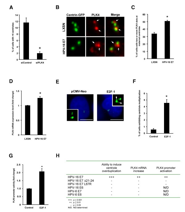
Background: Infection with high-risk human papillomaviruses (HPVs) such as HPV-16 is intimately associated with squamous cell carcinomas (SCCs) of the anogenital tract and a subset of oropharyngeal carcinomas. Such lesions, including pre-invasive precursors, frequently show multipolar mitoses and aneuploidy. The high-risk HPV-16-encoded E7 oncoprotein has been shown to rapidly induce centrosome abnormalities thereby causing the formation of supernumerary mitotic spindle poles and increasing the risk for chromosome missegregation. HPV-16 E7 has been found to rapidly induce centriole overduplication, in part, through the simultaneous formation of more than one daughter centriole at single maternal centrioles (centriole multiplication). The precise molecular mechanism that underlies HPV-16 E7-induced centriole multiplication, however, remains poorly understood. Findings: Here, we show that human keratinocytes engineered to stably express the HPV-16 E7 oncoprotein exhibit aberrant Polo-like kinase 4 (PLK4) protein expression at maternal centrioles. Real-time quantitative reverse transcriptase (qRT-PCR) analysis of these cells revealed an increase of PLK4 mRNA levels compared to control cells. Importantly, the ability of the HPV-16 E7 oncoprotein to induce centriole multiplication was found to correlate with its ability to activate the PLK4 promoter and to up-regulate PLK4 mRNA. Conclusions: These results highlight the critical role of PLK4 transcriptional deregulation in centriole multiplication in HPV-16 E7-expressing cells. Our findings encourage further experiments to test transcriptional inhibitors or small molecules targeting PLK4 to prevent centriole abnormalities, mitotic infidelity and malignant progression in HPV-associated neoplasms and other tumors in which PLK4 regulation is disrupted.
Overview
The claims and statistics of this paper haven’t been checked yet.
Run the check — free, no account needed. It reads the open-access text and checks the sources it cites.
Integrity signals
Registry
Cites
Statistics
Figures
Verification
Claims the paper makes
The claims and statistics of this paper haven’t been checked yet.
Run the check — free, no account needed. It reads the open-access text and checks the sources it cites.
Claims other papers make about this one
Each is checked against this paper’s own text: does it say what the citing paper says it does?
In the citing paper
“Abnormal centriole multiplication was also shown to correlate with up-regulation of Polo-like kinase 4 in HPV-16 E7-expressing cells [314]”
From The Not-So-Good, the Bad and the Ugly: HPV E5, E6 and E7 Oncoproteins in the Orchestration of Carcinogenesis (2021) · cites this paper as [314]
Self-check
Percentages, sums and reported differences re-computed from the paper’s own numbers.
The self-check needs the paper’s full text.
Citations
Cites
No references could be resolved yet.
Research works printed but not linked: 29
- zur Hausen H: Papillomaviruses and cancer: from basic studies to clinical application. Nat Rev Cancer. 2002, 2: 342-350. 10.1038/nrc798 · DOI
- Korzeniewski N, Spardy N, Duensing A, Duensing S: Genomic instability and cancer: Lessons learned from human papillomaviruses. Cancer Lett. 2011, 305: 113-122. 10.1016/j.canlet.2010.10.013 · DOI
- McLaughlin-Drubin ME, Munger K: The human papillomavirus E7 oncoprotein. Virology. 2009, 384: 335-344. 10.1016/j.virol.2008.10.006 · DOI
- Longworth MS, Laimins LA: The binding of histone deacetylases and the integrity of zinc finger-like motifs of the E7 protein are essential for the life cycle of human papillomavirus type 31. J Virol. 2004, 78: 3533-3541. 10.1128/JVI.78.7.3533-3541.2004 · DOI
- Brehm A, Nielsen SJ, Miska EA, McCance DJ, Reid JL, Bannister AJ, Kouzarides T: The E7 oncoprotein associates with Mi2 and histone deacetylase activity to promote cell growth. EMBO J. 1999, 18: 2449-2458. 10.1093/emboj/18.9.2449 · DOI · Paper page
- Hwang SG, Lee D, Kim J, Seo T, Choe J: Human papillomavirus type 16 E7 binds to E2F1 and activates E2F1-driven transcription in a retinoblastoma protein-independent manner. J Biol Chem. 2002, 277: 2923-2930. 10.1074/jbc.M109113200 · DOI · Paper page
- McLaughlin-Drubin ME, Huh KW, Munger K: Human papillomavirus type 16 E7 oncoprotein associates with E2F6. J Virol. 2008, 82: 8695-8705. 10.1128/JVI.00579-08 · DOI · Paper page
- Nguyen CL, Munger K: Direct association of the HPV16 E7 oncoprotein with cyclin A/CDK2 and cyclin E/CDK2 complexes. Virology. 2008, 380: 21-25. 10.1016/j.virol.2008.07.017 · DOI · Paper page
- He W, Staples D, Smith C, Fisher C: Direct activation of cyclin-dependent kinase 2 by human papillomavirus E7. J Virol. 2003, 77: 10566-10574. 10.1128/JVI.77.19.10566-10574.2003 · DOI · Paper page
- Duensing S, Munger K: Mechanisms of genomic instability in human cancer: insights from studies with human papillomavirus oncoproteins. Int J Cancer. 2004, 109: 157-162. 10.1002/ijc.11691 · DOI
- Nigg EA: Centrosome duplication: of rules and licenses. Trends Cell Biol. 2007, 17: 215-221. 10.1016/j.tcb.2007.03.003 · DOI
- Azimzadeh J, Bornens M: Structure and duplication of the centrosome. J Cell Sci. 2007, 120: 2139-2142. 10.1242/jcs.005231 · DOI
- Ganem NJ, Godinho SA, Pellman D: A mechanism linking extra centrosomes to chromosomal instability. Nature. 2009, 460: 278-282. 10.1038/nature08136 · DOI
- Duensing A, Spardy N, Chatterjee P, Zheng L, Parry J, Cuevas R, Korzeniewski N, Duensing S: Centrosome overduplication, chromosomal instability, and human papillomavirus oncoproteins. Environ Mol Mutagen. 2009, 50: 741-747. 10.1002/em.20478 · DOI
- Tsou MF, Wang WJ, George KA, Uryu K, Stearns T, Jallepalli PV: Polo kinase and separase regulate the mitotic licensing of centriole duplication in human cells. Dev Cell. 2009, 17: 344-354. 10.1016/j.devcel.2009.07.015 · DOI
- Kleylein-Sohn J, Westendorf J, Le Clech M, Habedanck R, Stierhof YD, Nigg EA: Plk4-induced centriole biogenesis in human cells. Dev Cell. 2007, 13: 190-202. 10.1016/j.devcel.2007.07.002 · DOI
- Duensing S, Duensing A, Crum CP, Munger K: Human papillomavirus type 16 E7 oncoprotein-induced abnormal centrosome synthesis is an early event in the evolving malignant phenotype. Cancer Res. 2001, 61: 2356-2360.
- Duensing A, Liu Y, Perdreau SA, Kleylein-Sohn J, Nigg EA, Duensing S: Centriole overduplication through the concurrent formation of multiple daughter centrioles at single maternal templates. Oncogene. 2007, 26: 6280-6288. 10.1038/sj.onc.1210456 · DOI
- Duensing A, Chin A, Wang L, Kuan SF, Duensing S: Analysis of centrosome overduplication in correlation to cell division errors in high-risk human papillomavirus (HPV)-associated anal neoplasms. Virology. 2008, 372: 157-164. 10.1016/j.virol.2007.10.030 · DOI
- Korzeniewski N, Zheng L, Cuevas R, Parry J, Chatterjee P, Anderton B, Duensing A, Munger K, Duensing S: Cullin 1 functions as a centrosomal suppressor of centriole multiplication by regulating polo-like kinase 4 protein levels. Cancer Res. 2009, 69: 6668-6675. 10.1158/0008-5472.CAN-09-1284 · DOI
- Piel M, Meyer P, Khodjakov A, Rieder CL, Bornens M: The respective contributions of the mother and daughter centrioles to centrosome activity and behavior in vertebrate cells. J Cell Biol. 2000, 149: 317-330. 10.1083/jcb.149.2.317 · DOI
- Habedanck R, Stierhof YD, Wilkinson CJ, Nigg EA: The Polo kinase Plk4 functions in centriole duplication. Nat Cell Biol. 2005, 7: 1140-1146. · DOI
- Cunha-Ferreira I, Rodrigues-Martins A, Bento I, Riparbelli M, Zhang W, Laue E, Callaini G, Glover DM, Bettencourt-Dias M: The SCF/Slimb ubiquitin ligase limits centrosome amplification through degradation of SAK/PLK4. Curr Biol. 2009, 19: 43-49. 10.1016/j.cub.2008.11.037 · DOI
- Fode C, Binkert C, Dennis JW: Constitutive expression of murine Sak-a suppresses cell growth and induces multinucleation. Mol Cell Biol. 1996, 16: 4665-4672. · DOI
- Rogers GC, Rusan NM, Roberts DM, Peifer M, Rogers SL: The SCF Slimb ubiquitin ligase regulates Plk4/Sak levels to block centriole reduplication. J Cell Biol. 2009, 184: 225-239. 10.1083/jcb.200808049 · DOI
- Karlseder J, Rotheneder H, Wintersberger E: Interaction of Sp1 with the growth- and cell cycle-regulated transcription factor E2F. Mol Cell Biol. 1996, 16: 1659-1667. · DOI
- Li J, Tan M, Li L, Pamarthy D, Lawrence TS, Sun Y: SAK, a new polo-like kinase, is transcriptionally repressed by p53 and induces apoptosis upon RNAi silencing. Neoplasia. 2005, 7: 312-323. 10.1593/neo.04325 · DOI
- Duensing S, Munger K: Human papillomavirus type 16 E7 oncoprotein can induce abnormal centrosome duplication through a mechanism independent of inactivation of retinoblastoma protein family members. J Virol. 2003, 77: 12331-12335. 10.1128/JVI.77.22.12331-12335.2003 · DOI · Paper page
- Duensing A, Liu Y, Spardy N, Bartoli K, Tseng M, Kwon JA, Teng X, Duensing S: RNA polymerase II transcription is required for human papillomavirus type 16 E7- and hydroxyurea-induced centriole overduplication. Oncogene. 2007, 26: 215-223. 10.1038/sj.onc.1209782 · DOI
Cited by
Basukala, O., & Banks, L. (2021) · Viruses · doi:10.3390/v13101892 · Cited by 137
Metadata
- Authors
- Korzeniewski, N., Treat, B., & Duensing, S.
- Venue
- Molecular Cancer
- Published
- 2011-05-24
- Volume
- 10
- Issue
- 1
- ISSN
- 1476-4598
- PMC
- PMC3120798
- Cited by
- 50
- References
- 0
- Language
- en
About these checks
Registry, retraction, Cites and statistics results are computed, not judged, with one exception: a printed reference that no index lists is read once by a model before it counts as a citation error. Claims are extracted by a model and kept only when their quote appears verbatim in the text; each cited source is then checked, with a model’s help, for whether it says what the claim relies on. Those verdicts can be wrong — read the claim’s quote and the cited source before relying on one.
Statistics and arithmetic are recomputed, not judged.
Confidence bands are shown only for fields calibrated on our annotated test set. None is calibrated yet, so no band is shown.
What counts as a claim ›How we measure confidence ›How we check this ›
Are you an author, or do you see a problem with this page? Request a review
