

The DOI matches its Crossref record; 38 of the 39 references it cites were found in a registry or an index.
Levels of plasma circulating cell free nuclear and mitochondrial DNA as potential biomarkers for breast tumors
Open accessAbstract
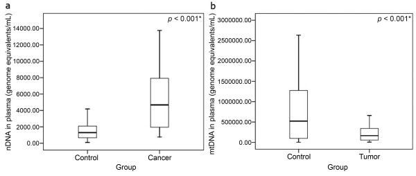
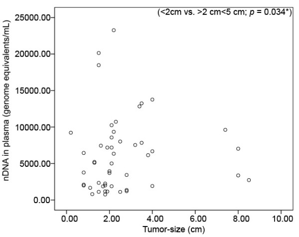
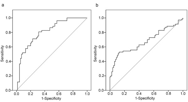
Background: With the aim to simplify cancer management, cancer research lately dedicated itself more and more to discover and develop non-invasive biomarkers. In this connection, circulating cell-free DNA (ccf DNA) seems to be a promising candidate. Altered levels of ccf nuclear DNA (nDNA) and mitochondrial DNA (mtDNA) have been found in several cancer types and might have a diagnostic value. Methods: Using multiplex real-time PCR we investigated the levels of ccf nDNA and mtDNA in plasma samples from patients with malignant and benign breast tumors, and from healthy controls. To evaluate the applicability of plasma ccf nDNA and mtDNA as a biomarker for distinguishing between the three study-groups we performed ROC (Receiver Operating Characteristic) curve analysis. We also compared the levels of both species in the cancer group with clinicopathological parameters. Results: While the levels of ccf nDNA in the cancer group were significantly higher in comparison with the benign tumor group (P < 0.001) and the healthy control group (P < 0.001), the level of ccf mtDNA was found to be significantly lower in the two tumor-groups (benign: P < 0.001; malignant: P = 0.022). The level of ccf nDNA was also associated with tumor-size (<2 cm vs. >2 cm<5 cm; 2250 vs. 6658; Mann-Whitney-U-Test: P = 0.034). Using ROC curve analysis, we were able to distinguish between the breast cancer cases and the healthy controls using ccf nDNA as marker (cut-off: 1866 GE/ml; sensitivity: 81%; specificity: 69%; P < 0.001) and between the tumor group and the healthy controls using ccf mtDNA as marker (cut-off: 463282 GE/ml; sensitivity: 53%; specificity: 87%; P < 0.001). Conclusion: Our data suggests that nuclear and mitochondrial ccf DNA have potential as biomarkers in breast tumor management. However, ccf nDNA shows greater promise regarding sensitivity and specificity.
Overview
The claims and statistics of this paper haven’t been checked yet.
Run the check — free, no account needed. It reads the open-access text and checks the sources it cites.
Integrity signals
Registry
Cites
Statistics
Figures
Verification
Claims the paper makes
The claims and statistics of this paper haven’t been checked yet.
Run the check — free, no account needed. It reads the open-access text and checks the sources it cites.
Claims other papers make about this one
Each is checked against this paper’s own text: does it say what the citing paper says it does?
The cited source says what the claim attributes to it.
In the citing paper
“As extracellular mtDNA is reportedly associated with various pathologies, including trauma25,26, burn injury27, cancer28 and rheumatoid arthritis17,29”
From Anti-mitochondrial autoantibodies in systemic lupus erythematosus and their association with disease manifestations (2019) · cites this paper as [28]
Self-check
Percentages, sums and reported differences re-computed from the paper’s own numbers.
The self-check needs the paper’s full text.
Citations
Cites
Circulating mutant DNA to assess tumor dynamics
Diehl, F., Schmidt, K., Choti, M. A., Romans, K. E., Goodman, S. N., Li, M., Thornton, K. A., Agrawal, N., Sokoll, L. J., Szabo, S., et al. (2008) · Nature Medicine · doi:10.1038/nm.1789 · Cited by 2,746
Understanding diagnostic tests 3: receiver operating characteristic curves
Akobeng, A. K. (2007) · Acta Paediatrica · doi:10.1111/j.1651-2227.2006.00178.x · Cited by 1,882
Berg, W. A. (2008) · JAMA · doi:10.1001/jama.299.18.2151 · Cited by 1,527
Multiple Biomarkers for the Prediction of First Major Cardiovascular Events and Death
Wang, T. J., Gona, P. N., Larson, M. G., Tofler, G. H., Levy, D. A., Newton‐Cheh, C., Jacques, P. F., Rifai, N., Selhub, J., Robins, S. J., et al. (2006) · New England Journal of Medicine · doi:10.1056/nejmoa055373 · Cited by 1,337
Mitochondrial DNA mutations and mitochondrial DNA depletion in gastric cancer
Wu, C., Yin, P., Hung, W., Li, A. F., Li, S., Chi, C., Wei, Y., & Lee, H. (2005) · Genes Chromosomes and Cancer · doi:10.1002/gcc.20213 · Cited by 381
Tumor Markers in Breast Cancer – European Group on Tumor Markers Recommendations
Molina, R., Barak, V., van Dalen, A., Duffy, M. J., EINARSSON, R., Gion, M., Goike, H. M., Lamerz, R., Nap, M., tormos, G. O. R. S. O. L. E., et al. (2005) · Tumor Biology · doi:10.1159/000089260 · Cited by 345
Mitochondrial DNA mutations and mitochondrial DNA depletion in breast cancer
Tseng, L., Yin, P., Chi, C., Hsu, C., Wu, C.-W., Lee, L.-M., Wei, Y., & Lee, H. (2006) · Genes Chromosomes and Cancer · doi:10.1002/gcc.20326 · Cited by 244
Cell-free DNA: measurement in various carcinomas and establishment of normal reference range
Wu, T., Zhang, D., Chia, J., Tsao, K., Sun, C., & Wu, J. T. (2002) · Clinica Chimica Acta · doi:10.1016/s0009-8981(02)00091-8 · Cited by 231
Cancer biomarkers: a systems approach
Hartwell, L., Mankoff, D. A., Paulovich, A. G., Ramsey, S. D., & Swisher, E. M. (2006) · Nature Biotechnology · doi:10.1038/nbt0806-905 · Cited by 202
Tumor‐specific changes in mtDNA content in human cancer
Mambo, E., Chatterjee, A., Xing, M., Tallini, G., Haugen, B. R., Yeung, S. J., Sukumar, S. V., & Sidransky, D. (2005) · International Journal of Cancer · doi:10.1002/ijc.21110 · Cited by 177
Cited by
Sorted by citations
Items 101–200 of 215
Neutrophil Extracellular Traps (NETs): Opportunities for Targeted Therapy
Volkov, D. V., Tetz, G. V., Rubtsov, Y. P., Stepanov, A. V., & Gabibov, A. G. (2021) · Acta Naturae · doi:10.32607/actanaturae.11503 · Cited by 30
Liu, Y., Shen, Q., Zhao, X. Q., Zou, M., Shao, S., Li, J., Ren, X., & Zhang, L. (2019) · Reproductive Biology and Endocrinology · doi:10.1186/s12958-019-0495-6 · Cited by 28
Predictive Biomarkers in Breast Cancer: Their Value in Neoadjuvant Chemotherapy
Fuksa, L., Mičuda, S., Grim, J., Ryška, A., & Hornychová, H. (2012) · Cancer Investigation · doi:10.3109/07357907.2012.725441 · Cited by 27
Koessler, T., Paradiso, V., Piscuoglio, S., Nienhold, R., Ho, L. K. F., Christinat, Y., Terracciano, L., Cathomas, G., WICKI, A. N., McKee, T., et al. (2020) · Laboratory Investigation · doi:10.1038/s41374-020-0459-7 · Cited by 26
Stawski, R. S., Walczak, K., Perdas, E., Włodarczyk, A., Sarniak, A., Kosielski, P., Meissner, P., Budlewski, T., Padula, G., & Nowak, D. (2019) · Scientific Reports · doi:10.1038/s41598-019-52409-w · Cited by 25
Current Trends in Cell-Free DNA Applications. Scoping Review of Clinical Trials
Stawski, R. S., Stec‐Martyna, E., Chmielecki, A., Nowak, D., & Perdas, E. (2021) · Biology · doi:10.3390/biology10090906 · Cited by 26
Wu, X., & Tanaka, H. (2015) · Oncotarget · doi:10.18632/oncotarget.5083 · Cited by 25
Kojer, K., Hering, T., Bazenet, C. E., Weiss, A., Herrmann, F., Taanman, J., & Orth, M. (2019) · Journal of Huntington s Disease · doi:10.3233/jhd-180324 · Cited by 25
Newell, C., Hume, S. L., Greenway, S. C., Podemski, L., Shearer, J., & Khan, A. (2018) · Molecular Genetics and Metabolism · doi:10.1016/j.ymgme.2018.10.002 · Cited by 24
Perdas, E., Stawski, R. S., Kaczka, K., Nowak, D., & Zubrzycka, M. (2019) · Scientific Reports · doi:10.1038/s41598-019-51000-7 · Cited by 24
A headlight on liquid biopsies: a challenging tool for breast cancer management
Massihnia, D., Perez, A., Bazan, V., Bronte, G., Castiglia, M., Fanale, D., Barraco, N., Cangemi, A. G., Piazza, F. D., Calò, V., et al. (2016) · Tumor Biology · doi:10.1007/s13277-016-4856-x · Cited by 23
Selective striatal mtDNA depletion in end-stage Huntington's disease R6/2 mice
Hering, T., Birth, N., Taanman, J., & Orth, M. (2015) · Experimental Neurology · doi:10.1016/j.expneurol.2015.02.004 · Cited by 23
Liao, H., & Li, H. (2020) · Cancer Management and Research · doi:10.2147/cmar.s249041 · Cited by 23
Cheng, Y.-H., Wang, C., Hsu, K., & Lee, G. (2022) · Analytical Chemistry · doi:10.1021/acs.analchem.1c04988 · Cited by 22
Post-mortem ventricular cerebrospinal fluid cell-free-mtDNA in neurodegenerative disease
Lowes, H., Kurzawa-Akanbi, M., Pyle, A., & Hudson, G. (2020) · Scientific Reports · doi:10.1038/s41598-020-72190-5 · Cited by 21
Clinical utility of liquid biopsy in breast cancer: A systematic review
Duque, G., Manterola, C., Otzen, T., Arias, C., Galindo, B., Mora, M., Guerrero, E., & García, N. D. B. (2021) · Clinical Genetics · doi:10.1111/cge.14077 · Cited by 21
Plasma circulating cell-free mitochondrial DNA in the assessment of Friedreich's ataxia
Dantham, S., Srivastava, A. K., Gulati, S., & Rajeswari, M. R. (2016) · Journal of the Neurological Sciences · doi:10.1016/j.jns.2016.04.016 · Cited by 22
Zeng, Z., Yi, Z., & Xu, B. (2025) · Cancer Letters · doi:10.1016/j.canlet.2025.217574 · Cited by 20
Liquid Biopsy in Solid Malignancy
Scarlotta, M., Şimşek, C., & Kim, A. K. (2019) · Genetic Testing and Molecular Biomarkers · doi:10.1089/gtmb.2018.0237 · Cited by 22
Choudhuri, S., Sharma, C., Banerjee, A., Kumar, S., Kumar, L., & Singh, N. (2013) · Molecular and Cellular Biochemistry · doi:10.1007/s11010-013-1863-8 · Cited by 20
Mitochondrial inheritance and cancer
Dong, J., Wong, L. C., & Mims, M. P. (2018) · Translational research · doi:10.1016/j.trsl.2018.06.004 · Cited by 19
Pisareva, E., Roch, B., Sánchez, C., Pastor, B., Mirandola, A., Diab‐Assaf, M., Mazard, T., Prévostel, C., Dache, Z. A. A., & Thierry, A. R. (2023) · Frontiers in Genetics · doi:10.3389/fgene.2023.1104732 · Cited by 17
Uzawa, K., Kasamatsu, A., BABA, T., Kimura, Y., Nakashima, D., Higo, M., Sakamoto, Y., Ogawara, K., Shiiba, M., & Tanzawa, H. (2015) · International Journal of Oncology · doi:10.3892/ijo.2015.3083 · Cited by 17
Cell-Free DNA: Potential Application in COVID-19 Diagnostics and Management
Stawski, R. S., Nowak, D., & Perdas, E. (2022) · Viruses · doi:10.3390/v14020321 · Cited by 17
Implementing circulating tumor DNA analysis in a clinical laboratory: A user manual
Koessler, T., Addeo, A., & Nouspikel, T. P. (2019) · Advances in clinical chemistry · doi:10.1016/bs.acc.2018.12.004 · Cited by 17
Smolle, M. A., Seidel, M. G., Kashofer, K., Liegl-Atzwanger, B., Sadoghi, P., Müller, D. A., & Leithner, A. (2025) · Archives of Orthopaedic and Trauma Surgery · doi:10.1007/s00402-024-05711-w · Cited by 16
Ma, G., Wang, J., Huang, H., Xu, H., Xu, J., Veeramootoo, J. S., Xia, T., & Wang, S. (2020) · Cancer Medicine · doi:10.1002/cam4.2906 · Cited by 16
Circulating Tumor Nucleic Acids: Perspective in Breast Cancer
Casciano, I., Vinci, A. D., Banelli, B., Brigati, C., Forlani, A., Allemanni, G., & Romani, M. (2010) · Breast Care · doi:10.1159/000310113 · Cited by 16
Mitochondrial D-loop mutations can be detected in sporadic malignant tumours in dogs
Ślaska, B., Surdyka, M., Brodzki, A., Nisztuk, S., Gurgul, A., Bugno‐Poniewierska, M., Śmiech, A., Różańska, D., & Orzelski, M. (2014) · Journal of Veterinary Research · doi:10.2478/bvip-2014-0096 · Cited by 16
Amniotic fluid cell‐free DNA in preterm prelabor rupture of membranes
Kacerovský, M., Vlková, B., Musilova, I. K., Andrýs, C., Plíšková, L., Žemličková, H., Stráník, J., Halada, P., Jacobsson, B., & Celec, P. (2018) · Prenatal Diagnosis · doi:10.1002/pd.5366 · Cited by 15
Platelets, Macrophages, and Thromboinflammation in Chagas Disease
Choudhuri, S., & Garg, N. J. (2022) · Journal of Inflammation Research · doi:10.2147/jir.s380896 · Cited by 14
Mitochondrial DNA Levels in Blood and Tissue Samples from Breast Cancer Patients of Different Stages
Xia, P., Wang, H., Geng, T., Xun, X., Zhou, W.-J., Jin, T., & Chen, C. (2014) · Asian Pacific Journal of Cancer Prevention · doi:10.7314/apjcp.2014.15.3.1339 · Cited by 14
Clinical significance of serum mitochondrial DNA in lung cancer
Hou, Y.-L., Chen, J.-J., Wu, Y.-F., Xue, C.-J., Li, F.-Z., Zheng, Q., & Chen, H. (2013) · Clinical Biochemistry · doi:10.1016/j.clinbiochem.2013.04.009 · Cited by 13
Wang, Y., Zhao, Y., Yu, M., Wei, L., Zhang, W., Wang, Z., & Yuan, Y. (2023) · Annals of Clinical and Translational Neurology · doi:10.1002/acn3.51901 · Cited by 14
Cabral, R. E. C. (2010) · The Open Lung Cancer Journal · doi:10.2174/1876819901003010038 · Cited by 12
Paolini, E., Longo, M., Corsini, A., & Dongiovanni, P. (2023) · International Journal of Molecular Sciences · doi:10.3390/ijms24032331 · Cited by 12
FREE EXTRACELLULAR DNA IN NORMAL STATE AND UNDER PATHOLOGICAL CONDITIONS
Va, K. (2014) · Medical Immunology (Russia) · doi:10.15789/1563-0625-2013-5-399-412 · Cited by 12
Cell-free tumor DNA: Emerging reality in oral squamous cell carcinoma
Babji, D., Nayak, R. S., Bhat, K. G., & Kotrashetti, V. (2019) · Journal of Oral and Maxillofacial Pathology · doi:10.4103/jomfp.jomfp_36_19 · Cited by 12
Sundquist, K., Sundquist, J., Hedelius, A., & Memon, A. A. (2020) · Molecular Carcinogenesis · doi:10.1002/mc.23261 · Cited by 11
Manueldas, S., Benterud, T., Rueegg, C. S., Garberg, H. T., Huun, M. U., Pankratov, L., Atneosen‐Åsegg, M., Solberg, R., Escobar, J., Saugstad, O. D., et al. (2018) · PLoS ONE · doi:10.1371/journal.pone.0206601 · Cited by 10
Wu, Y., Wan, S.-G., Long, Y., Ye, H., Yang, J., Luo, Y., Zhong, Y.-B., Li, X., Chen, H., & Wang, M. (2024) · Arthritis Research & Therapy · doi:10.1186/s13075-024-03438-y · Cited by 10
Circulating Nucleic Acids in Health and Disease
RYKOVA, E. Y., Laktionov, P. P., & Vlassov, V. V. (2010) · Nucleic acids and molecular biology · doi:10.1007/978-3-642-12617-8_7 · Cited by 9
Circulating Level of Myelin Basic Protein Predicts Postherpetic Neuralgia
Yang, X., Shen, Y., Ding, Z., Tian, Y., Hu, J., Guo, Q., & Huang, C. (2021) · Clinical Journal of Pain · doi:10.1097/ajp.0000000000000937 · Cited by 9
Nasr, R., Sleiman, F., Awada, Z., & Zgheib, N. K. (2016) · Pharmacogenomics · doi:10.2217/pgs-2016-0081 · Cited by 9
Altered Striatocerebellar Metabolism and Systemic Inflammation in Parkinson’s Disease
Yu, C.-C., Chen, M., Lu, C., Huang, Y., Chen, H.-L., Tsai, N., Wang, H., Yang, I.-H., Li, S.-H., & Lin, W.-C. (2016) · Oxidative Medicine and Cellular Longevity · doi:10.1155/2016/1810289 · Cited by 9
The rising tide of cell-free DNA profiling: from snapshot to temporal genome analysis
Bronkhorst, A. J., Ungerer, V., Oberhofer, A., & Holdenrieder, S. (2022) · Journal of Laboratory Medicine · doi:10.1515/labmed-2022-0030 · Cited by 10
Profiling disease and tissue-specific epigenetic signatures in cell-free DNA
Oberhofer, A., Bronkhorst, A. J., Ungerer, V., & Holdenrieder, S. (2022) · Journal of Laboratory Medicine · doi:10.1515/labmed-2022-0031 · Cited by 9
Mitochondrial DNA Mutations and Ageing
Whitehall, J. C., Smith, A. L. M., & Greaves, L. C. (2023) · Sub-cellular biochemistry/Subcellular biochemistry · doi:10.1007/978-3-031-21410-3_4 · Cited by 9
Martinsson, T. (2011) · International Journal of Oncology · doi:10.3892/ijo.2011.1120 · Cited by 9
Neutrophil extracellular traps in diseases of the female reproductive organs
Morawiec, M.-L., Kubina, R., Stępień, S., Smycz‐Kubańska, M., Królewska‐Daszczyńska, P., Ratajczak–Wrona, W., & Mielczarek‐Palacz, A. (2025) · Frontiers in Immunology · doi:10.3389/fimmu.2025.1589329 · Cited by 9
Habiburrahman, M., Sutopo, S., & Wardoyo, M. P. (2022) · Critical Reviews in Oncology/Hematology · doi:10.1016/j.critrevonc.2022.103897 · Cited by 8
Mitochondrial DNA instability in human cancers
Lin, C.-S., & Wang, L.-S. (2013) · Formosan Journal of Surgery · doi:10.1016/j.fjs.2013.04.002 · Cited by 8
Wielscher, M., Pulverer, W., Peham, J. R., Hofner, M., Rappaport, C., Singer, C., Jungbauer, C., Nöhammer, C., & Weinhäusel, A. (2011) · BMC Clinical Pathology · doi:10.1186/1472-6890-11-11 · Cited by 8
Plasma derived cell-free mitochondrial DNA originates mainly from circulating cell-free mitochondriaPreprint
Roch, B., Pisareva, E., Sanchez, C., Pastor, B., Tanos, R., Mirandola, A., Mazard, T., Dache, Z. A. A., & Thierry, A. R. (2021) · bioRxiv (Cold Spring Harbor Laboratory) · doi:10.1101/2021.09.03.458846 · Cited by 8
Tsirka, G., Zikopoulos, A., Papageorgiou, K., Kostoulas, C., Tsigkas, I., Moustakli, E., Kaltsas, A., Sarafi, E., Michaelidis, T. M., & Georgiou, I. A. (2023) · Genes · doi:10.3390/genes14071504 · Cited by 7
Khadka, P., Young, C. K. J., Sachidanandam, R., Brard, L., & Young, M. J. (2024) · Frontiers in Oncology · doi:10.3389/fonc.2024.1394699 · Cited by 8
Ali, Z., Waseem, S., Shahzadi, I., Bukhari, S., Anis, R. A., Ahmed, I., & Anees, M. (2021) · Molecular Biology Reports · doi:10.1007/s11033-021-06313-0 · Cited by 7
Huebner, H., Lubrich, H., Blum, S., Antoniadis, S., Lermann, J. H., Ekici, A. B., Fasching, P. A., Beckmann, M. W., Ruebner, M., & Burghaus, S. (2021) · Reproductive BioMedicine Online · doi:10.1016/j.rbmo.2021.08.004 · Cited by 7
Ma, X., Wang, Z., Wang, S., Tian, Y., Xie, B., Li, J., Ma, B., & Li, L. (2024) · Expert Review of Anticancer Therapy · doi:10.1080/14737140.2024.2312246 · Cited by 6
la Cruz, D. D. G.-D., Juárez‐Rojop, I. E., Tovilla‐Zárate, C. A., Nicolini, H., & Mendoza, A. D. G. (2024) · Neuropsychiatric Disease and Treatment · doi:10.2147/ndt.s460554 · Cited by 6
Safi, M., & Najib, A. R. (2020) · Experimental and Therapeutic Medicine · doi:10.3892/etm.2020.9497 · Cited by 6
Bitenc, M., Tune, B. G., Melheim, M., Atneosen‐Åsegg, M., Lai, X., Rajar, P., Solberg, R., & Baumbusch, L. O. (2022) · Molecular Biology Reports · doi:10.1007/s11033-022-08135-0 · Cited by 6
Sultana, G. N. N., Akter, F., Israfil, S. M. H., Ray, U. C., Jahan, R. A., Ali, M. S., Din, S. A., Rahman, S., Halim, R., & Alam, M. S. (2023) · Frontiers in Oncology · doi:10.3389/fonc.2023.1171412 · Cited by 6
Exploring the Role of Cell-Free Nucleic Acids and Peritoneal Dialysis: A Narrative Review
Morisi, N., Virzì, G. M., Ferrarini, M., Alfano, G., Zanella, M., Ronco, C., & Donati, G. (2024) · Genes · doi:10.3390/genes15050553 · Cited by 8
Cell-Free DNA Integrity: Applications
Ravaioli, S. (2018) · Methods in molecular biology · doi:10.1007/978-1-4939-8973-7_5 · Cited by 6
Han, B., Cai, G., Liu, Q., Yang, X., Guo, Z., Huang, L., Li, K., Ouyang, G., Yang, X., Ye, G., et al. (2021) · Clinica Chimica Acta · doi:10.1016/j.cca.2021.06.008 · Cited by 9
Dai, C., Cai, W., Hu, F., Lan, Y., Li, L., Chung, C., Caughey, B. W., Zhang, K., & Tang, X. (2015) · Current Molecular Medicine · doi:10.2174/1566524016666151123114401 · Cited by 6
Guil‐Luna, S., Sánchez‐Céspedes, R., Rivas‐Crespo, A., Fernández, M. D., Fernández-Sarmiento, J. A., Rodríguez‐Ariza, A., & Millán, Y. (2023) · Frontiers in Veterinary Science · doi:10.3389/fvets.2023.1157878 · Cited by 5
Lee, S. W., Oh, S.-Y., Park, H. J., Kim, T.-C., Park, Y. H., Kaer, L. V., & Hong, S. (2022) · International Journal of Biological Macromolecules · doi:10.1016/j.ijbiomac.2022.10.280 · Cited by 5
Mitochondrial Cation Signalling in the Control of Inflammatory Processes
Pain, P., Spinelli, F. R., & Gherardi, G. (2023) · International Journal of Molecular Sciences · doi:10.3390/ijms242316724 · Cited by 5
Wang, H., Liu, Z., Xie, J., Wang, Z., Zhou, X., Fang, Y., Pan, H., & Han, W. (2017) · Oncotarget · doi:10.18632/oncotarget.21827 · Cited by 6
Potential of Cell-free Circulating DNA in Diagnosis of Cancer
Kubaczková, V., Sedlaříková, L., Bešše, L., Almáši, M., Hájek, R., & Ševčı́ková, S. (2015) · Klinicka onkologie · doi:10.14735/amko2015251 · Cited by 4
Bioenergetics of Human Cancer Cells and Normal Cells during Proliferation and Differentiation
Mikirova, N. A. (2015) · Journal of Advances in Medicine and Medical Research · doi:10.9734/bjmmr/2015/17113 · Cited by 4
An Investigation into Cell-Free DNA in Different Common Cancers
Nafar, S., Hosseini, K., Shokrgozar, N., Farahmandi, A. Y., Alamdari-Palangi, V., Sichani, A. S., & Fallahi, J. (2023) · Molecular Biotechnology · doi:10.1007/s12033-023-00976-9 · Cited by 4
Bioenergetics of Human Cancer Cells and Normal Cells During Proliferation and Differentiation
González, M. J. (2017) · Cancer therapy & Oncology International Journal · doi:10.19080/ctoij.2017.03.555623 · Cited by 4
The Role of Cell-Free Circulating DNA in the Diagnosis and Prognosis of Breast Cancer
Hosseinalizadeh, H., Mahmoodpour, M., & Ebrahimi, A. (2021) · Annals of Cancer Research and Therapy · doi:10.4993/acrt.29.169 · Cited by 4
Strelkova, I. Y., Abdullaev, S. A., Снигирева, Г. П., Bezlepkin, V. G., & Gaziev, A. I. (2010) · Biomeditsinskaya Khimiya · doi:10.18097/pbmc20105604517 · Cited by 4
Response of Circulating Free Cellular DNA to Repeated Exercise in Men with Type 1 Diabetes Mellitus
Walczak, K., Grzybowska-Adamowicz, J., Stawski, R. S., Brzezińska, O. E., Zmysłowska, A., & Nowak, D. (2024) · Journal of Clinical Medicine · doi:10.3390/jcm13195859 · Cited by 3
ccfDNA analysis for the classification of adrenocortical adenomas
Xu, M., Tourigny, D. S., Lippert, J., Crastin, A., Appenzeller, S., Asia, M. F., Podstawka, O., Smith, G., Elhassan, Y., Skordilis, K., et al. (2025) · Journal of Endocrinological Investigation · doi:10.1007/s40618-025-02540-5 · Cited by 3
Quantification of concentration and assessment of EGFR mutation in circulating DNA
Mazurek, A. M., Pierzyna-Świtała, M., Giglok, M., Dworzecka, U., Suwiński, R., & Małusecka, E. (2015) · Cancer Biomarkers · doi:10.3233/cbm-150471 · Cited by 3
Circulating tumour DNA: a minimally invasive biomarker for tumour detection and stratification
Surani, A. A. (2016) · British Journal of Pharmacy · doi:10.5920/bjpharm.2016.07 · Cited by 3
Seminal cell–free DNA as a potential marker for in vitro fertility of Nellore bulls
Dode, M. A. N., Capobianco, N. E., Vargas, L. N., Mion, B., Kussano, N. R., Sprícigo, J. F. W., & Franco, M. M. (2024) · Journal of Assisted Reproduction and Genetics · doi:10.1007/s10815-024-03068-y · Cited by 3
Circulating Cell Free DNA as Blood Based Biomarker in Breast Cancer
J, S. O. L. (2015) · Molecular Biology · doi:10.4172/2168-9547.1000120 · Cited by 3
Mitochondrial DNA Copy Number Alterations in Human Cancers
Yu, M. (2012) · Encyclopedia of Life Sciences · doi:10.1002/9780470015902.a0024873 · Cited by 3
Circulating DNA in Cancer: An Overview
Singh, N., & Saraya, A. (2015) · Chemotherapy Open access · doi:10.4172/2167-7700.1000172 · Cited by 2
Complexities and pitfalls in analyzing and interpreting mitochondrial DNA content in human cancer
Nguyen, H. T. M., & LaFramboise, T. (2020) · Journal of genetics and genomics/Journal of Genetics and Genomics · doi:10.1016/j.jgg.2020.04.007 · Cited by 2
Stance of MRD in Non-Hodgkin’s Lymphoma and its upsurge in the novel era of cell-free DNA
Garg, S., Kumar, A., & Gupta, R. (2021) · Clinical & Translational Oncology · doi:10.1007/s12094-021-02635-4 · Cited by 2
Circulating free DNA: a new surrogate marker for minimal residual disease?
Fehm, T. N., & Banys-Paluchowski, M. J. (2011) · Breast Cancer Research and Treatment · doi:10.1007/s10549-011-1392-7 · Cited by 2
Sørensen, P. D., Andersen, R. F., Pallisgaard, N., Madsen, J. S., Jakobsen, E. H., & Brandslund, I. (2015) · Journal of Circulating Biomarkers · doi:10.5772/61320 · Cited by 2
Role of Horizontal Gene Transfer in Cancer Progression
Abril, A. G., & Notario, V. (2019) · Springer eBooks · doi:10.1007/978-3-030-21862-1_17 · Cited by 3
Mitochondria: A source of potential biomarkers for non-communicable diseases
Ichegiri, A., Kodolikar, K., Bagade, V., Selukar, M., & Dey, T. (2024) · Advances in clinical chemistry · doi:10.1016/bs.acc.2024.04.007 · Cited by 1
Randeu, H., Bronkhorst, A. J., Oberhofer, A., Worf, K., Uhlig, C., Polatoglou, E., Mayer, Z., Dorman, K., Zhang, D., Boeck, S. H., et al. (2026) · Cells · doi:10.3390/cells15060527 · Cited by 1
Isocratean Discourse Theory and Neo-Sophistic Pedagogy: Implications for the Composition Classroom.
Blair, K. L. (1992) · Cited by 1
Fader, K. A., Gosink, M. M., Xia, S., Lanz, T. A., Halsey, C., Vaidya, V. S., & Radi, Z. A. (2023) · Toxicology and Applied Pharmacology · doi:10.1016/j.taap.2023.116582 · Cited by 1
Rodríguez-Torres, D. E., Saucedo-Sariñana, A. M., Palacios-Ramírez, A., Godínez-Rodríguez, M. Y., Salas-González, E., Alcaraz-Wong, A. A., García‐Ortíz, J. E., & Reynoso, M. A. R. (2024) · Gaceta Médica de México · doi:10.24875/gmm.m24000917 · Cited by 1
Girolimetti, G., Guerra, F., Eusebi, L. H., Ricci, C., Marzetti, E., Picca, A., & Bucci, C. (2025) · Applied Sciences · doi:10.3390/app15084410 · Cited by 1
Longo, M., Paolini, E., Meroni, M., Ripolone, M., Napoli, L., Gentile, F., Cespiati, A., Maggioni, M., Alisi, A., Miele, L., et al. (2024) · bioRxiv (Cold Spring Harbor Laboratory) · doi:10.1101/2024.06.03.597155 · Cited by 1
Strelkova, I. Y., Abdullaev, S. A., Снигирева, Г. П., Bezlepkin, V. G., & Gaziev, A. I. (2011) · Biochemistry (Moscow) Supplement Series B Biomedical Chemistry · doi:10.1134/s1990750811010148
Lehner, J. (2014) · Electronic Theses of LMU Munich (Ludwig-Maximilians-Universität München) · doi:10.5282/edoc.17347
Hagelgans, B. N. A. (2015) · Journal of Cell Science & Therapy · doi:10.4172/2157-7013.1000210
Metadata
- Authors
- Kohler, C., Radpour, R., Barekati, Z., Asadollahi, R., Bitzer, J., Wight, E., Bürki, N., Diesch, C., Holzgreve, W., & Zhong, X. Y.
- Affiliations
- University of Basel (CH)
- Kantonsspital Baselland (CH)
- University Medical Center Freiburg (DE)
- Corresponding author
- Zhong, Xiao Yan
- Venue
- Molecular Cancer
- Published
- 2009-11-17
- Volume
- 8
- Issue
- 1
- Pages
- 105-105
- ISSN
- 1476-4598 (ISSN-L)
- PMC
- PMC2780981
- OpenAlex
- W2171143590
- Cited by
- 215
- References
- 39
- Citations by year
- 2026: 3 · 2025: 8 · 2024: 12 · 2023: 15 · 2022: 11 · 2021: 14 · 2020: 15 · 2019: 18 · 2018: 14 · 2017: 11 · 2016: 13 · 2015: 25 · 2014: 9 · 2013: 15 · 2012: 11
- FWCI
- 3.46
- Type
- journal article
- Language
- en
- Open access
- gold
- doi.org
- Licence
- cc-by
- Locations
- Molecular Cancer (publishedVersion, cc-by)
- PubMed Central (publishedVersion, cc-by)
- PubMed (publishedVersion)
- DOAJ (DOAJ: Directory of Open Access Journals) (submittedVersion)
- Funders
- National Science Foundation
- Schweizerischer Nationalfonds zur Förderung der Wissenschaftlichen Forschung (320000, 119722)
- Krebsliga Beider Basel
- Topics
- Life Sciences › Biochemistry, Genetics and Molecular Biology › Cancer Research › Cancer Genomics and Diagnostics
- Life Sciences › Biochemistry, Genetics and Molecular Biology › Molecular Biology › Advanced biosensing and bioanalysis techniques
- Health Sciences › Medicine › Pediatrics, Perinatology and Child Health › Prenatal Screening and Diagnostics
- Keywords
- cell-free DNA, cell-free mitochondrial DNA, breast cancer, benign breast tumors, plasma biomarkers, tumor size, sensitivity and specificity
- MeSH terms
- Breast Neoplasms, Cell Nucleus, Cell-Free System, Diagnosis, Differential, DNA, Mitochondrial, Female, Health, Humans, Lymph Nodes, Neoplasm Metastasis, Receptors, Estrogen, Receptors, Progesterone, ROC Curve, Biomarkers, Tumor, Cohort Studies, Case-Control Studies, Erb-b2 Receptor Tyrosine Kinases
About these checks
Registry, retraction, Cites and statistics results are computed, not judged, with one exception: a printed reference that no index lists is read once by a model before it counts as a citation error. Claims are extracted by a model and kept only when their quote appears verbatim in the text; each cited source is then checked, with a model’s help, for whether it says what the claim relies on. Those verdicts can be wrong — read the claim’s quote and the cited source before relying on one.
Statistics and arithmetic are recomputed, not judged.
Confidence bands are shown only for fields calibrated on our annotated test set. None is calibrated yet, so no band is shown.
What counts as a claim ›How we measure confidence ›How we check this ›
Are you an author, or do you see a problem with this page? Request a review
