

The DOI matches its Crossref record; 53 of the 54 references it cites were found in a registry or an index.
The resistance of intracellular mediators to doxorubicin and cisplatin are distinct in 3D and 2D endometrial cancer
Open accessAbstract
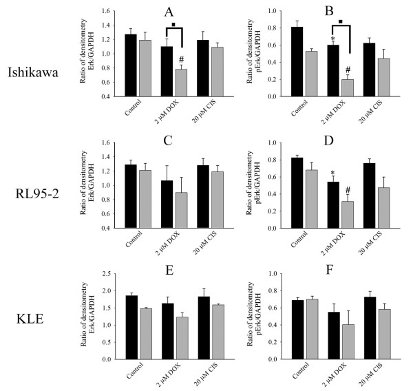
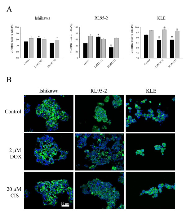
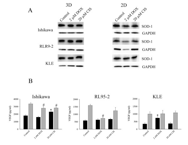
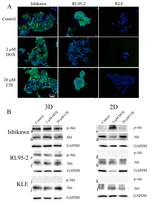
BACKGROUND: Advanced endometrial cancer often shows resistance to clinical chemotherapy although potencies of anticancer drugs in vitro are promising. The disparity suggests that in vivo microenvironments are not recapitulated by in vitro models used for preclinical testing. However, spheroids replicate some important properties of tumours in vivo. Therefore, for the first time, we compared effects of doxorubicin and cisplatin on 3D multicellular structures and 2D cell monolayers of endometrial cancer cells. METHODS: 3D multicellular structures were generated by culturing cancer cells on non-adherent surfaces; and for comparison cell monolayers were cultured on adherent culture plates. Ishikawa, RL95-2, and KLE cell lines were studied. Morphologies of 3D multicellular structures were examined. After 48 hours treatment with anticancer drugs, apoptosis, proliferation, glucose metabolism and vascular endothelial growth factor (VEGF) were analysed. Immunostaining of PCNA, Glut-1, p-Erk1/2, SOD-1 and p-Akt1/2/3 was also performed. RESULTS: Distinct 3D multicellular morphologies were formed by three different endometrial cancer cell lines. Doxorubicin induced less apoptosis in 3D multicellular structures of high grade cancer cells (RL95-2 and KLE cell lines) than in cell monolayers. Parallel alterations in Erk1/2 phosphorylation and cell proliferation might suggest they were linked and again doxorubicin had less effect on 3D multicellular structures than cell monolayers. On the other hand, there was no correlation between altered glucose metabolism and proliferation. The responses depended on cancer cell lines and were apparently not mediated by altered Glut-1 levels. The level of SOD-1 was high in 3D cell cultures. The effects on VEGF secretion were various and cancer cell line dependent. Importantly, both doxorubicin and cisplatin had selective paradoxical stimulatory effects on VEGF secretion. The microenvironment within 3D multicellular structures sustained Akt phosphorylation, consistent with it having a role in anchorage-independent pathways. CONCLUSIONS: The cancer cells responded to microenvironments in a distinctive manner. 3D multicellular structures exhibited greater resistance to the agents than 2D monolayers, and the differences between the culture formats were dependent on cancer cell lines. The effects of anticancer drugs on the intracellular mediators were not similar in 3D and 2D cultures. Therefore, using 3D cell models may have a significant impact on conclusions derived from screening drugs for endometrial carcinomas.
Overview
The claims and statistics of this paper haven’t been checked yet.
Run the check — free, no account needed. It reads the open-access text and checks the sources it cites.
Integrity signals
Registry
Cites
Statistics
Figures
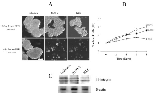
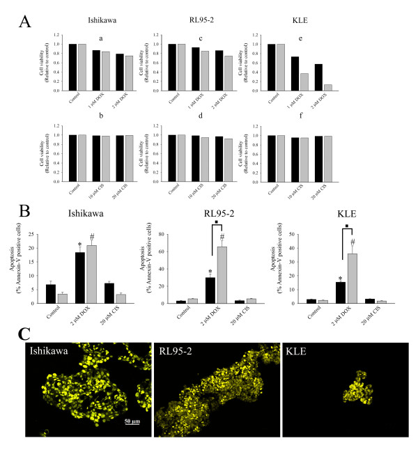
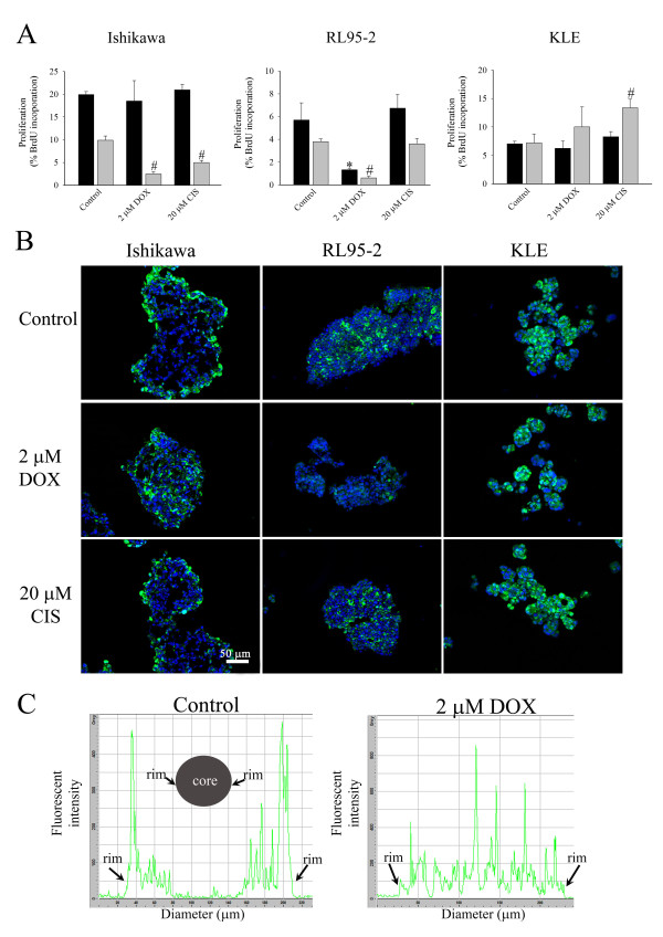
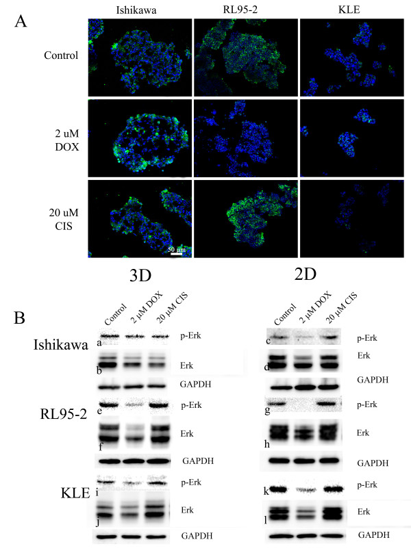
Verification
Claims the paper makes
The claims and statistics of this paper haven’t been checked yet.
Run the check — free, no account needed. It reads the open-access text and checks the sources it cites.
Claims other papers make about this one
Each is checked against this paper’s own text: does it say what the citing paper says it does?
The source backs part of the claim, not all of it — or backs it for a different population, condition or comparison.
In the citing paper
“The proliferation rates of cells cultured in 3D and 2D are usually different, and are cell line and matrix dependent.”
From Three-dimensional cell culture systems and their applications in drug discovery and cell-based biosensors (2014) · cites this paper as [27]
Experimental — accuracy not yet measuredThe source backs part of the claim, not all of it — or backs it for a different population, condition or comparison.
In the citing paper
“Chitcholtan et al . found that regions of hypoxia may exist due to lack of a transport system to remove waste from the center of the spheroid. 27”
From Three-dimensional cell culture systems and their applications in drug discovery and cell-based biosensors (2014) · cites this paper as [27]
Experimental — accuracy not yet measuredThe cited source says what the claim attributes to it.
In the citing paper
“Chitcholtan et al . observed the same pattern to a lesser degree with doxorubicin and KLE endometrial cells. 27”
From Three-dimensional cell culture systems and their applications in drug discovery and cell-based biosensors (2014) · cites this paper as [27]
The check wasn’t confident either way, so it gives no verdict — never disputed.
In the citing paper
“A variety of cell lines showed reduced proliferation rate in 3D cultures compared to those cultured in 2D. 17,27-31”
From Three-dimensional cell culture systems and their applications in drug discovery and cell-based biosensors (2014) · cites this paper as [27]
Experimental — accuracy not yet measured
Self-check
Percentages, sums and reported differences re-computed from the paper’s own numbers.
The self-check needs the paper’s full text.
Citations
Cites
1 retracted
Retraction: TGFbeta1 activates c-Jun and Erk1 via alphaVbeta6 integrin.
Luettich, K., & Schmidt, C. (2004) · Molecular Cancer · doi:10.1186/1476-4598-3-1 · Cited by 70
RetractedDrug penetration in solid tumours
Minchinton, A. I., & Tannock, I. F. (2006) · Nature reviews. Cancer · doi:10.1038/nrc1893 · Cited by 2,805
Majumder, P. K., Febbo, P. G., Bikoff, R., Berger, R., Xue, Q., McMahon, L. M., Manola, J., Brugarolas, J., McDonnell, T. J., Golub, T. R., et al. (2004) · Nature Medicine · doi:10.1038/nm1052 · Cited by 964
Engineering tumors with 3D scaffolds
Fischbach, C., Chen, R., Matsumoto, T., Schmelzle, T., Brugge, J. S., Polverini, P. J., & Mooney, D. J. (2007) · Nature Methods · doi:10.1038/nmeth1085 · Cited by 834
Rapid Generation of Single-Tumor Spheroids for High-Throughput Cell Function and Toxicity Analysis
Ivascu, A., & Kubbies, M. (2006) · SLAS DISCOVERY · doi:10.1177/1087057106292763 · Cited by 618
Epidemiology of Endometrial Cancer 2
Elwood, M., Cole, P. T., Rothman, K. J., & Kaplan, S. D. (1977) · JNCI Journal of the National Cancer Institute · doi:10.1093/jnci/59.4.1055 · Cited by 430
The epidemiology of endometrial cancer
Parazzini, F., Vecchia, C. L., Bocciolone, L., & Franceschi, S. (1991) · Gynecologic Oncology · doi:10.1016/0090-8258(91)90246-2 · Cited by 399
Dynamic analysis of hepatoma spheroid formation: roles of E-cadherin and β1-integrin
Lin, R.-Z., Chou, L.-F., Chien, C.-C. M., & Chang, H.-Y. (2006) · Cell and Tissue Research · doi:10.1007/s00441-005-0148-2 · Cited by 305
Sodek, K. L., Ringuette, M. J., & Brown, T. J. (2009) · International Journal of Cancer · doi:10.1002/ijc.24188 · Cited by 248
Oxygenation and differentiation in multicellular spheroids of human colon carcinoma.
Sutherland, R. M., Sordat, B., Bamat, J., Gabbert, H. E., Bourrat, B., & Mueller‐Klieser, W. (1986) · PubMed · Cited by 237
β1-Integrins Regulate the Formation and Adhesion of Ovarian Carcinoma Multicellular Spheroids
Casey, R. C., Burleson, K. M., Skubitz, K. M., Pambuccian, S. E., Oegema, T. R., Ruff, L. E., & Skubitz, A. P. N. (2001) · American Journal Of Pathology · doi:10.1016/s0002-9440(10)63058-1 · Cited by 236
Epidemiology of endometrial cancer
Purdie, D. M., & Green, A. C. (2001) · Best Practice & Research Clinical Obstetrics & Gynaecology · doi:10.1053/beog.2000.0180 · Cited by 235
VEGF release by MMP-9 mediated heparan sulphate cleavage induces colorectal cancer angiogenesis
Hawinkels, L. J. A. C., Zuidwijk, K., Verspaget, H. W., de Jonge‐Muller, E. S. M., van Duijn, W., Ferreira, V., Fontijn, R. D., David, G., Hommes, D. W., Lamers, C. B. H. W., et al. (2008) · European Journal of Cancer · doi:10.1016/j.ejca.2008.06.031 · Cited by 212
Molecular description of a 3D in vitro model for the study of epithelial ovarian cancer (EOC)
Zietarska, M., Maugard, C. M., Filali‐Mouhim, A., Alam‐Fahmy, M., Tonin, P. N., Provencher, D. M., & Mes‐Masson, A. (2007) · Molecular Carcinogenesis · doi:10.1002/mc.20315 · Cited by 209
Khaitan, D., Chandna, S., Arya, M., & Dwarakanath, B. (2006) · Journal of Translational Medicine · doi:10.1186/1479-5876-4-12 · Cited by 196
Hanging-drop multicellular spheroids as a model of tumour angiogenesis
Timmins, N., Dietmair, S., & Nielsen, L. K. (2004) · Angiogenesis · doi:10.1007/s10456-004-8911-7 · Cited by 177
Prediction of Broad Spectrum Resistance of Tumors towards Anticancer Drugs
Efferth, T., Konkimalla, V. B., Wang, Y., Sauerbrey, A., Meinhardt, S., Zintl, F., Mattern, J., & Volm, M. (2008) · Clinical Cancer Research · doi:10.1158/1078-0432.ccr-07-4525 · Cited by 168
Barbone, D., Yang, T., Morgan, J. R., Gaudino, G., & Broaddus, V. C. (2008) · Journal of Biological Chemistry · doi:10.1074/jbc.m709698200 · Cited by 149
Gagnon, V., Themsche, C. V., Turner, S., Leblanc, V., & Asselin, É. (2007) · APOPTOSIS · doi:10.1007/s10495-007-0165-6 · Cited by 123
Uterine serous and grade 3 endometrioid carcinomas
Boruta, D. M., Gehrig, P. A., Groben, P. A., Bae-Jump, V. L., Boggess, J. F., Fowler, W. C., & Le, L. V. (2004) · Cancer · doi:10.1002/cncr.20645 · Cited by 122
Chan, M. M.-Y., Soprano, K. J., Weinstein, K., & Fong, D. (2006) · Journal of Cellular Physiology · doi:10.1002/jcp.20569 · Cited by 114
AKT involvement in cisplatin chemoresistance of human uterine cancer cells
Gagnon, V., Mathieu, I., Sexton, É., Leblanc, K., & Asselin, É. (2004) · Gynecologic Oncology · doi:10.1016/j.ygyno.2004.06.023 · Cited by 112
Huszar, M., Pfeifer, M., Schirmer, U. B., Kiefel, H., Konecny, G. E., Ben‐Arie, A., Edler, L., Münch, M., Müller‐Holzner, E., Jerabek-Klestil, S., et al. (2009) · The Journal of Pathology · doi:10.1002/path.2673 · Cited by 110
St-Germain, M.-É., Gagnon, V., Parent, S., & Asselin, É. (2004) · Molecular Cancer · doi:10.1186/1476-4598-3-7 · Cited by 109
L'Espérance, S., Bachvarova, M., Tetu, B., Mes-Masson, A.-M., & Bachvarov, D. (2008) · BMC Genomics · doi:10.1186/1471-2164-9-99 · Cited by 108
Green, S. K., Francia, G., Isidoro, C., & Kerbel, R. S. (2004) · Molecular Cancer Therapeutics · doi:10.1158/1535-7163.149.3.2 · Cited by 107
Analysis of the p53 gene in human uterine carcinoma cell lines.
Yaginuma, Y., & Westphal, H. (1991) · PubMed · Cited by 106
Li, X., Lü, Y., Liang, K., Liu, B., & Fan, Z. (2005) · Breast Cancer Research · doi:10.1186/bcr1259 · Cited by 103
Castelbaum, A. J., Ying, L., Somkuti, S. G., Sun, J., Ilesanmi, A. O., & Lessey, B. A. (1997) · The Journal of Clinical Endocrinology & Metabolism · doi:10.1210/jcem.82.1.3658 · Cited by 101
Kwon, D. S., Kwon, C. H., Kim, J. H., Woo, J. S., Jung, J. S., & Kim, Y. K. (2006) · European Journal of Cell Biology · doi:10.1016/j.ejcb.2006.06.001 · Cited by 81
Valcárcel, M., Arteta, B., Jaureguibeitia, A., Lopategi, A., Martinez, I. Z., Mendoza, L., Muruzábal, F., Salado, C., & Vidal‐Vanaclocha, F. (2008) · Journal of Translational Medicine · doi:10.1186/1479-5876-6-57 · Cited by 80
Effects of sustained and intermittent paclitaxel therapy on tumor repopulation in ovarian cancer
Vassileva, V., Allen, C. J., & Piquette‐Miller, M. (2008) · Molecular Cancer Therapeutics · doi:10.1158/1535-7163.mct-07-2117 · Cited by 80
Humtsoe, J. O., & Kramer, R. H. (2009) · Oncogene · doi:10.1038/onc.2009.419 · Cited by 69
Thigpen, T., Vance, R. B., & Khansur, T. (1995) · PubMed · Cited by 62
Three‐dimensional in vitro cell biology models of ovarian and endometrial cancer
Grün, B., Benjamin, E. O., Sinclair, J., Timms, J. F., Jacobs, I. J., Gayther, S. A., & Dafou, D. (2009) · Cell Proliferation · doi:10.1111/j.1365-2184.2008.00579.x · Cited by 60
Wartenberg, M., Hescheler, J., Acker, H., Diedershagen, H., & Sauer, H. (1998) · Cytometry · doi:10.1002/(sici)1097-0320(19980201)31:2 · Cited by 55
Endometrial cancer incidence trends in Norway during 1953–2007 and predictions for 2008–2027
Lindemann, K. Y., Eskild, A., VATTEN, L. J., & Bray, F. (2010) · International Journal of Cancer · doi:10.1002/ijc.25267 · Cited by 53
A Switching Mechanism in Doxorubicin Bioactivation Can Be Exploited to Control Doxorubicin Toxicity
Finn, N. A., Findley, H. W., & Kemp, M. L. (2011) · PLoS Computational Biology · doi:10.1371/journal.pcbi.1002151 · Cited by 51
Medina, R., Meneses, A., Vera, J., Guzmán, C. A., Nualart, F., Rodriguez, F., García, M. D. L. Á., Kato, S., Espinoza, N., Monso, C., et al. (2004) · Journal of Endocrinology · doi:10.1677/joe.0.1820467 · Cited by 43
Nishimura, M. (2003) · Carcinogenesis · doi:10.1093/carcin/bgg121 · Cited by 39
Fayad, W., Brnjic, S., Berglind, D., Blixt, S., Shoshan, M. C., Berndtsson, M., Olofsson, M. H., & Linder, S. T. (2009) · International Journal of Cancer · doi:10.1002/ijc.24627 · Cited by 38
Cella, D. F., Huang, H. Q., Homesley, H. D., MONTAG, A. G., Salani, R., Geest, K. D., Lee, R., & Spirtos, N. M. (2010) · Gynecologic Oncology · doi:10.1016/j.ygyno.2010.08.022 · Cited by 38
Albitar, L., Carter, M. B., Davies, S., & Leslie, K. K. (2007) · Gynecologic Oncology · doi:10.1016/j.ygyno.2007.03.006 · Cited by 36
Kanayama, S., Yamada, Y., Kawaguchi, R., Tsuji, Y., Haruta, S., & Kobayashi, H. (2008) · Oncology Reports · doi:10.3892/or.19.1.117 · Cited by 35
Polo, M. L., Arnoni, V., Riggio, M., Wargon, V., Lanari, C., & Novaro, V. (2010) · PLoS ONE · doi:10.1371/journal.pone.0010786 · Cited by 30
Smith, J. A. (2003) · Gynecologic Oncology · doi:10.1016/j.ygyno.2003.09.037 · Cited by 25
Park, K. R., Inoue, T. K., Ueda, M., Hirano, T., Higuchi, T., Konishi, I., Fujiwara, H., & Fujii, S. (2000) · Molecular Human Reproduction · doi:10.1093/molehr/6.8.719 · Cited by 24
Ferrante, A., Rainaldi, G., Indovina, P., Indovina, P. L., & Santini, M. T. (2006) · International Journal of Oncology · doi:10.3892/ijo.28.1.111 · Cited by 21
Lee, S. Y., Jeong, E.-K., Jeon, H. M., Kim, C. H., & Kang, H. S. (2010) · PubMed · Cited by 19
Assessment of tumor cell repopulation after chemotherapy for advanced ovarian cancer: Pilot study
Davis, A. J., Chapman, W. B., Hedley, D. W., Oza, A. M., & Tannock, I. F. (2002) · Cytometry Part A · doi:10.1002/cyto.a.10001 · Cited by 16
Wada, Y., Shimada, K., Kimura, T., & Ushiyama, S. (2005) · Immunology Letters · doi:10.1016/j.imlet.2005.04.010 · Cited by 16
Activated stress response pathways within multicellular aggregates utilize an autocrine component
Jack, G. D., Cabrera, M. C., Manning, M. L., Slaughter, S. M., Potts, M., & Helm, R. F. (2006) · Cellular Signalling · doi:10.1016/j.cellsig.2006.10.005 · Cited by 14
Rantanen, V. T., Engblom, P., Raitanen, M., Hietanen, S., Haarala, M., Grénman, S., & Syrjänen, S. (2002) · European Journal of Cancer · doi:10.1016/s0959-8049(02)00119-3 · Cited by 13
Cited by
Sorted by citations
Edmondson, R., Broglie, J. J., Adcock, A. F., & Yang, L. (2014) · ASSAY and Drug Development Technologies · doi:10.1089/adt.2014.573 · Cited by 2,515
Three-Dimensional Cell Culture: A Breakthrough in Vivo
Antoni, D., Burckel, H., Josset, E., & Noël, G. (2015) · International Journal of Molecular Sciences · doi:10.3390/ijms16035517 · Cited by 1,049
Advanced Cell Culture Techniques for Cancer Drug Discovery
Lovitt, C. J., Shelper, T., & Avery, V. M. (2014) · Biology · doi:10.3390/biology3020345 · Cited by 292
Models of osteoarthritis: the good, the bad and the promising
Cope, P. J., Ourradi, K., Li, Y., & Sharif, M. (2018) · Osteoarthritis and Cartilage · doi:10.1016/j.joca.2018.09.016 · Cited by 265
Modeling Ewing sarcoma tumors in vitro with 3D scaffolds
Fong, E. L. S., Lamhamedi-Cherradi, S.-E., Burdett, E., Ramamoorthy, V., Lazar, A. J., Kasper, F. K., Farach‐Carson, M. C., Vishwamitra, D., Demicco, E. G., Menegaz, B. A., et al. (2013) · Proceedings of the National Academy of Sciences · doi:10.1073/pnas.1221403110 · Cited by 238
Metadata
- Authors
- Chitcholtan, K., Sykes, P. H., & Evans, J. J.
- Affiliations
- University of Otago (NZ)
- Christchurch Hospital (NZ)
- Corresponding author
- Chitcholtan, Kenny
- Venue
- Journal of Translational Medicine
- Published
- 2012-03-06
- Volume
- 10
- Issue
- 1
- Pages
- 38-38
- ISSN
- 1479-5876 (ISSN-L)
- PMC
- PMC3316127
- OpenAlex
- W2169461456
- Cited by
- 148
- References
- 54
- Citations by year
- 2026: 4 · 2025: 8 · 2024: 6 · 2023: 7 · 2022: 16 · 2021: 7 · 2020: 16 · 2019: 11 · 2018: 14 · 2017: 16 · 2016: 13 · 2015: 7 · 2014: 8 · 2013: 13 · 2012: 2
- FWCI
- 4.74
- Type
- journal article
- Language
- en
- Open access
- gold
- doi.org
- Licence
- Locations
- Journal of Translational Medicine (publishedVersion, cc-by)
- Europe PMC (PubMed Central) (publishedVersion, cc-by)
- PubMed Central (publishedVersion, cc-by)
- PubMed (publishedVersion)
- DOAJ (DOAJ: Directory of Open Access Journals) (submittedVersion)
- Topics
- Health Sciences › Medicine › Oncology › Cancer Cells and Metastasis
- Life Sciences › Biochemistry, Genetics and Molecular Biology › Cancer Research › Cancer, Hypoxia, and Metabolism
- Health Sciences › Medicine › Obstetrics and Gynecology › Endometrial and Cervical Cancer Treatments
- Keywords
- endometrial cancer, doxorubicin, cisplatin, chemoresistance, multicellular spheroids, tumor microenvironment, apoptosis, glucose metabolism, VEGF secretion, Akt phosphorylation, ERK1/2 phosphorylation, SOD1, anchorage-independent growth, cancer cell lines, Ishikawa
- MeSH terms
- Antioxidants, Cell Survival, Cisplatin, Densitometry, Deoxyglucose, Doxorubicin, Female, Glucose, Humans, 4-Chloro-7-nitrobenzofurazan, Phosphorylation, Superoxide Dismutase, Tumor Cells, Cultured, Endometrial Neoplasms, Apoptosis, Spheroids, Cellular, Drug Resistance, Neoplasm, Integrin beta1, Vascular Endothelial Growth Factor A, Intracellular Space, Tissue Culture Techniques, Extracellular Signal-Regulated MAP Kinases, Cell Proliferation, Proto-Oncogene Proteins c-akt, Glucose Transporter Type 1
About these checks
Registry, retraction, Cites and statistics results are computed, not judged, with one exception: a printed reference that no index lists is read once by a model before it counts as a citation error. Claims are extracted by a model and kept only when their quote appears verbatim in the text; each cited source is then checked, with a model’s help, for whether it says what the claim relies on. Those verdicts can be wrong — read the claim’s quote and the cited source before relying on one.
Statistics and arithmetic are recomputed, not judged.
Confidence bands are shown only for fields calibrated on our annotated test set. None is calibrated yet, so no band is shown.
What counts as a claim ›How we measure confidence ›How we check this ›
Are you an author, or do you see a problem with this page? Request a review
