

The DOI matches its Crossref record; 53 of the 54 references it cites were found in a registry or an index.
The resistance of intracellular mediators to doxorubicin and cisplatin are distinct in 3D and 2D endometrial cancer
Open accessAbstract
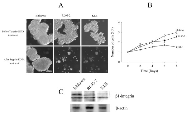
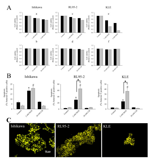
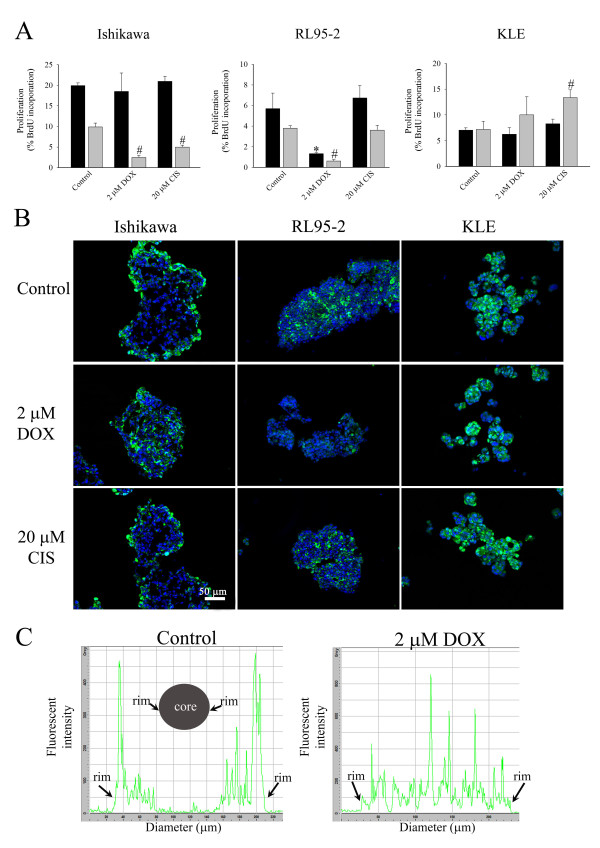
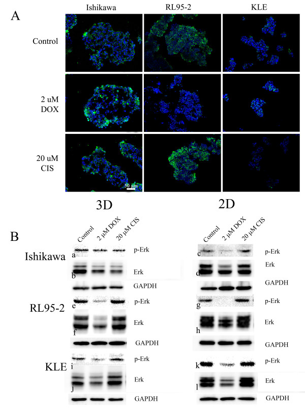
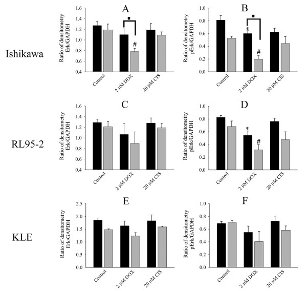
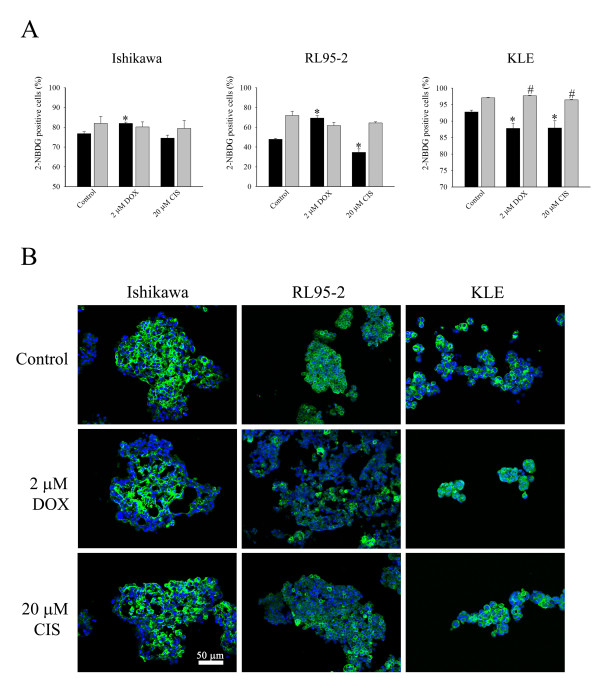
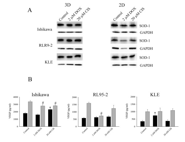
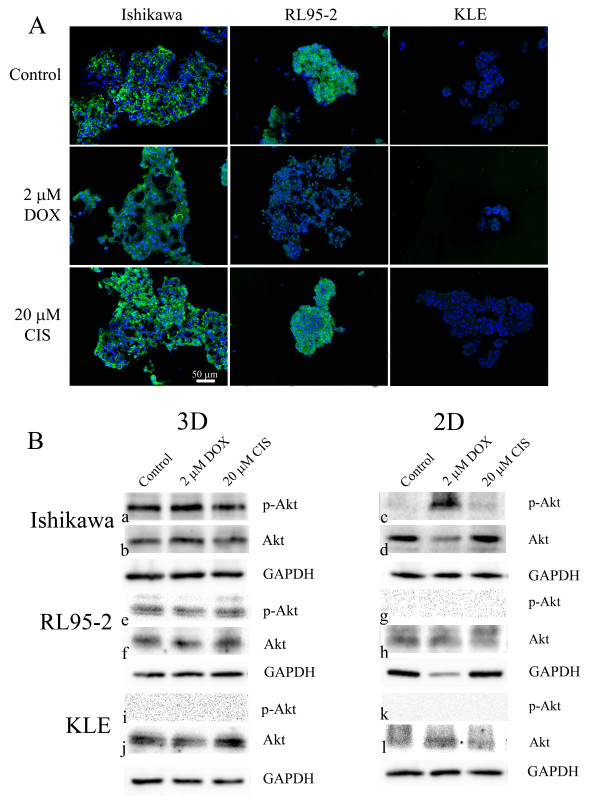
BACKGROUND: Advanced endometrial cancer often shows resistance to clinical chemotherapy although potencies of anticancer drugs in vitro are promising. The disparity suggests that in vivo microenvironments are not recapitulated by in vitro models used for preclinical testing. However, spheroids replicate some important properties of tumours in vivo. Therefore, for the first time, we compared effects of doxorubicin and cisplatin on 3D multicellular structures and 2D cell monolayers of endometrial cancer cells. METHODS: 3D multicellular structures were generated by culturing cancer cells on non-adherent surfaces; and for comparison cell monolayers were cultured on adherent culture plates. Ishikawa, RL95-2, and KLE cell lines were studied. Morphologies of 3D multicellular structures were examined. After 48 hours treatment with anticancer drugs, apoptosis, proliferation, glucose metabolism and vascular endothelial growth factor (VEGF) were analysed. Immunostaining of PCNA, Glut-1, p-Erk1/2, SOD-1 and p-Akt1/2/3 was also performed. RESULTS: Distinct 3D multicellular morphologies were formed by three different endometrial cancer cell lines. Doxorubicin induced less apoptosis in 3D multicellular structures of high grade cancer cells (RL95-2 and KLE cell lines) than in cell monolayers. Parallel alterations in Erk1/2 phosphorylation and cell proliferation might suggest they were linked and again doxorubicin had less effect on 3D multicellular structures than cell monolayers. On the other hand, there was no correlation between altered glucose metabolism and proliferation. The responses depended on cancer cell lines and were apparently not mediated by altered Glut-1 levels. The level of SOD-1 was high in 3D cell cultures. The effects on VEGF secretion were various and cancer cell line dependent. Importantly, both doxorubicin and cisplatin had selective paradoxical stimulatory effects on VEGF secretion. The microenvironment within 3D multicellular structures sustained Akt phosphorylation, consistent with it having a role in anchorage-independent pathways. CONCLUSIONS: The cancer cells responded to microenvironments in a distinctive manner. 3D multicellular structures exhibited greater resistance to the agents than 2D monolayers, and the differences between the culture formats were dependent on cancer cell lines. The effects of anticancer drugs on the intracellular mediators were not similar in 3D and 2D cultures. Therefore, using 3D cell models may have a significant impact on conclusions derived from screening drugs for endometrial carcinomas.
Overview
The claims and statistics of this paper haven’t been checked yet.
Run the check — free, no account needed. It reads the open-access text and checks the sources it cites.
Integrity signals
Registry
Cites
Statistics
Figures
Verification
Claims the paper makes
The claims and statistics of this paper haven’t been checked yet.
Run the check — free, no account needed. It reads the open-access text and checks the sources it cites.
Claims other papers make about this one
Each is checked against this paper’s own text: does it say what the citing paper says it does?
The source backs part of the claim, not all of it — or backs it for a different population, condition or comparison.
In the citing paper
“The proliferation rates of cells cultured in 3D and 2D are usually different, and are cell line and matrix dependent.”
From Three-dimensional cell culture systems and their applications in drug discovery and cell-based biosensors (2014) · cites this paper as [27]
Experimental — accuracy not yet measuredThe source backs part of the claim, not all of it — or backs it for a different population, condition or comparison.
In the citing paper
“Chitcholtan et al . found that regions of hypoxia may exist due to lack of a transport system to remove waste from the center of the spheroid. 27”
From Three-dimensional cell culture systems and their applications in drug discovery and cell-based biosensors (2014) · cites this paper as [27]
Experimental — accuracy not yet measuredThe cited source says what the claim attributes to it.
In the citing paper
“Chitcholtan et al . observed the same pattern to a lesser degree with doxorubicin and KLE endometrial cells. 27”
From Three-dimensional cell culture systems and their applications in drug discovery and cell-based biosensors (2014) · cites this paper as [27]
The check wasn’t confident either way, so it gives no verdict — never disputed.
In the citing paper
“A variety of cell lines showed reduced proliferation rate in 3D cultures compared to those cultured in 2D. 17,27-31”
From Three-dimensional cell culture systems and their applications in drug discovery and cell-based biosensors (2014) · cites this paper as [27]
Experimental — accuracy not yet measured
Self-check
Percentages, sums and reported differences re-computed from the paper’s own numbers.
The self-check needs the paper’s full text.
Citations
Cites
1 retracted
Retraction: TGFbeta1 activates c-Jun and Erk1 via alphaVbeta6 integrin.
Luettich, K., & Schmidt, C. (2004) · Molecular Cancer · doi:10.1186/1476-4598-3-1 · Cited by 70
RetractedDrug penetration in solid tumours
Minchinton, A. I., & Tannock, I. F. (2006) · Nature reviews. Cancer · doi:10.1038/nrc1893 · Cited by 2,805
Majumder, P. K., Febbo, P. G., Bikoff, R., Berger, R., Xue, Q., McMahon, L. M., Manola, J., Brugarolas, J., McDonnell, T. J., Golub, T. R., et al. (2004) · Nature Medicine · doi:10.1038/nm1052 · Cited by 964
Engineering tumors with 3D scaffolds
Fischbach, C., Chen, R., Matsumoto, T., Schmelzle, T., Brugge, J. S., Polverini, P. J., & Mooney, D. J. (2007) · Nature Methods · doi:10.1038/nmeth1085 · Cited by 834
Rapid Generation of Single-Tumor Spheroids for High-Throughput Cell Function and Toxicity Analysis
Ivascu, A., & Kubbies, M. (2006) · SLAS DISCOVERY · doi:10.1177/1087057106292763 · Cited by 618
Epidemiology of Endometrial Cancer 2
Elwood, M., Cole, P. T., Rothman, K. J., & Kaplan, S. D. (1977) · JNCI Journal of the National Cancer Institute · doi:10.1093/jnci/59.4.1055 · Cited by 430
The epidemiology of endometrial cancer
Parazzini, F., Vecchia, C. L., Bocciolone, L., & Franceschi, S. (1991) · Gynecologic Oncology · doi:10.1016/0090-8258(91)90246-2 · Cited by 399
Dynamic analysis of hepatoma spheroid formation: roles of E-cadherin and β1-integrin
Lin, R.-Z., Chou, L.-F., Chien, C.-C. M., & Chang, H.-Y. (2006) · Cell and Tissue Research · doi:10.1007/s00441-005-0148-2 · Cited by 305
Sodek, K. L., Ringuette, M. J., & Brown, T. J. (2009) · International Journal of Cancer · doi:10.1002/ijc.24188 · Cited by 248
Oxygenation and differentiation in multicellular spheroids of human colon carcinoma.
Sutherland, R. M., Sordat, B., Bamat, J., Gabbert, H. E., Bourrat, B., & Mueller‐Klieser, W. (1986) · PubMed · Cited by 237
Cited by
Sorted by citations
Edmondson, R., Broglie, J. J., Adcock, A. F., & Yang, L. (2014) · ASSAY and Drug Development Technologies · doi:10.1089/adt.2014.573 · Cited by 2,515
Three-Dimensional Cell Culture: A Breakthrough in Vivo
Antoni, D., Burckel, H., Josset, E., & Noël, G. (2015) · International Journal of Molecular Sciences · doi:10.3390/ijms16035517 · Cited by 1,049
Advanced Cell Culture Techniques for Cancer Drug Discovery
Lovitt, C. J., Shelper, T., & Avery, V. M. (2014) · Biology · doi:10.3390/biology3020345 · Cited by 292
Models of osteoarthritis: the good, the bad and the promising
Cope, P. J., Ourradi, K., Li, Y., & Sharif, M. (2018) · Osteoarthritis and Cartilage · doi:10.1016/j.joca.2018.09.016 · Cited by 265
Modeling Ewing sarcoma tumors in vitro with 3D scaffolds
Fong, E. L. S., Lamhamedi-Cherradi, S.-E., Burdett, E., Ramamoorthy, V., Lazar, A. J., Kasper, F. K., Farach‐Carson, M. C., Vishwamitra, D., Demicco, E. G., Menegaz, B. A., et al. (2013) · Proceedings of the National Academy of Sciences · doi:10.1073/pnas.1221403110 · Cited by 238
AlgiMatrix™ Based 3D Cell Culture System as an In-Vitro Tumor Model for Anticancer Studies
Godugu, C., Patel, A. R., Desai, U. S., Andey, T. A., Sams, A., & Singh, M. S. (2013) · PLoS ONE · doi:10.1371/journal.pone.0053708 · Cited by 239
Costa, E. C., de Melo‐Diogo, D., Moreira, A. F., Carvalho, M. P., & Correia, I. J. (2018) · Biotechnology Journal · doi:10.1002/biot.201700417 · Cited by 224
do Amaral, R. L. F., Miranda, M. A., Marcato, P. D., & Swiech, K. (2017) · Frontiers in Physiology · doi:10.3389/fphys.2017.00605 · Cited by 227
Jo, Y., Choi, N., Kim, K., Koo, H., Choi, J. H., & Kim, H. N. (2018) · Theranostics · doi:10.7150/thno.29098 · Cited by 188
Emerging Roles of Electrospun Nanofibers in Cancer Research
Chen, S., Boda, S. K., Batra, S. K., Li, X., & Xie, J. (2017) · Advanced Healthcare Materials · doi:10.1002/adhm.201701024 · Cited by 177
Chitcholtan, K., Asselin, E., Parent, S., Sykes, P. H., & Evans, J. J. (2013) · Experimental Cell Research · doi:10.1016/j.yexcr.2012.09.012 · Cited by 173
Self-citationDunne, L. W., Huang, Z., Meng, W., Fan, X., Zhang, N., Zhang, Q., & An, Z. (2014) · Biomaterials · doi:10.1016/j.biomaterials.2014.03.003 · Cited by 167
Using space-based investigations to inform cancer research on Earth
Becker, J. L., & Souza, G. R. (2013) · Nature reviews. Cancer · doi:10.1038/nrc3507 · Cited by 145
Lin, Z., Luo, G., Du, W., Kong, T., Liu, C., & Liu, Z. (2019) · Small · doi:10.1002/smll.201903899 · Cited by 134
Ruppen, J., Wildhaber, F. D., Strub, C., Hall, S. R. R., Schmid, R. A., Geiser, T., & Guenat, O. T. (2015) · Lab on a Chip · doi:10.1039/c5lc00454c · Cited by 129
3D Bioprinting of Tumor Models for Cancer Research
Kang, Y., Datta, P., Shanmughapriya, S., & Ozbolat, I. T. (2020) · ACS Applied Bio Materials · doi:10.1021/acsabm.0c00791 · Cited by 112
Flow perfusion effects on three-dimensional culture and drug sensitivity of Ewing sarcoma
Santoro, M., Lamhamedi-Cherradi, S.-E., Menegaz, B. A., Ludwig, J. A., & Mikos, A. G. (2015) · Proceedings of the National Academy of Sciences · doi:10.1073/pnas.1506684112 · Cited by 107
Two-Dimensional (2D) and Three-Dimensional (3D) Cell Culturing in Drug Discovery
Joseph, J. S., Malindisa, S. T., & Ntwasa, M. M. (2019) · IntechOpen eBooks · doi:10.5772/intechopen.81552 · Cited by 107
Yang, Y., Yang, X., Zou, J., Jia, C., Hu, Y., Du, H., & Wang, H. (2014) · Lab on a Chip · doi:10.1039/c4lc01065e · Cited by 102
Liquid-based three-dimensional tumor models for cancer research and drug discovery
Ham, S. L., Joshi, R., Thakuri, P. S., & Tavana, H. (2016) · Experimental Biology and Medicine · doi:10.1177/1535370216643772 · Cited by 91
From cells-on-a-chip to organs-on-a-chip: scaffolding materials for 3D cell culture in microfluidics
Terrell, J. A., Jones, C. G., Kabandana, G. K. M., & Chen, C. (2020) · Journal of Materials Chemistry B · doi:10.1039/d0tb00718h · Cited by 90
Small cell lung cancer: model of circulating tumor cell tumorospheres in chemoresistance
Klameth, L., Rath, B., Hochmaier, M., Moser, D., Redl, M., Mungenast, F., Gelles, K., Ulsperger, E., Zeillinger, R., & Hamilton, G. (2017) · Scientific Reports · doi:10.1038/s41598-017-05562-z · Cited by 87
Strategies for modelling endometrial diseases
Murphy, A. R., Campo, H., & Kim, J. J. (2022) · Nature Reviews Endocrinology · doi:10.1038/s41574-022-00725-z · Cited by 88
Antitumor drug delivery in multicellular spheroids by electropermeabilization
Gibot, L., Wasungu, L., Teissié, J., & Rols, M. (2013) · Journal of Controlled Release · doi:10.1016/j.jconrel.2013.01.021 · Cited by 87
Yang, Y., Hu, Y., & Wang, H. (2016) · Oxidative Medicine and Cellular Longevity · doi:10.1155/2016/5274084 · Cited by 76
Edmondson, R., Adcock, A. F., & Yang, L. (2016) · PLoS ONE · doi:10.1371/journal.pone.0158116 · Cited by 69
Structure establishment of three-dimensional (3D) cell culture printing model for bladder cancer
Kim, M. J., Chi, B. H., Yoo, J. J., Ju, Y. M., Whang, Y. M., & Chang, I. H. (2019) · PLoS ONE · doi:10.1371/journal.pone.0223689 · Cited by 69
Three-Dimensional 3D Culture Models in Gynecological and Breast Cancer Research
Salinas-Vera, Y. M., Valdés, J., Pérez‐Navarro, Y., Mandujano-Lázaro, G., Marchat, L. A., Ramos‐Payán, R., Núñez-Olvera, S. I., Pérez‐Plasencia, C., & López‐Camarillo, C. (2022) · Frontiers in Oncology · doi:10.3389/fonc.2022.826113 · Cited by 74
Anticancer Activity of Bee Venom Components against Breast Cancer
Kwon, N.-Y., Sung, S.-H., Sung, H.-K., & Park, J.-K. (2022) · Toxins · doi:10.3390/toxins14070460 · Cited by 59
Collins, A., Miles, G. J., Wood, J. P., MacFarlane, M., Pritchard, C. A., & Moss, E. L. (2019) · Gynecologic Oncology · doi:10.1016/j.ygyno.2019.11.020 · Cited by 61
Dubois, C., Dufour, R., Daumar, P., Aubel, C., Szczepaniak, C., Blavignac, C., Mounetou, E., Penault-Llorca, F. M., & Bamdad, M. (2017) · Oncotarget · doi:10.18632/oncotarget.20517 · Cited by 65
Engel, B. J., Constantinou, P. E., Sablatura, L. K., Doty, N. J., Carson, D. D., Farach‐Carson, M. C., Harrington, D. A., & Zarembinski, T. I. (2015) · Advanced Healthcare Materials · doi:10.1002/adhm.201500258 · Cited by 59
Spheroid Culture Differentially Affects Cancer Cell Sensitivity to Drugs in Melanoma and RCC Models
Filipiak-Duliban, A., Brodaczewska, K. K., Kajdasz, A., & Kiéda, C. (2022) · International Journal of Molecular Sciences · doi:10.3390/ijms23031166 · Cited by 59
Arai, K., Sakamoto, R., Kubota, D., & Kondo, T. (2013) · PROTEOMICS · doi:10.1002/pmic.201300053 · Cited by 57
Magnussen, G. I., Emilsen, E., Fleten, K. G., Engesæter, B. Ø., Nähse, V., Fjær, R., Slipicevic, A., & Flørenes, V. A. (2015) · BMC Cancer · doi:10.1186/s12885-015-1474-8 · Cited by 50
Tino, A. B., Chitcholtan, K., Sykes, P. H., & Garrill, A. (2016) · Journal of Ovarian Research · doi:10.1186/s13048-016-0293-0 · Cited by 46
Self-citationThree dimensional cultivation increases chemo- and radioresistance of colorectal cancer cell lines
Koch, J., Mönch, D., Maaß, A., Gromoll, C., Hehr, T., Leibold, T., Schlitt, H. J., Dahlke, M., & Renner, P. (2021) · PLoS ONE · doi:10.1371/journal.pone.0244513 · Cited by 50
Real-time monitoring of cisplatin cytotoxicity on three-dimensional spheroid tumor cells
An, S. S. A., Seo, O. W., Lee, J.-H., Hulme, J., & Baek, N. (2016) · Drug Design Development and Therapy · doi:10.2147/dddt.s108004 · Cited by 47
Turetta, M., Ben, F. D., Brisotto, G., Biscontin, E., Bulfoni, M., Cesselli, D., Colombatti, A., Scoles, G., Gigli, G., & del Mercato, L. L. (2018) · Current Medicinal Chemistry · doi:10.2174/0929867325666180605122633 · Cited by 46
Doddapaneni, R., Patel, K. R., Chowdhury, N., & Singh, M. S. (2017) · Scientific Reports · doi:10.1038/s41598-017-15531-1 · Cited by 39
Nunes, A. S., Costa, E. C., Barros, A. S., de Melo‐Diogo, D., & Correia, I. J. (2018) · Biotechnology Journal · doi:10.1002/biot.201800268 · Cited by 37
Organoids of the Female Reproductive Tract: Innovative Tools to Study Desired to Unwelcome Processes
Heremans, R., Jan, Ž., Timmerman, D., & Vankelecom, H. E. J. (2021) · Frontiers in Cell and Developmental Biology · doi:10.3389/fcell.2021.661472 · Cited by 36
A microfluidic gradient generator to simulate the oxygen microenvironment in cancer cell culture
Orcheston-Findlay, L., Hashemi, A., Garrill, A., & Nock, V. (2018) · Microelectronic Engineering · doi:10.1016/j.mee.2018.04.011 · Cited by 32
Mani, V., Lyu, Z., Kumar, V., Ercal, B., Chen, H., Malhotra, S. V., & Demirci, U. (2018) · Advanced Biosystems · doi:10.1002/adbi.201800223 · Cited by 32
Architectural control of metabolic plasticity in epithelial cancer cells
Al‐Masri, M., Paliotti, K., Tran, R., Halaoui, R., Lelarge, V., Chatterjee, S. J., Wang, L., Moraes, C., & McCaffrey, L. M. (2021) · Communications Biology · doi:10.1038/s42003-021-01899-4 · Cited by 33
Kedaria, D., & Vasita, R. (2017) · Journal of Tissue Engineering · doi:10.1177/2041731417718391 · Cited by 27
Rezakhani, L., Rahmati, S., Ghasemi, S., Alizadeh, M., & Alizadeh, A. (2022) · Chemistry and Physics of Lipids · doi:10.1016/j.chemphyslip.2022.105179 · Cited by 27
Wang, W., Cai, Q., Zhou, F., Liu, J., Jin, X., Ni, P., Lu, M., Wang, G., & Zhang, J. (2017) · Redox Biology · doi:10.1016/j.redox.2017.12.009 · Cited by 27
Cancer Wars: Revenge of the AMPs (Antimicrobial Peptides), a New Strategy against Colorectal Cancer
Răileanu, M., & Bacalum, M. (2023) · Toxins · doi:10.3390/toxins15070459 · Cited by 26
Fujiwara, D., Kato, K., Nohara, S., Iwanuma, Y., & Kajiyama, Y. (2013) · Biochemical and Biophysical Research Communications · doi:10.1016/j.bbrc.2013.04.008 · Cited by 25
Gentilin, E., Minoia, M., Bondanelli, M., Tagliati, F., Uberti, E. C. D., & Zatelli, M. C. (2016) · Endocrine · doi:10.1007/s12020-016-1085-4 · Cited by 25
3D patient-derived tumor models to recapitulate pediatric brain tumors In Vitro
Tang‐Schomer, M. D., Chandok, H., Wu, W.-B., Lau, C. C., Bookland, M. J., & George, J. (2022) · Translational Oncology · doi:10.1016/j.tranon.2022.101407 · Cited by 26
Microdevice-based mechanical compression on living cells
Önal, S., Alkaisi, M. M., & Nock, V. (2022) · iScience · doi:10.1016/j.isci.2022.105518 · Cited by 26
Assessing multiparametric drug response in tissue engineered tumor microenvironment models
Harris, A. R., Yuan, J. X., & Munson, J. M. (2017) · Methods · doi:10.1016/j.ymeth.2017.12.010 · Cited by 24
Dynamic in vitro models for tumor tissue engineering
Karami, D., Richbourg, N. R., & Sikavitsas, V. I. (2019) · Cancer Letters · doi:10.1016/j.canlet.2019.01.043 · Cited by 27
Evaluation of Cyclosaplin Efficacy Using a Silk Based 3D Tumor Model
Mishra, A., Mukhopadhyay, S. K., & Dey, S. (2019) · Biomolecules · doi:10.3390/biom9040123 · Cited by 24
Biomaterial strategies to replicate gynecological tissue
Cadena, I., Chen, A. L., Arvidson, A., & Fogg, K. C. (2020) · Biomaterials Science · doi:10.1039/d0bm01240h · Cited by 23
Winship, A., Sinderen, M. L. V., Rainczuk, K., & Dimitriadis, E. (2017) · Oncotarget · doi:10.18632/oncotarget.15187 · Cited by 22
3D Spheroids’ Sensitivity to Electric Field Pulses Depends on Their Size
Gibot, L., & Rols, M. (2013) · The Journal of Membrane Biology · doi:10.1007/s00232-013-9535-x · Cited by 27
Imaging of Intracellular pH in Tumor Spheroids Using Genetically Encoded Sensor SypHer2
Вадимовна, З. Е., Николаевна, Д. И., Mishina, N. M., Ignatova, N. I., Dudenkova, V. V., & Shirmanova, M. V. (2017) · Advances in experimental medicine and biology · doi:10.1007/978-3-319-67358-5_7 · Cited by 21
Ono, K., Sato, K., Nakamura, T., Yoshida, Y., Murata, S., Yoshida, K., Kanemoto, H., Umemori, K., Kawai, H., Obata, K., et al. (2022) · International Journal of Medical Sciences · doi:10.7150/ijms.74109 · Cited by 20
Glenister, A., Simone, M. I., & Hambley, T. W. (2019) · PLoS ONE · doi:10.1371/journal.pone.0217712 · Cited by 19
Tian, T., Li, C., Fu, X., Wang, S.-H., Lü, J., Guo, H., Yao, Y., Nan, K., & Yang, Y. (2018) · Journal of International Medical Research · doi:10.1177/0300060517740807 · Cited by 20
Hassan, W., Chitcholtan, K., Sykes, P. H., & Garrill, A. (2016) · Oncology and Therapy · doi:10.1007/s40487-016-0031-1 · Cited by 17
Self-citationTan, L. H., Sykes, P. H., Alkaisi, M. M., & Evans, J. J. (2017) · Biofabrication · doi:10.1088/1758-5090/aa5c9a · Cited by 18
Self-citationRavi, M., Kaviya, S. R., & Paramesh, V. (2014) · Cytotechnology · doi:10.1007/s10616-014-9795-z · Cited by 18
Cancer-on-a-chip for Drug Screening
Lee, I. (2019) · Current Pharmaceutical Design · doi:10.2174/1381612825666190206235233 · Cited by 17
Kamran, M. R., Zargan, J., Alikhani, H. K., & Hajinoormohammadi, A. (2019) · International Journal of Peptide Research and Therapeutics · doi:10.1007/s10989-019-09979-0 · Cited by 16
Zhang, G., Liang, Q., Wu, Y., & Wang, Y. (2025) · International Journal of Molecular Medicine · doi:10.3892/ijmm.2025.5626 · Cited by 17
Lattuada, D., Roda, B., Pignatari, C., Magni, R., Colombo, F., Cattaneo, A., Zattoni, A., Cetin, I., Reschiglian, P., & Bolis, G. (2012) · Analytical and Bioanalytical Chemistry · doi:10.1007/s00216-012-6337-4 · Cited by 15
Chemosensitivity and chemoresistance in endometriosis – differences for ectopic versus eutopic cells
Lavõgina, D., Samuel, K., Lavrits, A., Meltsov, A., Sõritsa, D., Kadastik, Ü., Peters, M., Rinken, A., & Salumets, A. (2019) · Reproductive BioMedicine Online · doi:10.1016/j.rbmo.2019.05.019 · Cited by 15
Fleten, K. G., Flørenes, V. A., Prasmickaite, L., Hill, O., Sykora, J., Mælandsmo, G. M., & Engesæter, B. (2016) · Cell Death Discovery · doi:10.1038/cddiscovery.2016.81 · Cited by 14
Issues with Cancer Spheroid Models in Therapeutic Drug Screening
Fröhlich, E. (2020) · Current Pharmaceutical Design · doi:10.2174/1381612826666200218094200 · Cited by 14
Roudsari, L. C., Jeffs, S. E., & West, J. L. (2017) · ACS Biomaterials Science & Engineering · doi:10.1021/acsbiomaterials.7b00011 · Cited by 14
Navigating Cancer Treatment: A Journey from 2D to 3D Cancer Models and Nanoscale Therapies
Kaur, N., Savitsky, M. J., Scutte, A., Mehta, R., Dridi, N., Sogbesan, T., Savannah, A., Lopez, D., Yang, D., & Ali, J. (2025) · ACS Pharmacology & Translational Science · doi:10.1021/acsptsci.5c00530 · Cited by 15
Trainito, C., Bayart, É., Subra, F., Français, O., & Pioufle, B. L. (2016) · The Journal of Membrane Biology · doi:10.1007/s00232-016-9880-7 · Cited by 15
Lee, S., NAM, J. K., Park, J. K., Lee, J., Min, D. S., & Kuh, H. (2014) · Oncology Reports · doi:10.3892/or.2014.3337 · Cited by 13
Nanocelluloses – Nanotoxicology, Safety Aspects and 3D Bioprinting
Chinga‐Carrasco, G., Rosendahl, J., & Catalán, J. (2022) · Advances in experimental medicine and biology · doi:10.1007/978-3-030-88071-2_7 · Cited by 13
Konings, G. F. J., Saarinen, N., Delvoux, B., Kooreman, L. F. S., Koskimies, P., Krakstad, C., Fasmer, K. E., Haldorsen, I. S., Zaffagnini, A., Häkkinen, M. R., et al. (2018) · International Journal of Molecular Sciences · doi:10.3390/ijms19092547 · Cited by 13
Lee, Y., Park, K.-S., Heo, S., Cho, M. K., & Lee, S.-H. (2023) · Oncology Reports · doi:10.3892/or.2023.8548 · Cited by 12
Lu, M., Zhou, F., Hao, K., Liu, J., Chen, Q., Ni, P., Zhou, H., Wang, G., & Zhang, J. (2014) · Biochemical Pharmacology · doi:10.1016/j.bcp.2014.11.010 · Cited by 12
Bilgin, S. (2024) · Molecular Biology Reports · doi:10.1007/s11033-024-09562-x · Cited by 11
Januszyk, P., Januszyk, K., Wierzbik-Strońska, M., Boroń, D., & Grabarek, B. O. (2020) · Current Pharmaceutical Biotechnology · doi:10.2174/1389201021666200629151008 · Cited by 11
Engineering human endometrial model systems in reproductive health and disease
Wang, Y., Guo, Y., Yang, Y., Ma, X., & Qin, J. (2025) · npj Biomedical Innovations. · doi:10.1038/s44385-025-00047-5 · Cited by 10
Bhaumik, S., Boyer, J. Z., Banerjee, C., Clark, S., Sebastiao, N., Vela, E. E., & Towne, P. (2020) · Journal of Cellular Biochemistry · doi:10.1002/jcb.29827 · Cited by 10
Two-dimensional and three-dimensional cell culture models in vitro: pros and cons
Galimova, E. S., & Galagudza, M. M. (2018) · Bulletin of Siberian Medicine · doi:10.20538/1682-0363-2018-3-188-196 · Cited by 10
Innovative Models to Assess Multiple Myeloma Biology and the Impact of Drugs
Ferrarini, M., Mazzoleni, G., Steimberg, N., Belloni, D., & Ferrero, E. (2013) · InTech eBooks · doi:10.5772/54312 · Cited by 12
Ravi, M., Sah, S., & Bhammar, R. (2013) · Journal of Cellular Physiology · doi:10.1002/jcp.24433 · Cited by 9
Pizzimenti, S. (2014) · Journal of Pediatric Oncology · doi:10.14205/2309-3021.2014.02.01.5 · Cited by 10
Proton NMR characterization of intact primary and metastatic melanoma cells in 2D & 3D cultures
Ramachandran, G. K., & Yeow, C. (2017) · Biological Research · doi:10.1186/s40659-017-0117-8 · Cited by 9
Trainito, C. (2015) · theses.fr (ABES) · doi:10.70675/aaec0da8z02d6z442czb0ddz578952a52dc8 · Cited by 9
Let’s Go 3D! New Generation of Models for Evaluating Drug Response and Resistance in Prostate Cancer
Petrić, T., & Sabol, M. (2023) · International Journal of Molecular Sciences · doi:10.3390/ijms24065293 · Cited by 8
Ionizing radiation induced biological effects in three-dimensional cell cultures
Pan, D., Xue, G., Zhu, J., & Hu, B. (2013) · RENDICONTI LINCEI · doi:10.1007/s12210-013-0260-2 · Cited by 8
Identification of Gastric Cancer Biomarkers Using 1H Nuclear Magnetic Resonance Spectrometry
Ramachandran, G. K., Yong, W. P., & Yeow, C. (2016) · PLoS ONE · doi:10.1371/journal.pone.0162222 · Cited by 8
Liu, Y., Yang, G. H., Wu, Y., Feng, Y., Liu, H., Cao, W., Xie, J., & Zhang, J. (2024) · International Journal of Molecular Sciences · doi:10.3390/ijms25021081 · Cited by 7
Zhuang, S., Ding, N., Zhang, T., & Yu, X. (2022) · Current Protein and Peptide Science · doi:10.2174/1389203723666220823152239 · Cited by 7
Sukphokkit, S., Kiatwuthinon, P., Kumkate, S., & Janvilisri, T. (2023) · Frontiers in Oncology · doi:10.3389/fonc.2022.999158 · Cited by 7
Li, Y., Qin, Z., Zhang, F., & Yang, S. (2021) · Biotechnology and Bioengineering · doi:10.1002/bit.28006 · Cited by 7
Going 3D – Cell Culture Approaches for Stem Cell Research and Therapy
Greiner, J. F. W., Kaltschmidt, B., Kaltschmidt, C., & Widera, D. (2013) · Current Tissue Engineering · doi:10.2174/2211542011302010003 · Cited by 6
Scaffold-mediated switching of lymphoma metabolism in culture
Bhatt, R., Ravi, D., Evens, A. M., & Parekkadan, B. (2022) · Cancer & Metabolism · doi:10.1186/s40170-022-00291-y · Cited by 6
Metadata
- Authors
- Chitcholtan, K., Sykes, P. H., & Evans, J. J.
- Affiliations
- University of Otago (NZ)
- Christchurch Hospital (NZ)
- Corresponding author
- Chitcholtan, Kenny
- Venue
- Journal of Translational Medicine
- Published
- 2012-03-06
- Volume
- 10
- Issue
- 1
- Pages
- 38-38
- ISSN
- 1479-5876 (ISSN-L)
- PMC
- PMC3316127
- OpenAlex
- W2169461456
- Cited by
- 148
- References
- 54
- Citations by year
- 2026: 4 · 2025: 8 · 2024: 6 · 2023: 7 · 2022: 16 · 2021: 7 · 2020: 16 · 2019: 11 · 2018: 14 · 2017: 16 · 2016: 13 · 2015: 7 · 2014: 8 · 2013: 13 · 2012: 2
- FWCI
- 4.74
- Type
- journal article
- Language
- en
- Open access
- gold
- doi.org
- Licence
- Locations
- Journal of Translational Medicine (publishedVersion, cc-by)
- Europe PMC (PubMed Central) (publishedVersion, cc-by)
- PubMed Central (publishedVersion, cc-by)
- PubMed (publishedVersion)
- DOAJ (DOAJ: Directory of Open Access Journals) (submittedVersion)
- Topics
- Health Sciences › Medicine › Oncology › Cancer Cells and Metastasis
- Life Sciences › Biochemistry, Genetics and Molecular Biology › Cancer Research › Cancer, Hypoxia, and Metabolism
- Health Sciences › Medicine › Obstetrics and Gynecology › Endometrial and Cervical Cancer Treatments
- Keywords
- endometrial cancer, doxorubicin, cisplatin, chemoresistance, multicellular spheroids, tumor microenvironment, apoptosis, glucose metabolism, VEGF secretion, Akt phosphorylation, ERK1/2 phosphorylation, SOD1, anchorage-independent growth, cancer cell lines, Ishikawa
- MeSH terms
- Antioxidants, Cell Survival, Cisplatin, Densitometry, Deoxyglucose, Doxorubicin, Female, Glucose, Humans, 4-Chloro-7-nitrobenzofurazan, Phosphorylation, Superoxide Dismutase, Tumor Cells, Cultured, Endometrial Neoplasms, Apoptosis, Spheroids, Cellular, Drug Resistance, Neoplasm, Integrin beta1, Vascular Endothelial Growth Factor A, Intracellular Space, Tissue Culture Techniques, Extracellular Signal-Regulated MAP Kinases, Cell Proliferation, Proto-Oncogene Proteins c-akt, Glucose Transporter Type 1
About these checks
Registry, retraction, Cites and statistics results are computed, not judged, with one exception: a printed reference that no index lists is read once by a model before it counts as a citation error. Claims are extracted by a model and kept only when their quote appears verbatim in the text; each cited source is then checked, with a model’s help, for whether it says what the claim relies on. Those verdicts can be wrong — read the claim’s quote and the cited source before relying on one.
Statistics and arithmetic are recomputed, not judged.
Confidence bands are shown only for fields calibrated on our annotated test set. None is calibrated yet, so no band is shown.
What counts as a claim ›How we measure confidence ›How we check this ›
Are you an author, or do you see a problem with this page? Request a review
