

The DOI matches its Crossref record; 62 of the 64 references it cites were found in a registry or an index.
A systematic study of the N-glycosylation sites of HIV-1 envelope protein on infectivity and antibody-mediated neutralization
Open accessAbstract
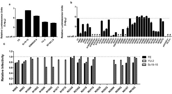
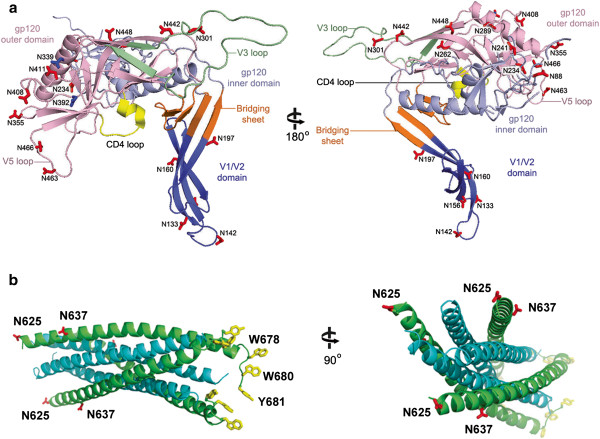
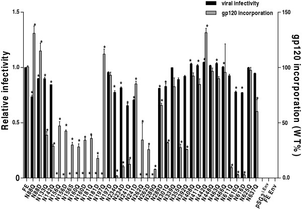
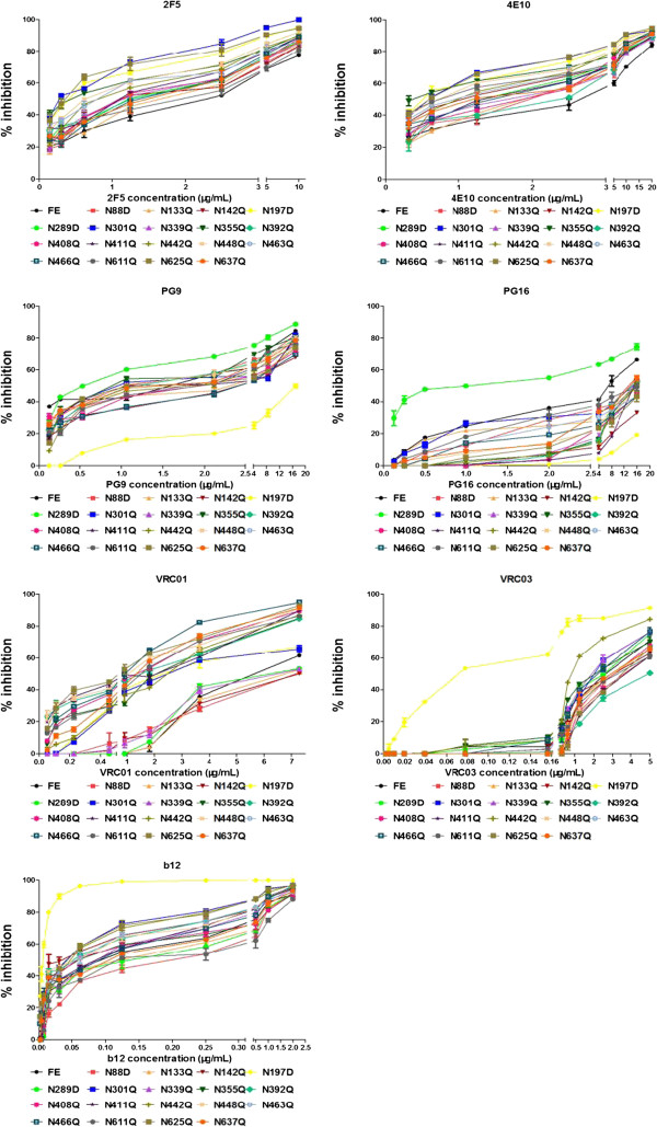
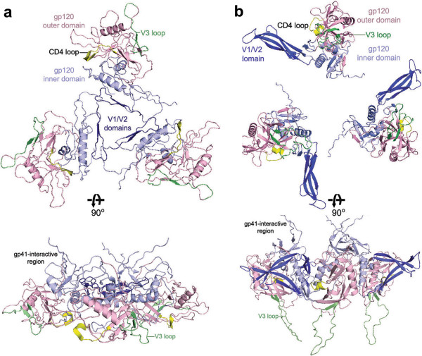
BACKGROUND: Glycans on the human immunodeficiency virus (HIV) envelope glycoprotein (Env) play an important role in viral infection and evasion of neutralization by antibodies. In this study, all 25 potential N-linked glycosylation sites (PNGS) on the HIV-1 CRF07_BC Env, FE, were mutated individually to study the effect of their removal on viral infectivity, virion production, and antibody-mediated neutralization. RESULTS: Removal of specific N-glycosylation sites has a significant effect on viral infectivity and antibody-mediated neutralization phenotype. Six of these glycosylation mutants located on the V1/V2 and C1/C2 domains lost infectivity. PNGS mutations located on V4/C4/V5 (except N392 on V4), were shown to increase viral infectivity. Furthermore, FE is much more dependent on specific glycans than clade B Env YU-2. On neutralization effect, PNGS mutations at N197 (C2), N301 (V3), N442 (C4) and N625 (gp41) rendered the virus more susceptible to neutralization by the monoclonal antibodies (MAbs) that recognize the CD4 binding site or gp41. Generally, mutations on V4/V5 loops, C2/C3/C4 regions and gp41 reduced the neutralization sensitivity to PG16. However, mutation of N289 (C2) made the virus more sensitive to both PG9 and PG16. Furthermore, we showed that mutations at N142 (V1), N355 (C3) and N463 (V5) conferred resistance to neutralization by anti-gp41 MAbs. We used the available structural information of HIV Env and homology modeling to provide a structural basis for the observed biological effects of these mutations. CONCLUSIONS: This report provides the first systematic experimental account of the biological role of the entire PNGS on an HIV-1 Env, which should provide valuable insights for understanding the function of Env in HIV infection cycle and for developing future anti-HIV strategies.
Overview
The claims and statistics of this paper haven’t been checked yet.
Run the check — free, no account needed. It reads the open-access text and checks the sources it cites.
Integrity signals
Registry
Cites
Statistics
Figures
Verification
Claims the paper makes
The claims and statistics of this paper haven’t been checked yet.
Run the check — free, no account needed. It reads the open-access text and checks the sources it cites.
Claims other papers make about this one
Each is checked against this paper’s own text: does it say what the citing paper says it does?
The source backs part of the claim, not all of it — or backs it for a different population, condition or comparison.
In the citing paper
“Unlike HIV-1 Env, which contains ~25 glycosylation sites [101], and forms a “glycan shield” protecting the virus from neutralizing antibodies [102], HTLV-1 Env contains only five sites for N-glycan attachment [103].”
From HIV-1 and HTLV-1 Transmission Modes: Mechanisms and Importance for Virus Spread (2022) · cites this paper as [101]
Experimental — accuracy not yet measured
Self-check
Percentages, sums and reported differences re-computed from the paper’s own numbers.
The self-check needs the paper’s full text.
Citations
Cites
Kwong, P. D., Wyatt, R. T., Robinson, J. E., Sweet, R. W., Sodroski, J., & Hendrickson, W. A. (1998) · Nature · doi:10.1038/31405 · Cited by 2,963
Antibody neutralization and escape by HIV-1
Wei, X., Decker, J. M., Wang, S., Hui, H., Kappes, J. C., Wu, X., Salazar-Gonzalez, J. F., Salazar, M. G., Kilby, J. M., Saag, M. S., et al. (2003) · Nature · doi:10.1038/nature01470 · Cited by 2,421
Self-citationBroad and Potent Neutralizing Antibodies from an African Donor Reveal a New HIV-1 Vaccine Target
Walker, L., Phogat, S., Chan-hui, P., Wagner, D., Pham, P., Goss, J. L., Wrin, T., Simek, M., Fling, S., Mitcham, J., et al. (2009) · Science · doi:10.1126/science.1178746 · Cited by 1,849
Atomic structure of the ectodomain from HIV-1 gp41
Weissenhorn, W., Dessen, A., Harrison, S. C., Skehel, J. J., & Wiley, D. C. (1997) · Nature · doi:10.1038/387426a0 · Cited by 1,625
The HIV-1 Envelope Glycoproteins: Fusogens, Antigens, and Immunogens
Wyatt, R. T., & Sodroski, J. (1998) · Science · doi:10.1126/science.280.5371.1884 · Cited by 1,605
Structural Basis for Broad and Potent Neutralization of HIV-1 by Antibody VRC01
Zhou, T., Georgiev, I. S., Wu, X., Yang, Z., Dai, K., Finzi, A., Kwon, Y. D., Scheid, J. F., Shi, W., Xu, L., et al. (2010) · Science · doi:10.1126/science.1192819 · Cited by 1,123
Li, M., Gao, F., Mascola, J. R., Stamatatos, L., Polonis, V. R., Koutsoukos, M., Voss, G., Goepfert, P., Gilbert, P. B., Greene, K., et al. (2005) · Journal of Virology · doi:10.1128/jvi.79.16.10108-10125.2005 · Cited by 1,077
A conserved neutralizing epitope on gp41 of human immunodeficiency virus type 1
Muster, T., Steindl, F., Purtscher, M., Trkola, A., Klima, A., Himmler, G., Rüker, F., & Katinger, H. W. D. (1993) · Journal of Virology · doi:10.1128/jvi.67.11.6642-6647.1993 · Cited by 1,011
Leonard, C. K., Spellman, M. W., Riddle, L. M., Harris, R. J., Thomas, J. N., & Gregory, T. (1990) · Journal of Biological Chemistry · doi:10.1016/s0021-9258(18)86956-3 · Cited by 986
Crystal Structure of a Neutralizing Human IgG Against HIV-1: A Template for Vaccine Design
Saphire, E. O., Parren, P. W. H. I., Pantophlet, R. A., Zwick, M. B., Morris, G. M., Rudd, P. M., Dwek, R. A., Stanfield, R. L., Burton, D. R., & Wilson, I. A. (2001) · Science · doi:10.1126/science.1061692 · Cited by 959
Research works printed but not linked: 1
- Human Retroviruses and AIDS 1989: A Compilation and Analysis of Nucleic Acid and Amino Acid Sequences. Edited by: Myers GRA, Josephs SF, Smith TF, Wong-Staal F, Berzofsky JA. 1989, Los Alamos, NM: Theoretical Biology and Biophysics Group, Los Alamos National Laboratory
Cited by
Sorted by citations
The Impact of Mutations in SARS-CoV-2 Spike on Viral Infectivity and Antigenicity
Li, Q., Wu, J., Nie, J., Zhang, L., Hao, H., Liu, S., Zhao, C., Zhang, Q., Liu, H., Nie, L., et al. (2020) · Cell · doi:10.1016/j.cell.2020.07.012 · Cited by 1,420
Self-citationCryo-EM Structure of a Fully Glycosylated Soluble Cleaved HIV-1 Envelope Trimer
Lyumkis, D., Julien, J., de Val, N., Cupo, A., Potter, C. S., Klasse, P.-J., Burton, D. R., Sanders, R. W., Moore, J. P., Carragher, B. O., et al. (2013) · Science · doi:10.1126/science.1245627 · Cited by 683
Exploitation of glycosylation in enveloped virus pathobiology
Watanabe, Y., Bowden, T. A., Wilson, I. A., & Crispin, M. (2019) · Biochimica et Biophysica Acta (BBA) - General Subjects · doi:10.1016/j.bbagen.2019.05.012 · Cited by 489
SARS-CoV-2 escape from a highly neutralizing COVID-19 convalescent plasma
Andreano, E., Piccini, G., Licastro, D., Casalino, L., Johnson, N. V., Paciello, I., Monego, S. D., Pantano, E., Manganaro, N., Manenti, A., et al. (2021) · Proceedings of the National Academy of Sciences · doi:10.1073/pnas.2103154118 · Cited by 429
SARS-CoV-2 escape in vitro from a highly neutralizing COVID-19 convalescent plasma
Andreano, E., Piccini, G., Licastro, D., Casalino, L., Johnson, N. V., Paciello, I., Monego, S. D., Pantano, E., Manganaro, N., Manenti, A., et al. (2020) · doi:10.1101/2020.12.28.424451
“The introduction of a glycan is a well-known immunogenic escape strategy described in influenza (29), HIV-1 and other viruses (30-32), although to our knowledge this finding presents the first patient-derived escape mutant utilizing this mechanism for SARS-CoV-2.”
Metadata
- Authors
- Wang, W., Nie, J., Prochnow, C., Truong, C., Jia, Z., Wang, S., Chen, X. S., & Wang, Y.
- Affiliations
- National Institutes for Food and Drug Control (CN)
- University of Southern California (US)
- Corresponding author
- Wang, Youchun
- Venue
- Retrovirology
- Published
- 2013-02-06
- Volume
- 10
- Issue
- 1
- Pages
- 14-14
- ISSN
- 1742-4690 (ISSN-L)
- PMC
- PMC3648360
- OpenAlex
- W2137366946
- Cited by
- 114
- References
- 64
- Citations by year
- 2026: 3 · 2025: 10 · 2024: 6 · 2023: 4 · 2022: 5 · 2021: 16 · 2020: 8 · 2019: 9 · 2018: 8 · 2017: 11 · 2016: 8 · 2015: 13 · 2014: 8 · 2013: 4
- FWCI
- 5.05
- Type
- journal article
- Language
- en
- Open access
- gold
- doi.org
- Licence
- Locations
- Retrovirology (publishedVersion, cc-by)
- Europe PMC (PubMed Central) (publishedVersion, cc-by)
- PubMed Central (publishedVersion, cc-by)
- PubMed (publishedVersion)
- Funders
- National Institutes of Health (GM087986)
- National Institute of General Medical Sciences (GM087986, R01GM087986)
- Topics
- Life Sciences › Immunology and Microbiology › Virology › HIV Research and Treatment
- Life Sciences › Biochemistry, Genetics and Molecular Biology › Molecular Biology › Glycosylation and Glycoproteins Research
- Physical Sciences › Chemistry › Organic Chemistry › Carbohydrate Chemistry and Synthesis
- Keywords
- HIV-1 envelope glycoprotein, N-glycosylation sites, viral infectivity, antibody neutralization, CRF07_BC, V1V2 loop, gp41, CD4 binding site, monoclonal antibodies, PG9, homology modeling, HIV-1 clade B
- MeSH terms
- Glycosylation, Humans, Neutralization Tests, Virus Replication, HIV Antibodies, HIV-1, env Gene Products, Human Immunodeficiency Virus, Antibodies, Neutralizing
About these checks
Registry, retraction, Cites and statistics results are computed, not judged, with one exception: a printed reference that no index lists is read once by a model before it counts as a citation error. Claims are extracted by a model and kept only when their quote appears verbatim in the text; each cited source is then checked, with a model’s help, for whether it says what the claim relies on. Those verdicts can be wrong — read the claim’s quote and the cited source before relying on one.
Statistics and arithmetic are recomputed, not judged.
Confidence bands are shown only for fields calibrated on our annotated test set. None is calibrated yet, so no band is shown.
What counts as a claim ›How we measure confidence ›How we check this ›
Are you an author, or do you see a problem with this page? Request a review
