

The DOI matches its Crossref record; 18 of the 18 references it cites were found in a registry or an index.
Enhancing brain-machine interface (BMI) control of a hand exoskeleton using electrooculography (EOG)
Open accessAbstract
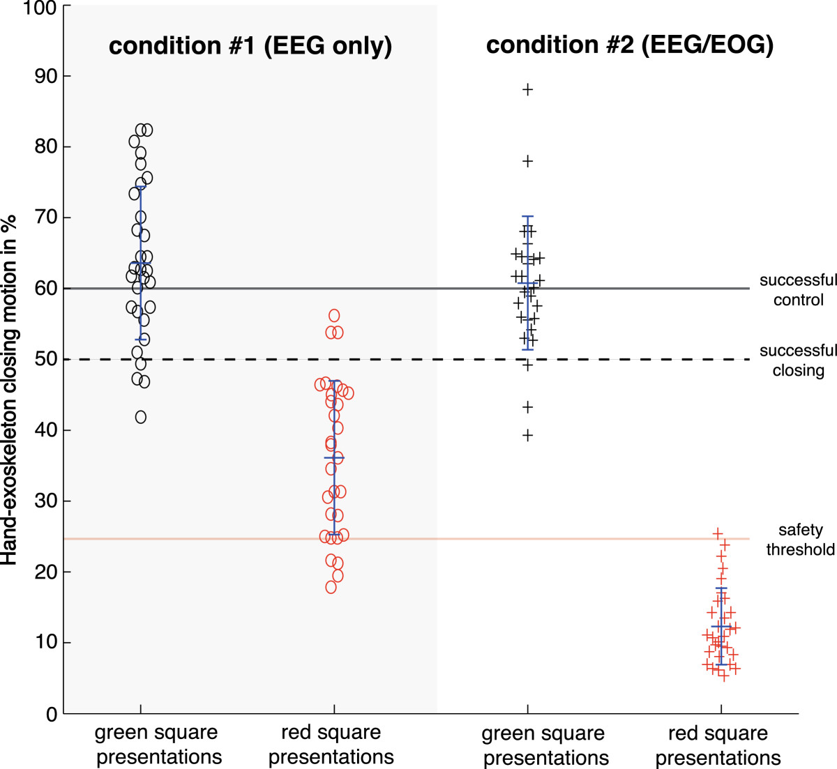
BACKGROUND: Brain-machine interfaces (BMIs) allow direct translation of electric, magnetic or metabolic brain signals into control commands of external devices such as robots, prostheses or exoskeletons. However, non-stationarity of brain signals and susceptibility to biological or environmental artifacts impede reliable control and safety of BMIs, particularly in daily life environments. Here we introduce and tested a novel hybrid brain-neural computer interaction (BNCI) system fusing electroencephalography (EEG) and electrooculography (EOG) to enhance reliability and safety of continuous hand exoskeleton-driven grasping motions. FINDINGS: 12 healthy volunteers (8 male, mean age 28.1 ± 3.63y) used EEG (condition #1) and hybrid EEG/EOG (condition #2) signals to control a hand exoskeleton. Motor imagery-related brain activity was translated into exoskeleton-driven hand closing motions. Unintended motions could be interrupted by eye movement-related EOG signals. In order to evaluate BNCI control and safety, participants were instructed to follow a visual cue indicating either to move or not to move the hand exoskeleton in a random order. Movements exceeding 25% of a full grasping motion when the device was not supposed to be moved were defined as safety violation. While participants reached comparable control under both conditions, safety was frequently violated under condition #1 (EEG), but not under condition #2 (EEG/EOG). CONCLUSION: EEG/EOG biosignal fusion can substantially enhance safety of assistive BNCI systems improving their applicability in daily life environments.
Overview
The claims and statistics of this paper haven’t been checked yet.
Free: it reads the open-access text and checks the first 5 claims that cite a source we can check. Create a free account to check every claim.
Integrity signals
Registry
Cites
Statistics
Figures


Verification
Claims the paper makes
The claims and statistics of this paper haven’t been checked yet.
Free: it reads the open-access text and checks the first 5 claims that cite a source we can check. Create a free account to check every claim.
Claims other papers make about this one
Each is checked against this paper’s own text: does it say what the citing paper says it does?
The check wasn’t confident either way, so it gives no verdict — never disputed.
In the citing paper
“Surface EMG is therefore, in principle, a low-cost and specific way of detecting what the patient wants the prosthetic to do.[400,401]”
From Flexible Electronics and Devices as Human–Machine Interfaces for Medical Robotics (2022) · cites this paper as [401]
Experimental — accuracy not yet measured
Self-check
Percentages, sums and reported differences re-computed from the paper’s own numbers.
The self-check needs the paper’s full text.
Citations
Cites
The assessment and analysis of handedness: The Edinburgh inventory
Oldfield, R. C. (1971) · Neuropsychologia · doi:10.1016/0028-3932(71)90067-4 · Cited by 35,900 · cited 1×
“All participants were right handed as evaluated by the Edinburgh Handedness Inventory [11], and gave written informed consent before the session.”
MethodMulticlass Brain–Computer Interface Classification by Riemannian Geometry
Barachant, A., Bonnet, S., Congedo, M., & Jutten, C. (2012) · IEEE Transactions on Biomedical Engineering · doi:10.1109/tbme.2011.2172210 · Cited by 878 · cited 1×
“based on Riemannian geometry [17], discriminative models [18] or machine learning are applied.”
MethodCombining brain-computer interfaces and assistive technologies: state-of-the-art and challenges
Millán, J. D. R. (2010) · Frontiers in Neuroscience · doi:10.3389/fnins.2010.00161 · Cited by 866 · cited 3×
“Real-time translation of brain activity into control signals of external devices, known today as brain-computer or brain-machine interfaces (BCI/BMI), can substantially enhance human-machine interaction (HMI) [1], e.g. allowing impaired individuals to operate assistive systems as hand prostheses or exoskeletons (Figur…”
BackgroundPfurtscheller, G., & Aranibar, A. (1979) · Electroencephalography and Clinical Neurophysiology · doi:10.1016/0013-4694(79)90063-4 · Cited by 758 · cited 1×
“To identify the optimal frequency for detection of motor-imagery related desynchronization of sensorimotor rhythms (SMR, 8-15Hz) of each participant, a power spectrum estimation (autoregressive model of order 16 using the Yule–Walker algorithm) was performed for each incoming sample, selecting the frequency that showe…”
MethodPfurtscheller, G., Solis-Escalante, T., Ortner, R., Linortner, P., & Muller-Putz, G. R. (2010) · IEEE Transactions on Neural Systems and Rehabilitation Engineering · doi:10.1109/tnsre.2010.2040837 · Cited by 276 · cited 1×
“A possible strategy to increase safety of brain-controlled assistive systems in daily life environments is to use a switch mechanism turning the BMI system off or into sleep mode when active brain control is not needed or desired [7,8].”
BackgroundBlankertz, B., Dornhege, G., Schafer, C., Krepki, R., Kohlmorgen, J., Muller, K.-R., Kunzmann, V., Losch, F., & Curio, G. (2003) · IEEE Transactions on Neural Systems and Rehabilitation Engineering · doi:10.1109/tnsre.2003.814456 · Cited by 226 · cited 1×
“of intelligent machine learning algorithms [5,6] or remarkable technical advances improving active BMI control [1,3], classification accuracy of most BMI systems is still insufficient for many assistive applications, particularly those related to motor control, where misclassification can lead to unwanted actions and…”
BackgroundBCI meeting 2005-workshop on BCI signal processing: feature extraction and translation
McFarland, D. J., Anderson, C. W., Muller, K.-R., Schlogl, A., & Krusienski, D. J. (2006) · IEEE Transactions on Neural Systems and Rehabilitation Engineering · doi:10.1109/tnsre.2006.875637 · Cited by 204 · cited 1×
“of intelligent machine learning algorithms [5,6] or remarkable technical advances improving active BMI control [1,3], classification accuracy of most BMI systems is still insufficient for many assistive applications, particularly those related to motor control, where misclassification can lead to unwanted actions and…”
BackgroundA Powered Finger–Thumb Wearable Hand Exoskeleton With Self-Aligning Joint Axes
Cempini, M., Cortese, M., & Vitiello, N. (2015) · IEEE/ASME Transactions on Mechatronics · doi:10.1109/tmech.2014.2315528 · Cited by 193 · cited 2×
“The illustrated device was developed by The BioRobotics Institute (Scuola Superiore Sant’Anna, Pisa, Italy) to perform opening and closing motions of a hand [2].”
BackgroundMethodSelf-citationAn asynchronous wheelchair control by hybrid EEG–EOG brain–computer interface
Wang, H., Li, Y., Long, J., Yu, T., & Gu, Z. (2014) · Cognitive Neurodynamics · doi:10.1007/s11571-014-9296-y · Cited by 132 · cited 1×
“to navigate a toy truck [9] or wheelchair [10].”
BackgroundSoekadar, S. R., Witkowski, M., Mellinger, J., Ramos‐Murguialday, A., Birbaumer, N., & Cohen, L. G. (2011) · IEEE Transactions on Neural Systems and Rehabilitation Engineering · doi:10.1109/tnsre.2011.2166809 · Cited by 87 · cited 3×
“To identify the optimal frequency for detection of motor-imagery related desynchronization of sensorimotor rhythms (SMR, 8-15Hz) of each participant, a power spectrum estimation (autoregressive model of order 16 using the Yule–Walker algorithm) was performed for each incoming sample, selecting the frequency that showe…”
BackgroundMethodSelf-citation
Cited by
Flexible Electronics and Devices as Human–Machine Interfaces for Medical Robotics
Heng, W., Solomon, S., & Gao, W. (2022) · Advanced Materials · doi:10.1002/adma.202107902 · Cited by 609
Hong, K.-S., & Khan, M. J. (2017) · Frontiers in Neurorobotics · doi:10.3389/fnbot.2017.00035 · Cited by 267
“Real-time video game control of Belkacem et al. (2015a) and exoskeleton control of Witkowski et al. (2014) have also been implemented in the form of hBCI using a thresholding scheme with the accuracies of 77.3 (for six commands) and 63.59% (for four commands), respectively.”
MethodInfluentialBrain–computer interface robotics for hand rehabilitation after stroke: a systematic review
Baniqued, P. D. E., Stanyer, E. C., Awais, M., Alazmani, A., Jackson, A. E., Mon-Williams, M. A., Mushtaq, F., & Holt, R. J. (2021) · Journal of NeuroEngineering and Rehabilitation · doi:10.1186/s12984-021-00820-8 · Cited by 246
Soekadar, S. R., Witkowski, M., Gómez, C., Opisso, E., Medina, J., Cortese, M., Cempini, M., Carrozza, M. C., Cohen, L. G., Birbaumer, N., et al. (2016) · Science Robotics · doi:10.1126/scirobotics.aag3296 · Cited by 242
Self-citationHuang, J., Huo, W., Xu, W., Mohammed, S., & Amirat, Y. (2015) · IEEE Transactions on Automation Science and Engineering · doi:10.1109/tase.2015.2466634 · Cited by 185
“Many bioelectrical signals can be used in the first class of methods, including the electroencephalography (EEG) [15], the electrooculography (EOG) [16], the electromyography (EMG) [17], [18], and so on.”
Method
Metadata
- Authors
- Witkowski, M., Cortese, M., Cempini, M., Mellinger, J., Vitiello, N., & Soekadar, S. R.
- Affiliations
- Universitätsklinikum Tübingen (DE)
- Scuola Superiore Sant'Anna (IT)
- Corresponding author
- Soekadar, Surjo R
- Venue
- Journal of NeuroEngineering and Rehabilitation
- Published
- 2014-12
- Volume
- 11
- Issue
- 1
- Pages
- 165-165
- ISSN
- 1743-0003 (ISSN-L)
- PMC
- PMC4274709
- OpenAlex
- W2142736086
- Semantic Scholar
- 22404083fde958ccb8363963bc57059c864a1cb2
- Cited by
- 83
- References
- 18
- Citations by year
- 2026: 1 · 2025: 3 · 2024: 8 · 2023: 7 · 2022: 8 · 2021: 14 · 2020: 9 · 2019: 9 · 2018: 12 · 2017: 2 · 2016: 7 · 2015: 3
- FWCI
- 2.23
- Type
- journal article
- Language
- en
- Open access
- gold
- doi.org
- Licence
- cc-by
- Locations
- Journal of NeuroEngineering and Rehabilitation (publishedVersion, cc-by)
- Europe PMC (PubMed Central) (publishedVersion, cc-by)
- PubMed Central (publishedVersion, cc-by)
- PubMed (publishedVersion)
- CINECA IRIS Institutional Research Information System (Sant'Anna School of Advanced Studies) (submittedVersion)
- Funders
- European Commission (2007–2013, FP7 2007-2013, FP7/2007, 288551)
- Deutsche Forschungsgemeinschaft (FP7/2007–2013, 01GQ0831)
- Eberhard Karls Universität Tübingen
- Bundesministerium für Bildung und Forschung (01GQ0831)
- Topics
- Life Sciences › Neuroscience › Cognitive Neuroscience › EEG and Brain-Computer Interfaces
- Physical Sciences › Engineering › Biomedical Engineering › Muscle activation and electromyography studies
- Physical Sciences › Engineering › Biomedical Engineering › Prosthetics and Rehabilitation Robotics
- Keywords
- brain-computer interface, hand exoskeleton, electrooculography, EEG, motor imagery, grasp control, assistive robotics, eye movement detection
- MeSH terms
- Exoskeleton Device, Adult, Brain, Electroencephalography, Electrooculography, Eye Movements, Female, Hand, Humans, Male, Movement, Reproducibility of Results, Artifacts, Brain-Computer Interfaces
About these checks
Registry, retraction, Cites and statistics results are computed, not judged, with one exception: a printed reference that no index lists is read once by a model before it counts as a citation error. Claims are extracted by a model and kept only when their quote appears verbatim in the text; each cited source is then checked, with a model’s help, for whether it says what the claim relies on. Those verdicts can be wrong — read the claim’s quote and the cited source before relying on one.
Statistics and arithmetic are recomputed, not judged.
Confidence bands are shown only for fields calibrated on our annotated test set. None is calibrated yet, so no band is shown.
What counts as a claim ›How we measure confidence ›How we check this ›
Are you an author, or do you see a problem with this page? Request a review
How to cite
Cite CoChat for the checks on this page, and the paper for its own findings.
- This page
- CoChat. (2026). Citation check: Enhancing brain-machine interface (BMI) control of a hand exoskeleton using electrooculography (EOG). https://app.cochat.ai/papers/10.1186/1743-0003-11-165
- The paper
- Witkowski, M., Cortese, M., Cempini, M., Mellinger, J., Vitiello, N., & Soekadar, S. R. (2014). Enhancing brain-machine interface (BMI) control of a hand exoskeleton using electrooculography (EOG). Journal of NeuroEngineering and Rehabilitation. https://doi.org/10.1186/1743-0003-11-165
Data sources
Bibliographic data: Semantic Scholar, OpenAlex, Crossref.
Contains information from the Semantic Scholar Academic Graph Datasets, made available under the ODC Attribution License (ODC-BY 1.0).
Semantic Scholar’s data is described in: Kinney, R., et al. (2023). The Semantic Scholar Open Data Platform. arXiv:2301.10140.
