

Patient specific ankle-foot orthoses using rapid prototyping
Open accessThe DOI matches its Crossref record; 12 of the 13 references it cites were found in a registry or an index.
Abstract
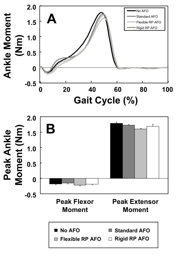
BACKGROUND: Prefabricated orthotic devices are currently designed to fit a range of patients and therefore they do not provide individualized comfort and function. Custom-fit orthoses are superior to prefabricated orthotic devices from both of the above-mentioned standpoints. However, creating a custom-fit orthosis is a laborious and time-intensive manual process performed by skilled orthotists. Besides, adjustments made to both prefabricated and custom-fit orthoses are carried out in a qualitative manner. So both comfort and function can potentially suffer considerably. A computerized technique for fabricating patient-specific orthotic devices has the potential to provide excellent comfort and allow for changes in the standard design to meet the specific needs of each patient. METHODS: In this paper, 3D laser scanning is combined with rapid prototyping to create patient-specific orthoses. A novel process was engineered to utilize patient-specific surface data of the patient anatomy as a digital input, manipulate the surface data to an optimal form using Computer Aided Design (CAD) software, and then download the digital output from the CAD software to a rapid prototyping machine for fabrication. RESULTS: Two AFOs were rapidly prototyped to demonstrate the proposed process. Gait analysis data of a subject wearing the AFOs indicated that the rapid prototyped AFOs performed comparably to the prefabricated polypropylene design. CONCLUSIONS: The rapidly prototyped orthoses fabricated in this study provided good fit of the subject's anatomy compared to a prefabricated AFO while delivering comparable function (i.e. mechanical effect on the biomechanics of gait). The rapid fabrication capability is of interest because it has potential for decreasing fabrication time and cost especially when a replacement of the orthosis is required.
Overview
The claims and statistics of this paper haven’t been checked yet.
Free: it reads the open-access text and checks the first 5 claims that cite a source we can check. Create a free account to check every claim.
Integrity signals
Registry
Cites
Statistics
Figures



Verification
Claims the paper makes
The claims and statistics of this paper haven’t been checked yet.
Free: it reads the open-access text and checks the first 5 claims that cite a source we can check. Create a free account to check every claim.
Claims other papers make about this one
Each is checked against this paper’s own text: does it say what the citing paper says it does?
The check wasn’t confident either way, so it gives no verdict — never disputed.
In the citing paper
“Auditory feedback may not only reallocate perceptual and cognitive workload, but also reduce distraction, since, unlike visual perception, auditory perception requires neither specific athlete orientation nor a focus of attention (Eldridge, 2006;Grond, Hermann, Verfaille, & Wanderley, 2010;Secoli, Milot, Rosati, & Reinkensmeyer, 2011).”
From Augmented visual, auditory, haptic, and multimodal feedback in motor learning: A review (2013) · cites this paper as Secoli et al., 2011
Experimental — accuracy not yet measured
Self-check
Percentages, sums and reported differences re-computed from the paper’s own numbers.
The self-check needs the paper’s full text.
Citations
Cites
A review of rapid prototyping (RP) techniques in the medical and biomedical sector
Webb, P. A. (2000) · Journal of Medical Engineering & Technology · doi:10.1080/03091900050163427 · Cited by 276 · cited 1×
“RP has been extensively used in medicine [6].”
Chua, C. K., Leong, K. F., Tan, K. H., Wiria, F. E., & Cheah, C. M. (2004) · Journal of Materials Science: Materials in Medicine · doi:10.1023/b:jmsm.0000046393.81449.a5 · Cited by 259
Noorani, R. (2006) · Cited by 228
Manufacture of Passive Dynamic Ankle–Foot Orthoses Using Selective Laser Sintering
Faustini, M. C., Neptune, R. R., Crawford, R. H., & Stanhope, S. J. (2008) · IEEE Transactions on Biomedical Engineering · doi:10.1109/tbme.2007.912638 · Cited by 209 · cited 1×
“[14].”
A preliminary investigation into the development of 3-D printing of prosthetic sockets
Herbert, N., Simpson, D., Spence, W. D., & Ion, W. (2005) · The Journal of Rehabilitation Research and Development · doi:10.1682/jrrd.2004.08.0134 · Cited by 181
Medical rapid prototyping applications and methods
Hieu, L. C., Zlatov, N., Vander Sloten, J., Bohez, E., Khanh, L., Binh, P. H., Oris, P., & Toshev, Y. (2005) · Assembly Automation · doi:10.1108/01445150510626415 · Cited by 150
Stereolithography for Craniofacial Surgery
Sinn, D. P., Cillo, J. E., & Miles, B. A. (2006) · Journal of Craniofacial Surgery · doi:10.1097/01.scs.0000230618.95012.1d · Cited by 83 · cited 9×
“Some of the advantages of SLA are its high accuracy, the ability to build clear models for examination, and - with some materials - sterilization for biocompatibility.”
InfluentialFacial prosthetic model fabrication using rapid prototyping tools
Kai, C. C., Meng, C. S., Lin, S. C., Lee, S.-T., & Aung, S. C. (2000) · Integrated Manufacturing Systems · doi:10.1108/09576060010303668 · Cited by 62
Bill Rogers, A., Bosker, G. W., Faustini, M. C., Walden, G., Neptune, R. R., & Crawford, R. (2007) · JPO Journal of Prosthetics and Orthotics · doi:10.1097/jpo.0b013e31815ea839 · Cited by 43
Tools for rapid prototyping in the biosciences
Zollikofer, C. P. E., & de León, M. S. P. (1995) · IEEE Computer Graphics and Applications · doi:10.1109/38.469515 · Cited by 38 · cited 1×
“Once the human anatomy has been recorded and a digital model has been created, the produced STL file instructs the RP machine about how to manufacture the intended medical model [4,5].”
Research works printed but not linked: 6
- Chelule K, Coole T, Chesire D: Fabrication of medical models from scan data via rapid prototyping techniques. In Proceedings of the 2000 Conference on Time Compression Technologies. Cardiff International Arena, UK; 2000.
- Milusheva S, Tochev D, Stefanova Y, Y Toshev Y: Virtual models and prototype of individual ankle foot orthosis. In Proceedings of ISB XXth Congress - ASB 29th Annual Meeting. Cleveland, Ohio; 2005:227.
- Alimed Inc.:Type C-90 Superior Posterior Leaf Spring. 2009. [ http://www.alimed.com ]
- 3D Systems Inc.:Products - SLA Systems - Viper Si2. 2009. [ http://www.3dsystems.com/products/sla/viper/datasheet.asp ]
- Rapidform Inc.:The Standard Software for 3D Scanning. 2009. [ http://www.rapidform.com/ ]
- 3D Systems Inc.:Products - Accura 40 SLA Resin. 2009. [ http://www.3dsystems.com/products/datafiles/accura/datasheets/DS-Accura_25_SL_material.pdf ]
Cited by
Augmented visual, auditory, haptic, and multimodal feedback in motor learning: A review
Sigrist, R., Rauter, G., Riener, R., & Wolf, P. (2013) · Psychonomic Bulletin & Review · doi:10.3758/s13423-012-0333-8 · Cited by 1,416
Gait Analysis Using Wearable Sensors
Tao, W., Liu, T., Zheng, R., & Feng, H. (2012) · Sensors · doi:10.3390/s120202255 · Cited by 1,165
Boyle, E. A., Hainey, T., Connolly, T. M., Gray, G., Earp, J., Ott, M., Lim, T., Ninaus, M., Ribeiro, C., & Pereira, J. (2016) · Computers & Education · doi:10.1016/j.compedu.2015.11.003 · Cited by 1,133
Current and emerging applications of 3D printing in medicine
Liaw, C.-Y., & Guvendiren, M. (2017) · Biofabrication · doi:10.1088/1758-5090/aa7279 · Cited by 604
Review of wearable thermoelectric energy harvesting: From body temperature to electronic systems
Nozariasbmarz, A., Collins, H., Dsouza, K., Polash, M. H., Hosseini, M., Hyland, M., Liu, J., Malhotra, A., Ortiz, F. M., Mohaddes, F., et al. (2020) · Applied Energy · doi:10.1016/j.apenergy.2019.114069 · Cited by 588
Metadata
- Authors
- Mavroidis, C., Ranky, R. G., Sivak, M. L., Patritti, B. L., DiPisa, J., Caddle, A., Gilhooly, K., Govoni, L., Sivak, S., Lancia, M., Drillio, R., & Bonato, P.
- Affiliations
- Northeastern University (US)
- Harvard University (US)
- Corresponding author
- Mavroidis, Constantinos
- Bonato, Paolo
- ORCID
- Bonato, Paolo
- Venue
- Journal of NeuroEngineering and Rehabilitation
- Published
- 2011-01-12
- Volume
- 8
- Issue
- 1
- Pages
- 1-1
- ISSN
- 1743-0003 (ISSN-L)
- PMC
- PMC3024934
- OpenAlex
- W2091296848
- Cited by
- 361
- References
- 13
- Citations by year
- 2026: 8 · 2025: 13 · 2024: 16 · 2023: 17 · 2022: 16 · 2021: 41 · 2020: 29 · 2019: 31 · 2018: 28 · 2017: 42 · 2016: 34 · 2015: 28 · 2014: 24 · 2013: 18 · 2012: 11
- FWCI
- 10.62
- Type
- journal article
- Language
- en
- Open access
- gold
- doi.org
- Licence
- cc-by
- creativecommons.org/licenses/by/2.0 (tdm)
- Locations
- Journal of NeuroEngineering and Rehabilitation (publishedVersion, cc-by)
- PubMed Central (publishedVersion, cc-by)
- PubMed (publishedVersion)
- Digital Access to Scholarship at Harvard (DASH) (Harvard University) (submittedVersion, cc-by)
- DOAJ (DOAJ: Directory of Open Access Journals) (submittedVersion)
- DSpace@MIT (Massachusetts Institute of Technology) (submittedVersion, cc-by-nc)
- BioMed Central eBooks
- Topics
- Physical Sciences › Engineering › Biomedical Engineering › Prosthetics and Rehabilitation Robotics
- Physical Sciences › Engineering › Biomedical Engineering › Anatomy and Medical Technology
- Physical Sciences › Engineering › Industrial and Manufacturing Engineering › Additive Manufacturing and 3D Printing Technologies
- Keywords
- ankle-foot orthosis, rapid prototyping, 3D laser scanning, prefabricated orthoses, gait analysis, computer-aided design
- MeSH terms
- Ankle, Biomechanical Phenomena, Equipment Design, Foot, Gait, Humans, Image Processing, Computer-Assisted, Orthotic Devices, Rehabilitation, Software, Reproducibility of Results, Biomimetic Materials, Mechanical Phenomena, Precision Medicine
About these checks
Registry, retraction, Cites and statistics results are computed, not judged, with one exception: a printed reference that no index lists is read once by a model before it counts as a citation error. Claims are extracted by a model and kept only when their quote appears verbatim in the text; each cited source is then checked, with a model’s help, for whether it says what the claim relies on. Those verdicts can be wrong — read the claim’s quote and the cited source before relying on one.
Statistics and arithmetic are recomputed, not judged.
Confidence bands are shown only for fields calibrated on our annotated test set. None is calibrated yet, so no band is shown.
What counts as a claim ›How we measure confidence ›How we check this ›
Are you an author, or do you see a problem with this page? Request a review
How to cite
Cite CoChat for the checks on this page, and the paper for its own findings.
- This page
- CoChat. (2026). Citation check: Patient specific ankle-foot orthoses using rapid prototyping. https://app.cochat.ai/papers/10.1186/1743-0003-8-1
- The paper
- Mavroidis, C., Ranky, R. G., Sivak, M. L., Patritti, B. L., DiPisa, J., Caddle, A., Gilhooly, K., Govoni, L., Sivak, S., Lancia, M., Drillio, R., & Bonato, P. (2011). Patient specific ankle-foot orthoses using rapid prototyping. Journal of NeuroEngineering and Rehabilitation. https://doi.org/10.1186/1743-0003-8-1
Data sources
Bibliographic data: Semantic Scholar, OpenAlex, Crossref.
Contains information from the Semantic Scholar Academic Graph Datasets, made available under the ODC Attribution License (ODC-BY 1.0).
Semantic Scholar’s data is described in: Kinney, R., et al. (2023). The Semantic Scholar Open Data Platform. arXiv:2301.10140.
