

The DOI matches its Crossref record; 1 of the 1 references it cites were found in a registry or an index.
The E5 protein of the human papillomavirus type 16 down-regulates HLA-I surface expression in calnexin-expressing but not in calnexin-deficient cells
Abstract
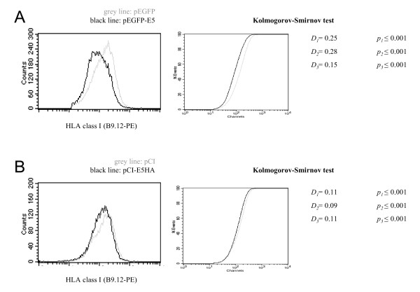
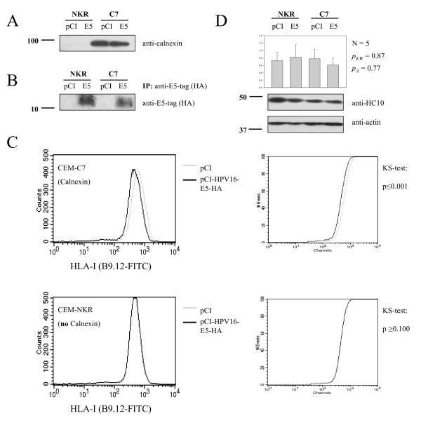
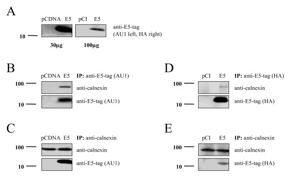
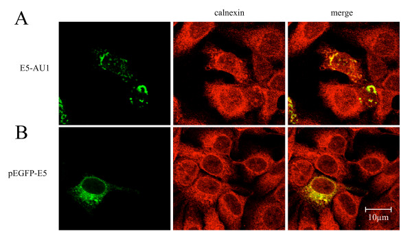
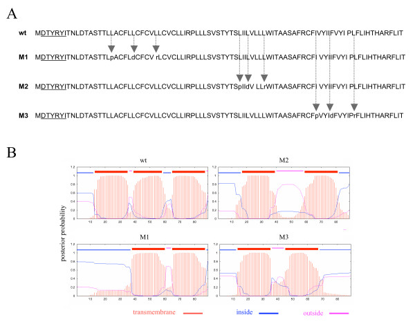
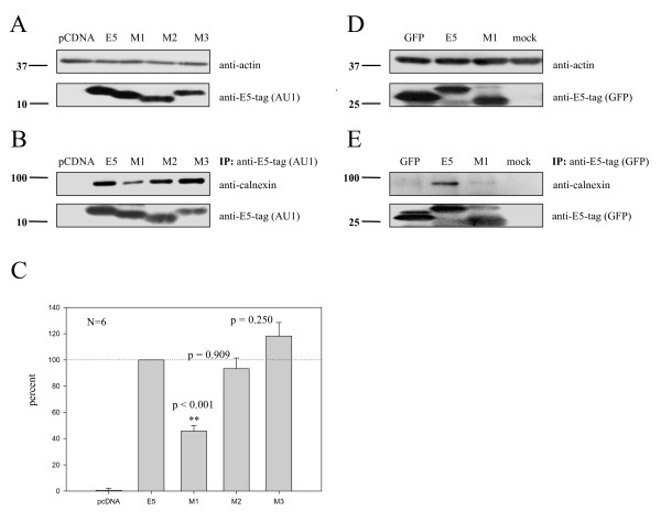
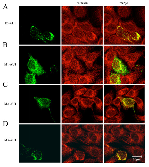
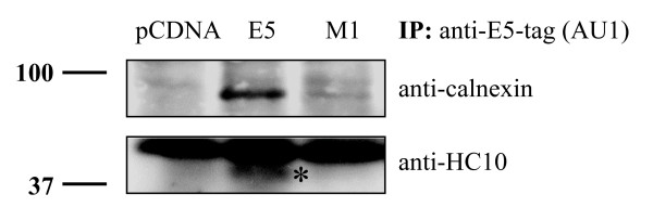
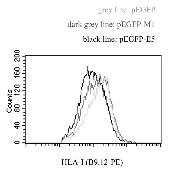
The human papillomavirus type 16 E5 protein (HPV16 E5) down-regulates surface expression of HLA-I molecules. The molecular mechanisms underlying this effect are so far unknown. Here we show that HPV16 E5 down-regulates HLA-I surface expression in calnexin-containing but not in calnexin-deficient cells. Immunoprecipitation experiments reveal that calnexin and HPV16E5 can be co-precipitated and that this association depends on the presence of a wild-type first hydrophobic region of E5. When an E5 mutant (M1) in which the first putative transmembrane helix had been disrupted was used for the transfections calnexin-E5 co-precipitation was strongly impaired. In addition, we show that the M1 mutant is only able to marginally down-regulate HLA-I surface expression compared to the wild-type protein. Besides, we demonstrate that E5 forms a ternary complex with calnexin and the heavy chain of HLA-I, which is mediated by the first hydrophobic region of the E5 protein. On the basis of our results we conclude that formation of this complex is responsible for retention of HLA-I molecules in the ER of the cells.
Overview
The claims and statistics of this paper haven’t been checked yet.
Run the check — free, no account needed. It reads the open-access text and checks the sources it cites.
Integrity signals
Registry
Cites
Statistics
Figures
Verification
Claims the paper makes
The claims and statistics of this paper haven’t been checked yet.
Run the check — free, no account needed. It reads the open-access text and checks the sources it cites.
Claims other papers make about this one
Each is checked against this paper’s own text: does it say what the citing paper says it does?
In the citing paper
“Furthermore, a ternary complex with calnexin, HPV-16 E5 and the heavy chain of HLA-I, via the first hydrophobic region of the E5 protein, has been suggested to be responsible for retention of HLA-I molecules in the ER of the cells [347].”
From The Not-So-Good, the Bad and the Ugly: HPV E5, E6 and E7 Oncoproteins in the Orchestration of Carcinogenesis (2021) · cites this paper as [347]
Self-check
Percentages, sums and reported differences re-computed from the paper’s own numbers.
The self-check needs the paper’s full text.
Citations
Cites
The human papilloma virus-16 E7-oncoprotein is able to bind to the retinoblastoma gene product
Dyson, N., Howley, P. M., Münger, K., & Harlow, E. (1989) · Science · doi:10.1126/science.2537532 · Cited by 3,017 · cited 1×
“E6 and E7 modulate apoptosis by binding and inactivating p53 and the product of tumour suppressor gene Rb1 respectively [10, 11 ], thereby deregulating the cell cycle.”
Background
Research works printed but not linked: 55
- DiMaio D, Liao JB: Human papillomaviruses and cervical cancer. Adv Virus Res. 2006, 66: 125-159. · DOI
- Munoz N, Bosch FX, de Sanjose S, Herrero R, Castellsague X, Shah KV, Snijders PJ, Meijer CJ: Epidemiologic classification of human papillomavirus types associated with cervical cancer. N Engl J Med. 2003, 348: 518-527. 10.1056/NEJMoa021641. · DOI · Paper page
- Franco EL, Schlecht NF, Saslow D: The epidemiology of cervical cancer. Cancer J. 2003, 9: 348-359. · DOI
- Walboomers JM, Jacobs MV, Manos MM, Bosch FX, Kummer JA, Shah KV, Snijders PJ, Peto J, Meijer CJ, Munoz N: Human papillomavirus is a necessary cause of invasive cervical cancer worldwide. J Pathol. 1999, 189: 12-19. 10.1002/(SICI)1096-9896(199909)189:1<12::AID-PATH431>3.0.CO;2-F. · DOI
- Clifford GM, Smith JS, Aguado T, Franceschi S: Comparison of HPV type distribution in high-grade cervical lesions and cervical cancer: a meta-analysis. Br J Cancer. 2003, 89: 101-105. 10.1038/sj.bjc.6601024. · DOI
- Bosch FX, Lorincz A, Munoz N, Meijer CJ, Shah KV: The causal relation between human papillomavirus and cervical cancer. J Clin Pathol. 2002, 55: 244-265. · DOI
- Stoler MH, Broker TR: In situ hybridization detection of human papillomavirus DNAs and messenger RNAs in genital condylomas and a cervical carcinoma. Hum Pathol. 1986, 17: 1250-1258. · DOI
- Crum CP, Symbula M, Ward BE: Topography of early HPV 16 transcription in high-grade genital precancers. Am J Pathol. 1989, 134: 1183-1188.
- Garnett TO, Duerksen-Hughes PJ: Modulation of apoptosis by human papillomavirus (HPV) oncoproteins. Arch Virol. 2006, 151: 2321-2335. 10.1007/s00705-006-0821-0. · DOI
- Werness BA, Levine AJ, Howley PM: Association of human papillomavirus types 16 and 18 E6 proteins with p53. Science. 1990, 248: 76-79. 10.1126/science.2157286. · DOI · Paper page
- Kabsch K, Alonso A: The human papillomavirus type 16 E5 protein impairs TRAIL- and FasL-mediated apoptosis in HaCaT cells by different mechanisms. J Virol. 2002, 76: 12162-12172. 10.1128/JVI.76.23.12162-12172.2002. · DOI
- Bravo IG, Crusius K, Alonso A: The E5 protein of human papillomavirus type 16 modulates composition and dynamics of membrane lipids in keratinocyte membranes. Arch Virol. 2005, 150: 231-246. 10.1007/s00705-004-0420-x. · DOI
- Sasagawa T, Inoue M, Tanizawa O, Yutsudo M, Hakura A: Identification of antibodies against human papillomavirus type 16 E6 and E7 proteins in sera of patients with cervical neoplasias. Jpn J Cancer Res. 1992, 83: 705-713. · DOI
- Muller M, Viscidi RP, Sun Y, Guerrero E, Hill PM, Shah F, Bosch FX, Munoz N, Gissmann L, Shah KV: Antibodies to HPV-16 E6 and E7 proteins as markers for HPV-16-associated invasive cervical cancer. Virology. 1992, 187: 508-514. 10.1016/0042-6822(92)90453-V. · DOI
- Dillner J: Mapping of linear epitopes of human papillomavirus type 16: the E1, E2, E4, E5, E6 and E7 open reading frames. Int J Cancer. 1990, 46: 703-711. 10.1002/ijc.2910460426. · DOI
- Kochel HG, Sievert K, Monazahian M, Mittelstadt-Deterding A, Teichmann A, Thomssen R: Antibodies to human papillomavirus type-16 in human sera as revealed by the use of prokaryotically expressed viral gene products. Virology. 1991, 182: 644-654. 10.1016/0042-6822(91)90605-B. · DOI
- Auvinen E, Crusius K, Steuer B, Alonso A: Human papillomavirus type 16 E5 protein (Review). Int J Oncol. 1997, 11: 1297-1304.
- O'Brien PM, Saveria Campo M: Evasion of host immunity directed by papillomavirus-encoded proteins. Virus Res. 2002, 88: 103-117. 10.1016/S0168-1702(02)00123-5. · DOI
- Zhang B, Li P, Wang E, Brahmi Z, Dunn KW, Blum JS, Roman A: The E5 protein of human papillomavirus type 16 perturbs MHC class II antigen maturation in human foreskin keratinocytes treated with interferon-gamma. Virology. 2003, 310: 100-108. 10.1016/S0042-6822(03)00103-X. · DOI · Paper page
- Ashrafi GH, Haghshenas M, Marchetti B, Campo MS: E5 protein of human papillomavirus 16 downregulates HLA class I and interacts with the heavy chain via its first hydrophobic domain. Int J Cancer. 2006, 119: 2105-2112. 10.1002/ijc.22089. · DOI · Paper page
- Ashrafi GH, Haghshenas MR, Marchetti B, O'Brien PM, Campo MS: E5 protein of human papillomavirus type 16 selectively downregulates surface HLA class I. Int J Cancer. 2005, 113: 276-283. 10.1002/ijc.20558. · DOI · Paper page
- Cromme FV, Snijders PJ, van den Brule AJ, Kenemans P, Meijer CJ, Walboomers JM: MHC class I expression in HPV 16 positive cervical carcinomas is post-transcriptionally controlled and independent from c-myc overexpression. Oncogene. 1993, 8: 2969-2975.
- Marchetti B, Ashrafi GH, Tsirimonaki E, O'Brien PM, Campo MS: The bovine papillomavirus oncoprotein E5 retains MHC class I molecules in the Golgi apparatus and prevents their transport to the cell surface. Oncogene. 2002, 21: 7808-7816. 10.1038/sj.onc.1205885. · DOI
- Ortmann B, Androlewicz MJ, Cresswell P: MHC class I/beta 2-microglobulin complexes associate with TAP transporters before peptide binding. Nature. 1994, 368: 864-867. 10.1038/368864a0. · DOI
- Ahluwalia N, Bergeron JJ, Wada I, Degen E, Williams DB: The p88 molecular chaperone is identical to the endoplasmic reticulum membrane protein, calnexin. J Biol Chem. 1992, 267: 10914-10918. · DOI
- Degen E, Cohen-Doyle MF, Williams DB: Efficient dissociation of the p88 chaperone from major histocompatibility complex class I molecules requires both beta 2-microglobulin and peptide. J Exp Med. 1992, 175: 1653-1661. 10.1084/jem.175.6.1653. · DOI
- Ritz U, Momburg F, Pilch H, Huber C, Maeurer MJ, Seliger B: Deficient expression of components of the MHC class I antigen processing machinery in human cervical carcinoma. Int J Oncol. 2001, 19: 1211-1220.
- Ayala-Torres S, Moller PC, Johnson BH, Thompson EB: Characteristics of 25-hydroxycholesterol-induced apoptosis in the human leukemic cell line CEM. Exp Cell Res. 1997, 235: 35-47. 10.1006/excr.1997.3630. · DOI
- Scott JE, Dawson JR: MHC class I expression and transport in a calnexin-deficient cell line. J Immunol. 1995, 155: 143-148. · DOI
- Howell DN, Andreotti PE, Dawson JR, Cresswell P: Natural killing target antigens as inducers of interferon: studies with an immunoselected, natural killing-resistant human T lymphoblastoid cell line. J Immunol. 1985, 134: 971-976. · DOI
- Bravo IG, Alonso A: Mucosal human papillomaviruses encode four different E5 proteins whose chemistry and phylogeny correlate with malignant or benign growth. J Virol. 2004, 78: 13613-13626. 10.1128/JVI.78.24.13613-13626.2004. · DOI · Paper page
- Oetke C, Auvinen E, Pawlita M, Alonso A: Human papillomavirus type 16 E5 protein localizes to the Golgi apparatus but does not grossly affect cellular glycosylation. Arch Virol. 2000, 145: 2183-2191. 10.1007/s007050070048. · DOI
- Gieswein CE, Sharom FJ, Wildeman AG: Oligomerization of the E5 protein of human papillomavirus type 16 occurs through multiple hydrophobic regions. Virology. 2003, 313: 415-426. 10.1016/S0042-6822(03)00296-4. · DOI · Paper page
- Rodriguez MI, Finbow ME, Alonso A: Binding of human papillomavirus 16 E5 to the 16 kDa subunit c (proteolipid) of the vacuolar H+-ATPase can be dissociated from the E5-mediated epidermal growth factor receptor overactivation. Oncogene. 2000, 19: 3727-3732. 10.1038/sj.onc.1203718. · DOI
- Yang DH, Wildeman AG, Sharom FJ: Overexpression, purification, and structural analysis of the hydrophobic E5 protein from human papillomavirus type 16. Protein Expr Purif. 2003, 30: 1-10. 10.1016/S1046-5928(03)00049-4. · DOI · Paper page
- Sonnhammer EL, von Heijne G, Krogh A: A hidden Markov model for predicting transmembrane helices in protein sequences. Proc Int Conf Intell Syst Mol Biol. 1998, 6: 175-182.
- Moller S, Croning MD, Apweiler R: Evaluation of methods for the prediction of membrane spanning regions. Bioinformatics. 2001, 17: 646-653. 10.1093/bioinformatics/17.7.646. · DOI
- Neefjes JJ, Momburg F, Hammerling GJ: Selective and ATP-dependent translocation of peptides by the MHC-encoded transporter. Science. 1993, 261: 769-771. 10.1126/science.8342042. · DOI
- Rebai N, Malissen B: Structural and genetic analyses of HLA class I molecules using monoclonal xenoantibodies. Tissue Antigens. 1983, 22: 107-117. · DOI
- Ashrafi GH, Tsirimonaki E, Marchetti B, O'Brien PM, Sibbet GJ, Andrew L, Campo MS: Down-regulation of MHC class I by bovine papillomavirus E5 oncoproteins. Oncogene. 2002, 21: 248-259. 10.1038/sj.onc.1205008. · DOI
- Cartin W, Alonso A: The human papillomavirus HPV2a E5 protein localizes to the Golgi apparatus and modulates signal transduction. Virology. 2003, 314: 572-579. 10.1016/S0042-6822(03)00509-9. · DOI
- Foley GE, Lazarus H, Farber S, Uzman BG, Boone BA, McCarthy RE: Continuous Culture of Human Lymphoblasts from Peripheral Blood of a Child with Acute Leukemia. Cancer. 1965, 18: 522-529. 10.1002/1097-0142(196504)18:4<522::AID-CNCR2820180418>3.0.CO;2-J. · DOI
- Loch S, Tampe R: Viral evasion of the MHC class I antigen-processing machinery. Pflugers Arch. 2005, 451: 409-417. 10.1007/s00424-005-1420-8. · DOI
- Vossen MT, Westerhout EM, Soderberg-Naucler C, Wiertz EJ: Viral immune evasion: a masterpiece of evolution. Immunogenetics. 2002, 54: 527-542. 10.1007/s00251-002-0493-1. · DOI
- Zhou J, Liu WJ, Peng SW, Sun XY, Frazer I: Papillomavirus capsid protein expression level depends on the match between codon usage and tRNA availability. J Virol. 1999, 73: 4972-4982. · DOI
- Leder C, Kleinschmidt JA, Wiethe C, Muller M: Enhancement of capsid gene expression: preparing the human papillomavirus type 16 major structural gene L1 for DNA vaccination purposes. J Virol. 2001, 75: 9201-9209. 10.1128/JVI.75.19.9201-9209.2001. · DOI
- Disbrow GL, Sunitha I, Baker CC, Hanover J, Schlegel R: Codon optimization of the HPV-16 E5 gene enhances protein expression. Virology. 2003, 311: 105-114. 10.1016/S0042-6822(03)00129-6. · DOI · Paper page
- Stam NJ, Spits H, Ploegh HL: Monoclonal antibodies raised against denatured HLA-B locus heavy chains permit biochemical characterization of certain HLA-C locus products. J Immunol. 1986, 137: 2299-2306. · DOI
- Seliger B, Ritz U, Ferrone S: Molecular mechanisms of HLA class I antigen abnormalities following viral infection and transformation. Int J Cancer. 2006, 118: 129-138. 10.1002/ijc.21312. · DOI
- Yewdell JW, Bennink JR: Mechanisms of viral interference with MHC class I antigen processing and presentation. Annu Rev Cell Dev Biol. 1999, 15: 579-606. 10.1146/annurev.cellbio.15.1.579. · DOI
- Vambutas A, DeVoti J, Pinn W, Steinberg BM, Bonagura VR: Interaction of human papillomavirus type 11 E7 protein with TAP-1 results in the reduction of ATP-dependent peptide transport. Clin Immunol. 2001, 101: 94-99. 10.1006/clim.2001.5094. · DOI
- Alonso A, Reed J: Modelling of the human papillomavirus type 16 E5 protein. Biochim Biophys Acta. 2002, 1601: 9-18. · DOI
- Bravo I, Alonso A, Auvinen E: Human papillomavirus type 16 E5 protein. Papillomavirus Report. 2004, 15: 1-6. 10.1179/095741904225004233. · DOI
- Cid-Arregui A, Juarez V, zur Hausen H: A synthetic E7 gene of human papillomavirus type 16 that yields enhanced expression of the protein in mammalian cells and is useful for DNA immunization studies. J Virol. 2003, 77: 4928-4937. 10.1128/JVI.77.8.4928-4937.2003. · DOI
- Samorski R, Gissmann L, Osen W: Codon optimized expression of HPV 16 E6 renders target cells susceptible to E6-specific CTL recognition. Immunol Lett. 2006, 107: 41-49. 10.1016/j.imlet.2006.07.003. · DOI
Cited by
Basukala, O., & Banks, L. (2021) · Viruses · doi:10.3390/v13101892 · Cited by 137
Metadata
- Authors
- Gruener, M., Bravo, I. G., Momburg, F., Alonso, A., & Tomakidi, P.
- Venue
- Virology Journal
- Published
- 2007-10-30
- Volume
- 4
- Issue
- 1
- ISSN
- 1743-422X
- PMC
- PMC2164959
- Cited by
- 53
- References
- 1
- Language
- en
About these checks
Registry, retraction, Cites and statistics results are computed, not judged, with one exception: a printed reference that no index lists is read once by a model before it counts as a citation error. Claims are extracted by a model and kept only when their quote appears verbatim in the text; each cited source is then checked, with a model’s help, for whether it says what the claim relies on. Those verdicts can be wrong — read the claim’s quote and the cited source before relying on one.
Statistics and arithmetic are recomputed, not judged.
Confidence bands are shown only for fields calibrated on our annotated test set. None is calibrated yet, so no band is shown.
What counts as a claim ›How we measure confidence ›How we check this ›
Are you an author, or do you see a problem with this page? Request a review
