

The DOI matches its Crossref record; 43 of the 43 references it cites were found in a registry or an index.
Tetralogy of Fallot
Open accessAbstract
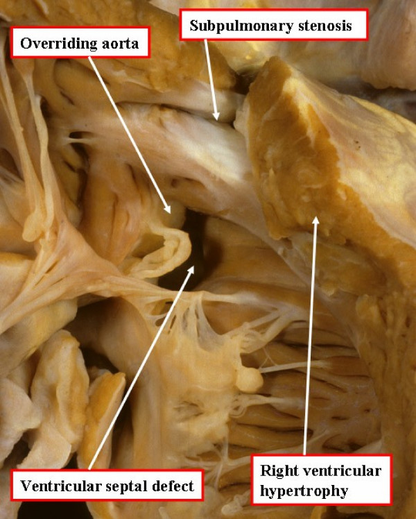
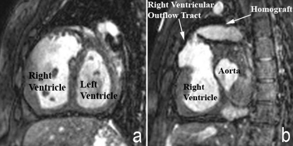
Tetralogy of Fallot is a congenital cardiac malformation that consists of an interventricular communication, also known as a ventricular septal defect, obstruction of the right ventricular outflow tract, override of the ventricular septum by the aortic root, and right ventricular hypertrophy. This combination of lesions occurs in 3 of every 10,000 live births, and accounts for 7-10% of all congenital cardiac malformations. Patients nowadays usually present as neonates, with cyanosis of varying intensity based on the degree of obstruction to flow of blood to the lungs. The aetiology is multifactorial, but reported associations include untreated maternal diabetes, phenylketonuria, and intake of retinoic acid. Associated chromosomal anomalies can include trisomies 21, 18, and 13, but recent experience points to the much more frequent association of microdeletions of chromosome 22. The risk of recurrence in families is 3%. Useful diagnostic tests are the chest radiograph, electrocardiogram, and echocardiogram. The echocardiogram establishes the definitive diagnosis, and usually provides sufficient information for planning of treatment, which is surgical. Approximately half of patients are now diagnosed antenatally. Differential diagnosis includes primary pulmonary causes of cyanosis, along with other cyanotic heart lesions, such as critical pulmonary stenosis and transposed arterial trunks. Neonates who present with ductal-dependent flow to the lungs will receive prostaglandins to maintain ductal patency until surgical intervention is performed. Initial intervention may be palliative, such as surgical creation of a systemic-to-pulmonary arterial shunt, but the trend in centres of excellence is increasingly towards neonatal complete repair. Centres that undertake neonatal palliation will perform the complete repair at the age of 4 to 6 months. Follow-up in patients born 30 years ago shows a rate of survival greater than 85%. Chronic issues that now face such adults include pulmonary regurgitation, recurrence of pulmonary stenosis, and ventricular arrhythmias. As the strategies for surgical and medical management have progressed, the morbidity and mortality of those born with tetralogy of Fallot in the current era is expected to be significantly improved.
Overview
The claims and statistics of this paper haven’t been checked yet.
Run the check — free, no account needed. It reads the open-access text and checks the sources it cites.
Integrity signals
Registry
Cites
Statistics
Figures
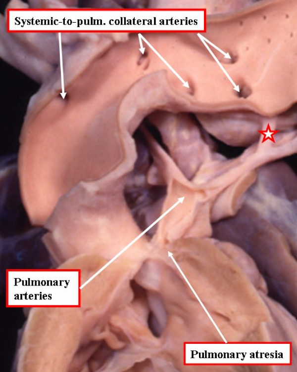
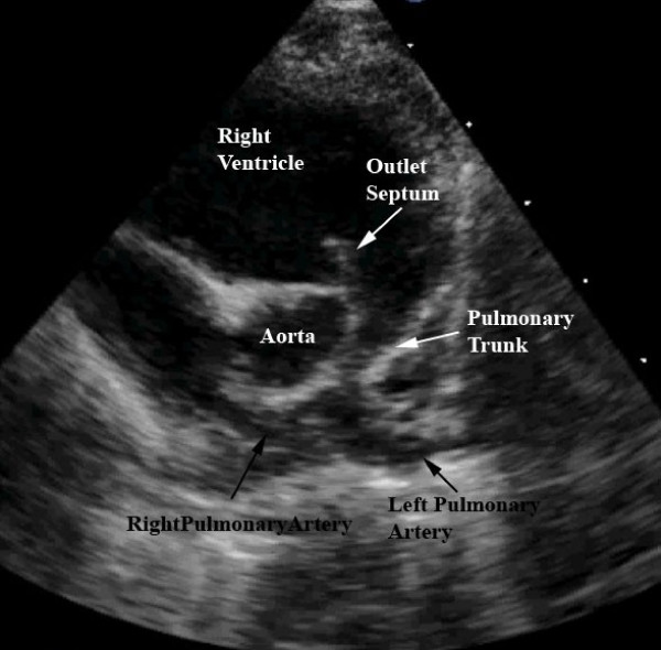
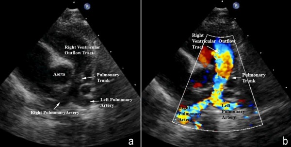
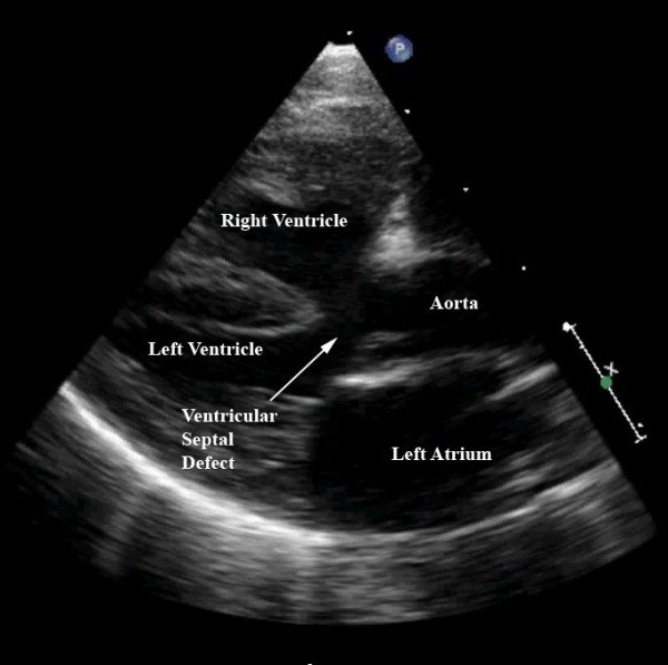
Verification
Claims the paper makes
The claims and statistics of this paper haven’t been checked yet.
Run the check — free, no account needed. It reads the open-access text and checks the sources it cites.
Claims other papers make about this one
Each is checked against this paper’s own text: does it say what the citing paper says it does?
The source backs part of the claim, not all of it — or backs it for a different population, condition or comparison.
In the citing paper
“This single lesion is responsible for all the main components of the disease: a <50% override of the aorta over the muscular ventricular septum, muscular obstruction of the RVOT, and right ventricle hypertrophy (RVH) [6].”
From Multimodality imaging assessment of tetralogy of Fallot: From diagnosis to long-term follow-up (2023) · cites this paper as [6]
Experimental — accuracy not yet measuredThe source backs part of the claim, not all of it — or backs it for a different population, condition or comparison.
In the citing paper
“Developed countries like the United States (US) reported a birth prevalence of 3.9 per 10,000 live births, with an estimated 1,660 babies born with TOF each year [2, 11]. In the US, TOF also represents approximately 6.7% of all CHDs [2].”
From Pooled prevalence and subgroup variations of Tetralogy of Fallot among children and adolescents with congenital heart defect in Sub-Saharan Africa: A systematic review and meta-analysis (2025) · cites this paper as [11]
Experimental — accuracy not yet measuredThe cited source says what the claim attributes to it.
In the citing paper
“Even though several anatomical types of VSD have been described, the typical VSD in TOF is an anterior malalignment type of outlet VSD, with the conal septum anteriorly diverging from the muscular septum.”
From Multimodality imaging assessment of tetralogy of Fallot: From diagnosis to long-term follow-up (2023) · cites this paper as [6]
Self-check
Percentages, sums and reported differences re-computed from the paper’s own numbers.
The self-check needs the paper’s full text.
Citations
Cites
Long-term outcome in patients undergoing surgical repair of tetralogy of Fallot
Murphy, J. G., Gersh, B. J., Mair, D. D., Fuster, V., McGoon, M. D., Ilstrup, D. M., McGoon, D. C., Kirklin, J. W., & Danielson, G. K. (1993) · New England Journal of Medicine · doi:10.1056/nejm199308263290901 · Cited by 998
Mechanoelectrical Interaction in Tetralogy of Fallot
Gatzoulis, M. Α., Till, J. A., Somerville, J., & Redington, A. N. (1995) · Circulation · doi:10.1161/01.cir.92.2.231 · Cited by 781
Nollert, G., Fischlein, T., Bouterwek, S., Böhmer, C., Klinner, W., & Reichart, B. (1997) · Journal of the American College of Cardiology · doi:10.1016/s0735-1097(97)00318-5 · Cited by 761
Heart Disease in Infants, Children and Adolescents
(1969) · Archives of Disease in Childhood · doi:10.1136/adc.44.234.287-a · Cited by 629
Oosterhof, T. H., van Straten, A., Vliegen, H. W., Meijboom, F. J., van Dijk, A. P. J., Spijkerboer, A. M., Bouma, B. J., Zwinderman, A. H., Hazekamp, M. G., Roos, A., et al. (2007) · Circulation · doi:10.1161/circulationaha.106.659664 · Cited by 610
Percutaneous Pulmonary Valve Implantation
Lurz, P. C., Coats, L., Khambadkone, S., Nordmeyer, J., Boudjemline, Y., Schievano, S., Muthurangu, V., Lee, T. Y., Parenzan, G., Derrick, G. P., et al. (2008) · Circulation · doi:10.1161/circulationaha.107.735779 · Cited by 481
The Effect of Chronic or Intermittent Hypoxia on Cognition in Childhood: A Review of the Evidence
Bass, J. L., Corwin, M. J., Gozal, D., Moore, C., Nishida, H., Parker, S. J., Schonwald, A., Wilker, R. E., Stehle, S., & Kinane, T. B. (2004) · PEDIATRICS · doi:10.1542/peds.2004-0227 · Cited by 452
Limperopoulos, C., Majnemer, A., Shevell, M. I., Rohlicek, C., Rosenblatt, B., Tchervenkov, C., & Darwish, H. Z. (2002) · The Journal of Pediatrics · doi:10.1067/mpd.2002.125227 · Cited by 323
Schievano, S., Coats, L., Migliavacca, F., Norman, W., Frigiola, A., Deanfield, J. E., Bonhoeffer, P., & Taylor, A. M. (2007) · Journal of Cardiovascular Magnetic Resonance · doi:10.1080/10976640601187596 · Cited by 243
Long-term results after early primary repair of tetralogy of Fallot.
Bacha, E. A., Scheule, A. M., Zurakowski, D., Erickson, L. C., Hung, J., Lang, P., Mayer, J. E., del Nido, P. J., & Jonas, R. A. (2001) · The Journal of Thoracic and Cardiovascular Surgery · doi:10.1067/mtc.2001.115156 · Cited by 240
Research works printed but not linked: 3
- Perry LW, Neill CA, Ferencz C, EUROCAT Working Party on. Congenital Heart Disease: Perspective in Pediatric Cardiology. Epidemiology of congenital heart disease, the Baltimore-Washington Infant Study 1981–89 Armonk, NY: Futura; 1993:33-62.
- Paediatric Cardiology 2nd edition. Edited by: Anderson RH, Baker EJ, Macartney FJ. London: Churchill Livingstone; 2002:1213-1280.
- Pediatric ECG Interpretation. Edited by: Deal BJ, Johnsrude CL, Buck SH. Malden, Massachussetts: Blackwell Publishing; 2004:88-122.
Cited by
Sorted by citations
Hot Topics in Tetralogy of Fallot
Villafañe, J., Feinstein, J. A., Jenkins, K. J., Vincent, R. N., Walsh, E. P., Dubin, A. M., Geva, T., Towbin, J. A., Cohen, M. S., Fraser, C. D., et al. (2013) · Journal of the American College of Cardiology · doi:10.1016/j.jacc.2013.07.100 · Cited by 232
Whole exome sequencing reveals the major genetic contributors to nonsyndromic tetralogy of Fallot
Page, D. J., Miossec, M. J., Williams, S. G., Monaghan, R. M., Fotiou, E., Cordell, H. J., Sutcliffe, L., Topf, A., Bourgey, M., Bourque, G., et al. (2019) · Circulation Research · doi:10.1161/circresaha.118.313250 · Cited by 189
Tetralogy of Fallot: Yesterday and Today
Starr, J. P. (2009) · World Journal of Surgery · doi:10.1007/s00268-009-0296-8 · Cited by 155
Notch Signaling in Cardiac Development and Disease
MacGrogan, D., Nus, M., & de la Pompa, J. L. (2010) · Current topics in developmental biology/Current Topics in Developmental Biology · doi:10.1016/s0070-2153(10)92011-5 · Cited by 96
Fetal MRI detects early alterations of brain development in tetralogy of fallot
Schellen, C., Ernst, S., Gruber, G. M., Mlczoch, E., Weber, M., Brugger, P. C., Ulm, B., Langs, G., Salzer-Muhar, U., Prayer, D., et al. (2015) · American Journal of Obstetrics and Gynecology · doi:10.1016/j.ajog.2015.05.046 · Cited by 79
Genetic Origins of Tetralogy of Fallot
Morgenthau, A., & Frishman, W. H. (2017) · Cardiology in Review · doi:10.1097/crd.0000000000000170 · Cited by 80
Notch signaling in cardiac valve development and disease
MacGrogan, D., Luna‐Zurita, L., & de la Pompa, J. L. (2011) · Birth Defects Research Part A Clinical and Molecular Teratology · doi:10.1002/bdra.20815 · Cited by 78
Miller, J. R., Stephens, E. H., Goldstone, A. B., Glatz, A. C., Kane, L. L., Arsdell, G. S. V., Stellin, G., Barron, D. J., d’Udekem, Y., Benson, L. N., et al. (2022) · Journal of Thoracic and Cardiovascular Surgery · doi:10.1016/j.jtcvs.2022.07.025 · Cited by 76
Tadokoro, M., Kanai, R., Taketani, T., Uchio, Y., Yamaguchi, S., & Ohgushi, H. (2009) · The Journal of Pediatrics · doi:10.1016/j.jpeds.2008.12.021 · Cited by 69
Cardiovascular imaging approach in pre and postoperative tetralogy of Fallot
Apostolopoulou, S. C., Manginas, A., Kelekis, N. L., & Noutsias, M. (2019) · BMC Cardiovascular Disorders · doi:10.1186/s12872-018-0996-9 · Cited by 60
Monaghan, R. M., Page, D. J., Østergaard, P., & Keavney, B. D. (2020) · Cardiovascular Research · doi:10.1093/cvr/cvaa291 · Cited by 69
Wilson, R., Ross, O., & Griksaitis, M. J. (2019) · BJA Education · doi:10.1016/j.bjae.2019.07.003 · Cited by 56
Identification of TBX5 mutations in a series of 94 patients with Tetralogy of Fallot
Baban, A., Postma, A. V., Marini, M., Trocchio, G., Santilli, A., Pelegrini, M., Sirleto, P., Lerone, M., Albanese, S. B., Barnett, P., et al. (2014) · American Journal of Medical Genetics Part A · doi:10.1002/ajmg.a.36783 · Cited by 56
Major Aortopulmonary Collateral Arteries
Alex, A., Ayyappan, A. P., Valakkada, J., Kramadhari, H., Sasikumar, D., & Menon, S. (2022) · Radiology Cardiothoracic Imaging · doi:10.1148/ryct.210157 · Cited by 48
Notch Signaling in Cardiac Development and Disease
de la Pompa, J. L. (2009) · Pediatric Cardiology · doi:10.1007/s00246-008-9368-z · Cited by 46
Birth Defects in Gaza: Prevalence, Types, Familiarity and Correlation with Environmental Factors
Naim, A., Dalies, H. A., Balawi, M. E., Salem, E. S., Meziny, K. A., Shawwa, R. E., Minutolo, R., & Manduca, P. (2012) · International Journal of Environmental Research and Public Health · doi:10.3390/ijerph9051732 · Cited by 41
Management of Infantile Spasms During the COVID-19 Pandemic
Grinspan, Z. M., Mytinger, J. R., Baumer, F. M., Ciliberto, M. A., Cohen, B. H., Dlugos, D., Harini, C., Hussain, S. A., Joshi, S. M., Keator, C. G., et al. (2020) · Journal of Child Neurology · doi:10.1177/0883073820933739 · Cited by 40
The clinical anatomy of the right ventricle
Mureşian, H. (2014) · Clinical Anatomy · doi:10.1002/ca.22484 · Cited by 39
Potential role of an athlete-focused echocardiogram in sports eligibility
Palermi, S., Serio, A., Vecchiato, M., Sirico, F., Gambardella, F., Ricci, F., Iodice, F., Radmilovic, J., Russo, V., & D’Andrea, A. (2021) · World Journal of Cardiology · doi:10.4330/wjc.v13.i8.271 · Cited by 40
Cocomello, L., Meloni, M., Rapetto, F., Baquedano, M., Ordoñez, M. V., Biglino, G., Bucciarelli‐Ducci, C., Parry, A., Stoica, S. C., & Caputo, M. (2019) · Journal of the American Heart Association · doi:10.1161/jaha.119.013654 · Cited by 39
Arya, B., Levasseur, S. M., Woldu, K. L., Glickstein, J. S., Andrews, H., & Williams, I. A. (2013) · Pediatric Cardiology · doi:10.1007/s00246-013-0857-3 · Cited by 37
Congenital heart diseases and anaesthesia
Desurkar, V., & Junghare, S. W. (2017) · Indian Journal of Anaesthesia · doi:10.4103/ija.ija_415_17 · Cited by 37
A Review of Tetralogy of Fallot and Postoperative Management
Forman, J., Beech, R., Slugantz, L., & Donnellan, A. (2019) · Critical Care Nursing Clinics of North America · doi:10.1016/j.cnc.2019.05.003 · Cited by 36
Charles, S., & Natarajan, J. (2023) · Medinformatics · doi:10.47852/bonviewmedin32021554 · Cited by 35
Genetic insights into non-syndromic Tetralogy of Fallot
Althali, N. J., & Hentges, K. E. (2022) · Frontiers in Physiology · doi:10.3389/fphys.2022.1012665 · Cited by 32
Urzúa, B., Martínez, C., Ortega-Pinto, A. V., Adorno-Farías, D., Morales‐Bozo, I., Riadi, G., Lilian Jara, S., Plaza, A., Lefimil, C., Lozano, C., et al. (2015) · Archives of Oral Biology · doi:10.1016/j.archoralbio.2015.06.016 · Cited by 32
Martins, I. F., Doles, I. C., Bravo‐Valenzuela, N. J., dos Santos, A. D. O. R., & Varella, M. S. P. (2018) · Brazilian Journal of Cardiovascular Surgery · doi:10.21470/1678-9741-2018-0019 · Cited by 33
Reuter, M. S., Chaturvedi, R. R., Jobling, R. K., Pellecchia, G., Hamdan, O., Sung, W. W. L., Nalpathamkalam, T., Attaluri, P., Silversides, C. K., Wald, R. M., et al. (2021) · Circulation Genomic and Precision Medicine · doi:10.1161/circgen.121.003410 · Cited by 30
Altered microRNA and target gene expression related to Tetralogy of Fallot
Grunert, M., Appelt, S., Dunkel, I., Berger, F. F., & Sperling, S. (2019) · Scientific Reports · doi:10.1038/s41598-019-55570-4 · Cited by 27
Avesani, M., Borrelli, N., Krupickova, S., Sabatino, J., Donne, G. D., Ibrahim, A., Piccinelli, E., Josen, M., Michielon, G., Fraisse, A., et al. (2020) · International Journal of Cardiology · doi:10.1016/j.ijcard.2020.07.026 · Cited by 26
Role of connexins in human congenital heart disease: the chicken and egg problem
Salameh, A., Blanke, K., & Dähnert, I. (2013) · Frontiers in Pharmacology · doi:10.3389/fphar.2013.00070 · Cited by 26
You, G., Zu, B., Wang, B., Fu, Q., & Li, F. (2020) · Frontiers in Genetics · doi:10.3389/fgene.2020.00552 · Cited by 26
Arterial Stiffness in Congenital Heart Disease
Sandhu, K., Pepe, S., Smolich, J. J., Cheung, M. M. H., & Mynard, J. P. (2021) · Heart Lung and Circulation · doi:10.1016/j.hlc.2021.07.018 · Cited by 25
Lin, Q., Tam, P. K. H., & Tang, C. S.-M. (2023) · Frontiers in Pediatrics · doi:10.3389/fped.2023.1203289 · Cited by 25
Variations of pulmonary arteries and other associated defects in Tetralogy of Fallot
Sheikh, A. M., Kazmi, U., & Syed, N. H. (2014) · SpringerPlus · doi:10.1186/2193-1801-3-467 · Cited by 25
Tetralogy of Fallot in men: quality of life, family, education, and employment
Bygstad, E., Pedersen, L. C. V. M., Pedersen, T. A., & Hjortdal, V. E. (2011) · Cardiology in the Young · doi:10.1017/s1047951111001934 · Cited by 24
Hruşcă, A., Răchişan, A. L., Gach, P., Pico, H., Sorensen, C., Bonello, B., Ovaert, C., Petit, P., Fouilloux, V., Macé, L. G., et al. (2016) · Diagnostic and Interventional Imaging · doi:10.1016/j.diii.2016.03.010 · Cited by 25
Lashkarinia, S. S., Pişkin, Ş., Bozkaya, T. A., Salihoğlu, E., Yerebakan, C., & Pekkan, K. (2018) · Annals of Biomedical Engineering · doi:10.1007/s10439-018-2043-5 · Cited by 25
Holst, L. M., Kronborg, J. B., Jepsen, J. R. M., Christensen, J. Ø., Vejlstrup, N. G., Juul, K., Bjerre, J. V., Bilenberg, N., & Ravn, H. B. (2020) · Cardiology in the Young · doi:10.1017/s1047951119003184 · Cited by 24
Woldu, K. L., Arya, B., BACHA, E. A., & Williams, I. A. (2014) · The Annals of Thoracic Surgery · doi:10.1016/j.athoracsur.2014.05.034 · Cited by 24
Boumpouli, M., Sauvage, E., Capelli, C., Schievano, S., & Kazakidi, A. (2021) · Frontiers in Cardiovascular Medicine · doi:10.3389/fcvm.2021.703717 · Cited by 25
Twite, M. D., & Ing, R. J. (2012) · Seminars in Cardiothoracic and Vascular Anesthesia · doi:10.1177/1089253211434749 · Cited by 25
Computed tomography in tetralogy of Fallot: Pre- and postoperative imaging evaluation
Zucker, E. J. (2022) · Pediatric Radiology · doi:10.1007/s00247-021-05179-5 · Cited by 23
Hirono, K., Sekine, M., Shiba, N., Hayashi, S., Nakaoka, H., Ibuki, K., Saito, K., Watanabe, K., Ozawa, S. W., Higuma, T., et al. (2013) · Circulation Journal · doi:10.1253/circj.cj-13-1048 · Cited by 23
Midgestation Fetal Pulmonary Annulus Size Is Predictive of Outcome in Tetralogy of Fallot
Friedman, K. G., Balasubramanian, S., & Tworetzky, W. (2013) · Congenital Heart Disease · doi:10.1111/chd.12120 · Cited by 23
Salameh, A., Dhein, S., Mewes, M., Sigusch, S., Kiefer, P., Vollroth, M., Seeger, J., & Dähnert, I. (2019) · Saudi Journal of Biological Sciences · doi:10.1016/j.sjbs.2019.04.003 · Cited by 22
Chetboul, V., Pitsch, I., Tissier, R., Gouni, V., Misbach, C., Tréhiou-Sechi, E., Petit, A. M. P., Damoiseaux, C., Pouchelon, J., Desquilbet, L., et al. (2016) · Journal of the American Veterinary Medical Association · doi:10.2460/javma.249.8.909 · Cited by 21
Brain abscess: A rare complication in a child with tetralogy of Fallot
Lakhani, M., Memon, R. S., & Khan, F. (2020) · IDCases · doi:10.1016/j.idcr.2020.e00954 · Cited by 21
Probing Human Cardiovascular Congenital Disease Using Transgenic Mouse Models
Snider, P. L., & Conway, S. J. (2011) · Progress in molecular biology and translational science · doi:10.1016/b978-0-12-384878-9.00003-0 · Cited by 20
Donor supply for partial heart transplantation in the United States
Quintao, R., Kwon, J. H., Bishara, K., & Rajab, T. K. (2023) · Clinical Transplantation · doi:10.1111/ctr.15060 · Cited by 20
Pişkin, Ş., Ünal, G., Arnaz, A., Sarıoğlu, T., & Pekkan, K. (2017) · Cardiovascular Engineering and Technology · doi:10.1007/s13239-017-0302-5 · Cited by 20
Athanasiadis, D. I., Mylonas, K. S., Kasparian, K., Ziogas, I. A., Vlachopoulou, D., Sfyridis, P. G., Schizas, D., Spartalis, E. D., Nikiteas, N. I., Hemmati, P., et al. (2019) · Pediatric Cardiology · doi:10.1007/s00246-019-02133-z · Cited by 20
Echocardiographic Evaluation of Tetralogy of Fallot
Swamy, P. M., Bharadwaj, A. S., Varadarajan, P., & Pai, R. G. (2014) · Echocardiography · doi:10.1111/echo.12437 · Cited by 19
Transcatheter-Based Interventions for Tetralogy of Fallot Across All Age Groups
Flores‐Umanzor, E., Alshehri, B. M., Keshvara, R., WILSON, W. M., Osten, M., Benson, L. N., Abrahamyan, L., & Horlick, E. M. (2024) · JACC: Cardiovascular Interventions · doi:10.1016/j.jcin.2024.02.009 · Cited by 21
Xie, Y., Ma, A., Wang, B., Peng, R., Jing, Y., Wang, D., Finnell, R. H., Qiao, B., Wang, Y., Wang, H., et al. (2019) · Clinical Science · doi:10.1042/cs20180842 · Cited by 17
Bittel, D. C., Zhou, X.-G., Kibiryeva, N., Fiedler, S. D., O’Brien, J. E., Marshall, J. A., Yu, S., & Liu, H. (2014) · PLoS ONE · doi:10.1371/journal.pone.0087472 · Cited by 17
Multimodality imaging assessment of tetralogy of Fallot: From diagnosis to long-term follow-up
Moscatelli, S., Pergola, V., Motta, R., Fortuni, F., Borrelli, N., Sabatino, J., Leo, I., Avesani, M., Montanaro, C., Surkova, E., et al. (2023) · Children · doi:10.3390/children10111747 · Cited by 17
Tissue engineering: Relevance to neonatal congenital heart disease
Blum, K. M., Mirhaidari, G. J. M., & Breuer, C. K. (2021) · Seminars in Fetal and Neonatal Medicine · doi:10.1016/j.siny.2021.101225 · Cited by 17
Rare variants in KDR, encoding VEGF Receptor 2, are associated with tetralogy of Fallot
Škorić‐Milosavljević, D., Lahrouchi, N., Bosada, F. M., Dombrowsky, G., Williams, S. G., Lesurf, R., Tjong, F. V. Y., Walsh, R., Bouchikhi, I. E., Breckpot, J., et al. (2021) · Genetics in Medicine · doi:10.1038/s41436-021-01212-y · Cited by 17
Role of CT in the Pre- and Postoperative Assessment of Conotruncal Anomalies
Kumar, P., & Bhatia, M. (2022) · Radiology: Cardiothoracic Imaging · doi:10.1148/ryct.210089 · Cited by 16
Rykiel, G., López, C. S., Riesterer, J. L., Fries, I., Deosthali, S., Courchaine, K., Maloyan, A., Thornburg, K. L. R., & Rugonyi, S. (2020) · eLife · doi:10.7554/elife.58138 · Cited by 16
Identification of Copy Number Variations in Isolated Tetralogy of Fallot
Aguayo‐Gómez, A., Arteaga‐Vázquez, J., Svyryd, Y., Calderón-Colmenero, J. E., Zamora-González, C., Vargas‐Alarcón, G., & Mutchinick, O. M. (2015) · Pediatric Cardiology · doi:10.1007/s00246-015-1210-9 · Cited by 16
Congenital Heart Disease in Adults with Autosomal Dominant Polycystic Kidney Disease
Chedid, M., Hanna, C., Zaatari, G., Mkhaimer, Y. G., Prajwal, R., Rangel, L. J., Zubidat, D., Kaidbay, D.-H. N., Irazabal, M. V., Connolly, H. M., et al. (2022) · American Journal of Nephrology · doi:10.1159/000522377 · Cited by 15
Mogra, R. (2013) · Australasian Journal of Ultrasound in Medicine · doi:10.1002/j.2205-0140.2013.tb00243.x · Cited by 15
Govil, S., Mauger, C., Hegde, S. R., Occleshaw, C. J., Yu, X., Perry, J. C., Young, A. A., Omens, J. H., & McCulloch, A. D. (2023) · Scientific Reports · doi:10.1038/s41598-023-28358-w · Cited by 15
Histologic alterations in tetralogy of Fallot
Alpat, Ş., Yilmaz, M., Onder, S. C., Sargon, M. F., Güvener, M., Doğan, R., Demircin, M., & Paşaoğlu, İ. (2016) · Journal of Cardiac Surgery · doi:10.1111/jocs.12873 · Cited by 15
Origins and consequences of congenital heart defects affecting the right ventricle
Woudstra, O. I., Ahuja, S., Bokma, J. P., Bouma, B. J., Mulder, B. J. M., & Christoffels, V. M. (2017) · Cardiovascular Research · doi:10.1093/cvr/cvx155 · Cited by 22
Arrhythmogenic Remodeling of the Left Ventricle in a Porcine Model of Repaired Tetralogy of Fallot
Dubes, V., Benoîst, D., Roubertie, F., Gilbert, S., Constantin, M., Charron, S., Elbes, D., Vieillot, D., Quesson, B., Cochet, H., et al. (2018) · Circulation Arrhythmia and Electrophysiology · doi:10.1161/circep.117.006059 · Cited by 15
Management of tetralogy of Fallot in the pediatric intensive care unit
Hammett, O., & Griksaitis, M. J. (2023) · Frontiers in Pediatrics · doi:10.3389/fped.2023.1104533 · Cited by 14
Oswal, N., Christov, G., Sridharan, S., Khambadkone, S., Bull, C., & Sullivan, I. D. (2013) · Cardiology in the Young · doi:10.1017/s1047951113000644 · Cited by 14
Computing group cardinality constraint solutions for logistic regression problems
Zhang, Y., Kwon, D., & Pohl, K. M. (2016) · Medical Image Analysis · doi:10.1016/j.media.2016.05.011 · Cited by 14
Powell, A. W., Mays, W. A., Knecht, S. K., & Chin, C. C. (2018) · Cardiology in the Young · doi:10.1017/s1047951118001920 · Cited by 14
Dysregulation of Notch signaling in cardiac mesenchymal cells of patients with tetralogy of Fallot
Александрович, К. И., Docshin, P. M., Kostina, A. S., Kiselev, A. M., Ignatieva, E. V., Головкин, А. С., Pervunina, T. M., Grekhov, E. V., Гордеев, М. Л., Kostareva, A. A., et al. (2020) · Pediatric Research · doi:10.1038/s41390-020-0760-6 · Cited by 14
Kakucs, Z., Heidenhoffer, E., & Pop, M. (2022) · Journal of Clinical Medicine · doi:10.3390/jcm11195500 · Cited by 15
Lazar, J., & Hylarides, M. J. (2017) · BMJ Case Reports · doi:10.1136/bcr-2016-218351 · Cited by 13
Tetralogy of Fallot: stent palliation or neonatal repair?
Banjoko, A., Seyedzenouzi, G., Ashton, J., Hedayat, F., Smith, N. N., Nixon, H., Tarmahomed, A., Ashry, A., & Harky, A. (2021) · Cardiology in the Young · doi:10.1017/s1047951121000846 · Cited by 13
Primary repair of symptomatic neonates with tetralogy of Fallot with or without pulmonary atresia
Lee, C., Kwak, J. G., & Lee, C. (2014) · Korean Journal of Pediatrics · doi:10.3345/kjp.2014.57.1.19 · Cited by 13
Elhamine, F., Iorga, B. I., Krüger, M., Hunger, M. S., Eckhardt, J., Sreeram, N., Bennink, G. B. W. E., Brockmeier, K., Pfitzer, G., & Stehle, R. (2016) · Journal of the American Heart Association · doi:10.1161/jaha.116.003699 · Cited by 13
Weichert, J., & Weichert, A. (2021) · Echocardiography · doi:10.1111/echo.15037 · Cited by 13
Mead, T. J., Bhutada, S., Foulcer, S. J., Peruzzi, N., Nelson, C. M., Seifert, D. E., Larkin, J. D., Tran‐Lundmark, K., Filmus, J., & Apte, S. (2024) · Matrix Biology · doi:10.1016/j.matbio.2024.05.003 · Cited by 13
The burden of Fallot’s tetralogy among Nigerian children
Animasahun, B. A., Madise-Wobo, A. D., Falase, B. A., & Omokhodion, S. I. (2016) · Cardiovascular Diagnosis and Therapy · doi:10.21037/cdt.2016.05.04 · Cited by 11
Unrepaired Tetralogy of Fallot in an 85-Year-Old Man
Gorla, R., Macchi, A., Franzoni, I., Rosa, I., Buzzetti, F., Pavon, A. G., & Margonato, A. (2012) · Congenital Heart Disease · doi:10.1111/j.1747-0803.2012.00642.x · Cited by 11
Intranasal Midazolam for the Emergency Management of Hypercyanotic Spells in Tetralogy of Fallot
Montero, J. V., Nieto, E. M., Vallejo, I. R., & Montero, S. V. (2015) · Pediatric Emergency Care · doi:10.1097/pec.0000000000000403 · Cited by 11
An Adult Patient With a Tetralogy of Fallot Case
Alkashkari, W., Al-Husayni, F., Almaqati, A. S., Alrahimi, J. S., & Albugami, S. (2020) · Cureus · doi:10.7759/cureus.11658 · Cited by 11
Chassagnon, G., Lefort, B., Méot, M., Carpentier, E., Sirinelli, D., Chantepie, A., & Morel, B. (2017) · Journal of the American Heart Association · doi:10.1161/jaha.117.006921 · Cited by 10
Gu, Q., Chen, X., Xiao, Y.-B., Chen, L., Xue-feng, W., Fang, J., Chen, B., & Hao, J. (2014) · Pediatric Cardiology · doi:10.1007/s00246-014-0868-8 · Cited by 9
Yitsege, G., Stokes, B. A., Sabatino, J. A., Sugrue, K. F., Bányai, G., Paronett, E. M., Karpinski, B. A., Maynard, T. M., LaMantia, A., & Zohn, I. E. (2020) · Birth Defects Research · doi:10.1002/bdr2.1709 · Cited by 9
Anomalías congénitas de las arterias pulmonares: espectro de hallazgos en tomografía computarizada
Bueno, J., Flors, L., & Mejía, M. (2016) · Radiología · doi:10.1016/j.rx.2016.09.008 · Cited by 9
Zhuang, B., Hu, Y., Fan, X., Li, M., Zhu, J., Liu, H., Cao, L. H., Liang, D., Zhang, J., Yu, Z., et al. (2017) · Journal of Cellular Biochemistry · doi:10.1002/jcb.26204 · Cited by 8
Right ventricular contractile reserve in tetralogy of Fallot patients with pulmonary regurgitation
Kingsley, C., Ahmad, S. S., Pappachan, J., Khambekar, S., Smith, T., Gardiner, D., Shambrook, J. S., Baskar, S., Moore, R. A., & VELDTMAN, G. R. (2018) · Congenital Heart Disease · doi:10.1111/chd.12569 · Cited by 8
Coomans, I., Kinder, S. D., Belleghem, H. V., Groote, K. D., Panzer, J., Wilde, H. D., Mosquera, L. M., François, K., Bové, T., Martens, T., et al. (2020) · PLoS ONE · doi:10.1371/journal.pone.0244312 · Cited by 8
Kang, Y., Wang, D., Xu, J., Zhang, X., Wang, W., Gao, J., Wang, W., You, T., & Tian, J. (2022) · Frontiers in Cardiovascular Medicine · doi:10.3389/fcvm.2022.888258 · Cited by 9
Cherif, M., Caputo, M., Nakaoka, Y., Angelini, G. D., & Ghorbel, M. T. (2015) · BioMed Research International · doi:10.1155/2015/718492 · Cited by 8
Tetralogy of Fallot in Spain: a nationwide registry-based mortality study across 36 years
Llamosas‐Falcón, L., Bermejo, E., Sánchez-Díaz, G., Villaverde‐Hueso, A., de la Paz, M. P., & Alonso‐Ferreira, V. (2019) · Orphanet Journal of Rare Diseases · doi:10.1186/s13023-019-1056-y · Cited by 9
Tetralogy of Fallot: Imaging of common and uncommon associations by multidetector CT
Zakaria, R. H., Barsoum, N. R., Asaad, R. E., El-Basmy, A. A., & Azab, A. O. (2011) · The Egyptian Journal of Radiology and Nuclear Medicine · doi:10.1016/j.ejrnm.2011.07.003 · Cited by 8
Izumi, Y., Takanashi, S., Kitamura, M., Takamisawa, I., Saito, M., Otaki, Y., Iwakura, T., & Takayama, M. (2024) · Journal of Cardiology · doi:10.1016/j.jjcc.2024.07.002 · Cited by 7
Children With Tetralogy of Fallot in an Urban Centre in Africa
Animasahun, B. A., Madise-Wobo, A. D., Omokhodion, S., & Njokanma, O. F. (2015) · Journal of Cardiovascular and Thoracic Research · doi:10.15171/jcvtr.2015.36 · Cited by 7
Amirghofran, A. A., Badr, J., & Jannati, M. (2018) · BMC Surgery · doi:10.1186/s12893-018-0338-y · Cited by 7
Risk Factors and Outcomes of Tetralogy of Fallot: From Fetal to Neonatal Life
Silva, J. A., Neves, A. L., Flôr-de-Lima, F., Soares, P., & Guimarães, H. (2019) · Pediatric Cardiology · doi:10.1007/s00246-019-02239-4 · Cited by 7
Zhou, N., Liu, L., Zou, R., Zou, M., Zhang, M., Cao, F., Liu, W., Yuan, H., Huang, G., Ma, L., et al. (2022) · Frontiers in Cardiovascular Medicine · doi:10.3389/fcvm.2021.780123 · Cited by 7
Metadata
- Authors
- Bailliard, F., & Anderson, R. H.
- Affiliations
- University of North Carolina at Chapel Hill (US)
- ORCID
- Anderson, Robert H
- Venue
- Orphanet Journal of Rare Diseases
- Published
- 2009-01-13
- Volume
- 4
- Issue
- 1
- Pages
- 2-2
- ISSN
- 1750-1172 (ISSN-L)
- PMC
- PMC2651859
- OpenAlex
- W3204567248
- Cited by
- 345
- References
- 42
- Citations by year
- 2026: 27 · 2025: 31 · 2024: 31 · 2023: 44 · 2022: 26 · 2021: 22 · 2020: 24 · 2019: 25 · 2018: 16 · 2017: 13 · 2016: 19 · 2015: 16 · 2014: 10 · 2013: 11 · 2012: 12
- FWCI
- 1.22
- Type
- journal article
- Language
- en
- Open access
- gold
- doi.org
- Licence
- Locations
- Orphanet Journal of Rare Diseases (publishedVersion, cc-by)
- PubMed Central (publishedVersion, cc-by)
- PubMed (publishedVersion)
- Carolina Digital Repository (University of North Carolina at Chapel Hill) (submittedVersion)
- DOAJ (DOAJ: Directory of Open Access Journals) (submittedVersion, cc-by-sa)
- Topics
- Health Sciences › Medicine › Cardiology and Cardiovascular Medicine › Congenital Heart Disease Studies
- Life Sciences › Biochemistry, Genetics and Molecular Biology › Molecular Biology › Congenital heart defects research
- Health Sciences › Medicine › Pediatrics, Perinatology and Child Health › Cardiovascular Conditions and Treatments
- Keywords
- tetralogy of Fallot, congenital heart disease, ventricular septal defect, right ventricular outflow tract obstruction, cyanosis, prenatal diagnosis, echocardiography, neonatal palliation, prostaglandin E2, systemic-to-pulmonary shunt, pulmonary regurgitation, ventricular arrhythmias, adult congenital heart disease, Down syndrome, trisomy 18, trisomy 13, phenylketonuria, maternal diabetes, retinoic acid
- MeSH terms
- Adult, Genetic Counseling, Heart Defects, Congenital, Humans, Infant, Infant, Newborn, Prognosis, Tetralogy of Fallot, Ultrasonography
About these checks
Registry, retraction, Cites and statistics results are computed, not judged, with one exception: a printed reference that no index lists is read once by a model before it counts as a citation error. Claims are extracted by a model and kept only when their quote appears verbatim in the text; each cited source is then checked, with a model’s help, for whether it says what the claim relies on. Those verdicts can be wrong — read the claim’s quote and the cited source before relying on one.
Statistics and arithmetic are recomputed, not judged.
Confidence bands are shown only for fields calibrated on our annotated test set. None is calibrated yet, so no band is shown.
What counts as a claim ›How we measure confidence ›How we check this ›
Are you an author, or do you see a problem with this page? Request a review
