

The DOI matches its Crossref record; 85 of the 85 references it cites were found in a registry or an index.
Tau oligomers impair memory and induce synaptic and mitochondrial dysfunction in wild-type mice
Open accessAbstract
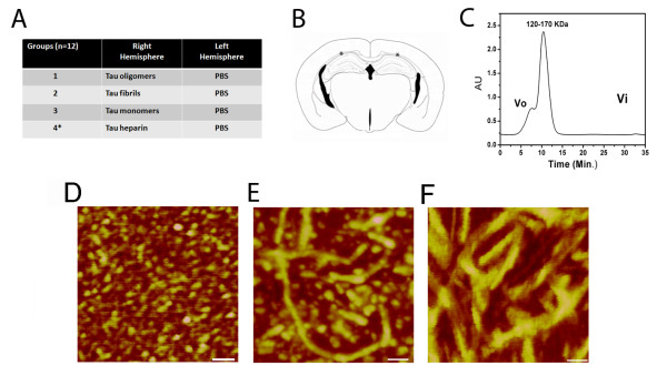
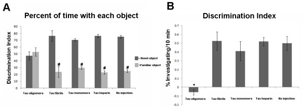
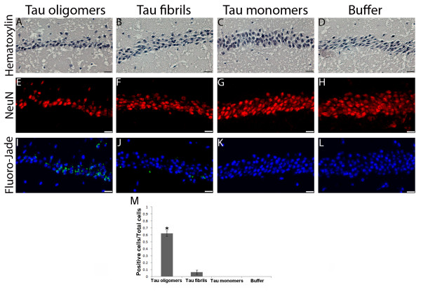
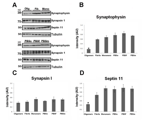
Background: The correlation between neurofibrillary tangles of tau and disease progression in the brains of Alzheimer's disease (AD) patients remains an area of contention. Innovative data are emerging from biochemical, cell-based and transgenic mouse studies that suggest that tau oligomers, a pre-filament form of tau, may be the most toxic and pathologically significant tau aggregate. Results: Here we report that oligomers of recombinant full-length human tau protein are neurotoxic in vivo after subcortical stereotaxic injection into mice. Tau oligomers impaired memory consolidation, whereas tau fibrils and monomers did not. Additionally, tau oligomers induced synaptic dysfunction by reducing the levels of synaptic vesicle-associated proteins synaptophysin and septin-11. Tau oligomers produced mitochondrial dysfunction by decreasing the levels of NADH-ubiquinone oxidoreductase (electron transport chain complex I), and activated caspase-9, which is related to the apoptotic mitochondrial pathway. Conclusions: This study identifies tau oligomers as an acutely toxic tau species in vivo, and suggests that tau oligomers induce neurodegeneration by affecting mitochondrial and synaptic function, both of which are early hallmarks in AD and other tauopathies. These results open new avenues for neuroprotective intervention strategies of tauopathies by targeting tau oligomers.
Overview
The claims and statistics of this paper haven’t been checked yet.
Run the check — free, no account needed. It reads the open-access text and checks the sources it cites.
Integrity signals
Registry
Cites
Statistics
Figures
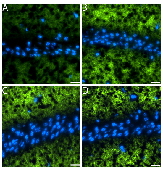
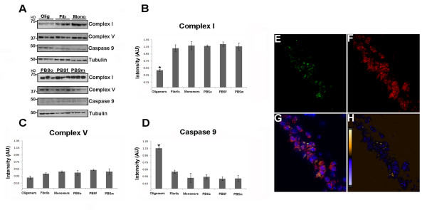
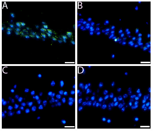
Verification
Claims the paper makes
The claims and statistics of this paper haven’t been checked yet.
Run the check — free, no account needed. It reads the open-access text and checks the sources it cites.
Claims other papers make about this one
Each is checked against this paper’s own text: does it say what the citing paper says it does?
In the citing paper
“our laboratory and others found that smaller, soluble tau oligomers (TauO) —not NFTs—are the most toxic tau species. 9 , 10 , 11”
From Senescence, brain inflammation, and oligomeric tau drive cognitive decline in Alzheimer's disease: Evidence from clinical and preclinical studies (2024) · cites this paper as [10]
Self-check
Percentages, sums and reported differences re-computed from the paper’s own numbers.
The self-check needs the paper’s full text.
Citations
Cites
Neuropathological stageing of Alzheimer-related changes.
Braak, H., & Braak, E. (1991) · Acta Neuropathologica · doi:10.1007/bf00308809 · Cited by 16,123 · cited 1×
“Most research has focused on NFTs because of the reported correlation between NFTs and disease progression in the brains of AD patients [8-10].”
BackgroundSoluble protein oligomers in neurodegeneration: lessons from the Alzheimer's amyloid β-peptide
Haass, C., & Selkoe, D. J. (2007) · Nature Reviews Molecular Cell Biology · doi:10.1038/nrm2101 · Cited by 4,744
Terry, R. D., Masliah, E., Salmon, D. P., Butters, N., DeTeresa, R., Hill, R., Hansen, L. A., & Katzman, R. (1991) · Annals of Neurology · doi:10.1002/ana.410300410 · Cited by 4,397
Braak, H., Alafuzoff, I., Arzberger, T., Kretzschmar, H., & Del Tredici, K. (2006) · Acta Neuropathologica · doi:10.1007/s00401-006-0127-z · Cited by 3,172
Lee, V. M.-Y., Goedert, M., & Trojanowski, J. Q. (2001) · Annual Review of Neuroscience · doi:10.1146/annurev.neuro.24.1.1121 · Cited by 2,480
Synapse Loss and Microglial Activation Precede Tangles in a P301S Tauopathy Mouse Model
Yoshiyama, Y., Higuchi, M., Zhang, B., Huang, S.-M., Iwata, N., Saido, T. C., Maeda, J., Suhara, T., Trojanowski, J. Q., & Lee, V. M.-Y. (2007) · Neuron · doi:10.1016/j.neuron.2007.01.010 · Cited by 2,208
Tau-mediated neurodegeneration in Alzheimer’s disease and related disorders
Ballatore, C., Lee, V. M.-Y., & Trojanowski, J. Q. (2007) · Nature Reviews Neuroscience · doi:10.1038/nrn2194 · Cited by 2,174
Tau Suppression in a Neurodegenerative Mouse Model Improves Memory Function
SantaCruz, K., Lewis, J., Spires, T., Paulson, J., Kotilinek, L., Ingelsson, M., Guimaraes, A., DeTure, M., Ramsden, M., McGowan, E., et al. (2005) · Science · doi:10.1126/science.1113694 · Cited by 2,014
Transmission and spreading of tauopathy in transgenic mouse brain
Clavaguera, F., Bolmont, T., Crowther, R. A., Abramowski, D., Frank, S., Probst, A., Fraser, G., Stalder, A. K., Beibel, M., Staufenbiel, M., et al. (2009) · Nature Cell Biology · doi:10.1038/ncb1901 · Cited by 1,803
The distribution of tau in the mammalian central nervous system.
Binder, L. I., Frankfurter, A., & Rebhun, L. I. (1985) · The Journal of cell biology · doi:10.1083/jcb.101.4.1371 · Cited by 1,588
Cited by
Sorted by citations
Items 201–300 of 636
mPOS is a novel mitochondrial trigger of cell death – implications for neurodegeneration
Coyne, L. P., & Chen, X. J. (2017) · FEBS Letters · doi:10.1002/1873-3468.12894 · Cited by 51
Zheng, J., Akbari, M., Schirmer, C., Reynaert, M., Loyens, A., Lefebvre, B., Buée, L., Croteau, D. L., Galas, M., & Bohr, V. A. (2020) · Acta Neuropathologica Communications · doi:10.1186/s40478-020-00896-8 · Cited by 54
Myricetin as a Promising Molecule for the Treatment of Post-Ischemic Brain Neurodegeneration
Pluta, R. M., Januszewski, S., & Czuczwar, S. J. (2021) · Nutrients · doi:10.3390/nu13020342 · Cited by 49
Glucocorticoid-driven mitochondrial damage stimulates Tau pathology
Du, F., Yu, Q., Swerdlow, R. H., & Waites, C. L. (2023) · Brain · doi:10.1093/brain/awad127 · Cited by 52
Brody, D. L., Benetatos, J., Bennett, R. E., Klemenhagen, K. C., & Donald, C. L. M. (2015) · Molecular and Cellular Neuroscience · doi:10.1016/j.mcn.2015.02.005 · Cited by 52
Potentiating tangle formation reduces acute toxicity of soluble tau species in the rat
d’Orange, M., Aurégan, G., Cheramy, D., Gaudin, M., Lieger, S., Guillermier, M., Stimmer, L., Joséphine, C., Hérard, A., Gaillard, M., et al. (2017) · Brain · doi:10.1093/brain/awx342 · Cited by 57
Ying, Y., Zhang, Y., Ma, C., Li, M., Tang, C.-Y., Yang, Y.-F., Zeng, J., Huang, X., Yi, J., Wang, X., et al. (2019) · Journal of Agricultural and Food Chemistry · doi:10.1021/acs.jafc.9b02954 · Cited by 51
Prospects for strain-specific immunotherapy in Alzheimer’s disease and tauopathies
Bittar, A., Sengupta, U., & Kayed, R. (2018) · npj Vaccines · doi:10.1038/s41541-018-0046-8 · Cited by 50
Self-citationShu, X., Zhang, Y., Li, M., Huang, X., Yang, Y., Zeng, J., Zhao, Y., Wang, X., Zhang, W., & Ying, Y. (2019) · Neuropharmacology · doi:10.1016/j.neuropharm.2019.04.018 · Cited by 51
Vacchi, E., Kaelin‐Lang, A., & Melli, G. (2020) · International Journal of Molecular Sciences · doi:10.3390/ijms21145030 · Cited by 51
Cerebrospinal Fluid Aβ42 Levels: When Physiological Become Pathological State
Martorana, A., Lorenzo, F. D., Belli, L., Sancesario, G., Toniolo, S., Sallustio, F., Sancesario, G. M., & Koch, G. (2015) · CNS Neuroscience & Therapeutics · doi:10.1111/cns.12476 · Cited by 51
Behavioral Abnormalities in Knockout and Humanized Tau Mice
Gonçalves, R. A., Wijesekara, N., Fraser, P. E., & Felice, F. G. D. (2020) · Frontiers in Endocrinology · doi:10.3389/fendo.2020.00124 · Cited by 52
Schroeder, S. K., Joly‐Amado, A., Gordon, M. N., & Morgan, D. (2015) · Journal of Neuroimmune Pharmacology · doi:10.1007/s11481-015-9637-6 · Cited by 51
The evolving landscape of Alzheimer’s disease therapy: From Aβ to tau
Courade, J., Zetterberg, H., Höglinger, G. U., & Dewachter, I. (2025) · Cell · doi:10.1016/j.cell.2025.11.033 · Cited by 57
Arendash, G. W., Cao, C., Abulaban, H., Baranowski, R., Wisniewski, G., Becerra, L. R., Andel, R., Lin, X., Zhang, X., Wittwer, D., et al. (2019) · Journal of Alzheimer s Disease · doi:10.3233/jad-190367 · Cited by 52
Tau Aggregation Inhibiting Peptides as Potential Therapeutics for Alzheimer Disease
Aillaud, I., & Funke, S. A. (2022) · Cellular and Molecular Neurobiology · doi:10.1007/s10571-022-01230-7 · Cited by 51
Zn2+ Aggravates Tau Aggregation and Neurotoxicity
Li, X., Du, X., & Ni, J. (2019) · International Journal of Molecular Sciences · doi:10.3390/ijms20030487 · Cited by 48
Abskharon, R. N. N., Pan, H., Sawaya, M. R., Seidler, P. M., Olivares, E. J., Chen, Y., Murray, K. A., Zhang, J., Lantz, C., Bentzel, M., et al. (2023) · Proceedings of the National Academy of Sciences · doi:10.1073/pnas.2300258120 · Cited by 50
Immunity and Alzheimer's disease: immunological perspectives on the development of novel therapies
Liu, Y.-H., Zeng, F., Wang, Y., Zhou, H., Giunta, B. N., Tan, J., & Wang, Y. (2013) · Drug Discovery Today · doi:10.1016/j.drudis.2013.07.020 · Cited by 48
Gaikwad, S., Puangmalai, N., Sonawane, M., Montalbano, M., Price, R., Iyer, M. S., Ray, A., Moreno, S., & Kayed, R. (2024) · Science Translational Medicine · doi:10.1126/scitranslmed.adj5958 · Cited by 46
Self-citationUmeda, T., Yamashita, T., Kimura, T., Ohnishi, K., Takuma, H., Ozeki, T., Takashima, A., Tomiyama, T., & Mori, H. (2013) · American Journal Of Pathology · doi:10.1016/j.ajpath.2013.03.015 · Cited by 46
Soluble tau aggregates inhibit synaptic long-term depression and amyloid β-facilitated LTD in vivo
Ondrejčák, T., Hu, N., Qi, Y., Klyubin, I. V., Corbett, G. T., Fraser, G. M., Perkinton, M. S., Walsh, D. M., Billinton, A., & Rowan, M. J. (2019) · Neurobiology of Disease · doi:10.1016/j.nbd.2019.03.022 · Cited by 47
Walker, S., Ullman, O., & Stultz, C. M. (2012) · Journal of Biological Chemistry · doi:10.1074/jbc.m111.336107 · Cited by 46
Central role for p62/SQSTM1 in the elimination of toxic tau species in a mouse model of tauopathy
Ono, M., Komatsu, M., Ji, B., Takado, Y., Shimojo, M., Minamihisamatsu, T., Warabi, E., Yanagawa, T., Matsumoto, G., Aoki, I., et al. (2022) · Aging Cell · doi:10.1111/acel.13615 · Cited by 45
Kim, J. H., Kim, E.-K., Choi, W. H., Lee, J., Lee, J. H., Lee, H., Kim, D., Suh, Y. H., & Lee, M. J. (2016) · Molecular Pharmaceutics · doi:10.1021/acs.molpharmaceut.6b00165 · Cited by 48
Tau Oligomer Pathology in Nucleus Basalis Neurons During the Progression of Alzheimer Disease
Tiernan, C. T., Mufson, E. J., Kanaan, N. M., & Counts, S. (2018) · Journal of Neuropathology & Experimental Neurology · doi:10.1093/jnen/nlx120 · Cited by 46
The prion-like transmission of tau oligomers via exosomes
Jackson, N. A., Guerrero‐Munoz, M. J., & Castillo‐Carranza, D. L. (2022) · Frontiers in Aging Neuroscience · doi:10.3389/fnagi.2022.974414 · Cited by 46
Self-citationTau Oligomers as Pathogenic Seeds: Preparation and Propagation In Vitro and In Vivo
Gerson, J. E., Sengupta, U., & Kayed, R. (2016) · Methods in molecular biology · doi:10.1007/978-1-4939-6598-4_9 · Cited by 43
Self-citationMotta, C., Finardi, A., Toniolo, S., Lorenzo, F. D., Scaricamazza, E., Loizzo, S., Mercuri, N. B., Furlan, R., Koch, G., & Martorana, A. (2020) · Journal of Alzheimer s Disease · doi:10.3233/jad-191250 · Cited by 44
Living with the enemy: from protein-misfolding pathologies we know, to those we want to know
Emwas, A.-H. M., Alghrably, M., Dhahri, M., Sharfalddin, A. A., Alsiary, R., Jaremko, M., Faa, G., Campagna, M., Congiu, T., Piras, M., et al. (2021) · Ageing Research Reviews · doi:10.1016/j.arr.2021.101391 · Cited by 43
Postsynaptic Proteome of Non-Demented Individuals with Alzheimer’s Disease Neuropathology
Zolochevska, O., Bjorklund, N., Woltjer, R., Wiktorowicz, J. E., & Taglialatela, G. (2018) · Journal of Alzheimer’s Disease · doi:10.3233/jad-180179 · Cited by 42
Grassi, D., Diaz-Perez, N., Volpicelli‐Daley, L. A., & Lasmézas, C. I. (2018) · Neurobiology of Disease · doi:10.1016/j.nbd.2018.11.015 · Cited by 42
Corsetti, V., Borreca, A., Latina, V., Giacovazzo, G., Pignataro, A., Krashia, P., Natale, F., Cocco, S., Rinaudo, M., Malerba, F., et al. (2020) · Brain Communications · doi:10.1093/braincomms/fcaa039 · Cited by 42
The structure and phase of tau: from monomer to amyloid filament
Zeng, Y., Yang, J., Zhang, B., Gao, M., Su, Z., & Huang, Y. (2020) · Cellular and Molecular Life Sciences · doi:10.1007/s00018-020-03681-x · Cited by 45
Li, L., Liang, J., & Fu, H. (2020) · Neuroscience & Biobehavioral Reviews · doi:10.1016/j.neubiorev.2020.10.020 · Cited by 42
Exosomal and vesicle‐free tau seeds—propagation and convergence in endolysosomal permeabilization
Polanco, J. C., & Götz, J. (2021) · FEBS Journal · doi:10.1111/febs.16055 · Cited by 43
Tau acetylates and stabilizes β‐catenin thereby promoting cell survival
Liu, E., Zhou, Q., Xie, A., Li, X., Li, M., Ye, J., Li, S., Ke, D., Wang, Q., Xu, Z., et al. (2020) · EMBO Reports · doi:10.15252/embr.201948328 · Cited by 40
Andrade-Talavera, Y., & Rodríguez-Moreno, A. (2021) · Frontiers in Molecular Neuroscience · doi:10.3389/fnmol.2021.696476 · Cited by 39
Böken, D., Cox, D., Burke, M., Lam, J. Y. L., Katsinelos, T., Danial, J. S. H., Fertan, E., McEwan, W. A., Rowe, J. B., & Klenerman, D. (2024) · Angewandte Chemie International Edition · doi:10.1002/anie.202317756 · Cited by 38
Rescue from tau-induced neuronal dysfunction produces insoluble tau oligomers
Cowan, C. M., Quraishe, S., Hands, S. L., Sealey, M. A., Mahajan, S., Allan, D. W., & Mudher, A. K. (2015) · Scientific Reports · doi:10.1038/srep17191 · Cited by 41
Azure C Targets and Modulates Toxic Tau Oligomers
Cascio, F. L., & Kayed, R. (2018) · ACS Chemical Neuroscience · doi:10.1021/acschemneuro.7b00501 · Cited by 41
Self-citationChatterjee, S., Ambegaokar, S. S., Jackson, G. R., & Mudher, A. K. (2019) · Frontiers in Neuroscience · doi:10.3389/fnins.2019.00801 · Cited by 40
Self-citationSengupta, U., Puangmalai, N., Bhatt, N. N., García, S., Zhao, Y., & Kayed, R. (2020) · Molecular Neurobiology · doi:10.1007/s12035-020-01913-6 · Cited by 38
Self-citationMarcatti, M., Fracassi, A., Montalbano, M., Natarajan, C., Krishnan, B., Kayed, R., & Taglialatela, G. (2022) · Cellular and Molecular Life Sciences · doi:10.1007/s00018-022-04255-9 · Cited by 38
Self-citationOkuda, M., Hijikuro, I., Fujita, Y., Wu, X., Nakayama, S., Sakata, Y., Noguchi, Y., Ogo, M., Akasofu, S., Ito, Y., et al. (2015) · PLoS ONE · doi:10.1371/journal.pone.0117511 · Cited by 38
Green, C., Sydow, A., Vogel, S. N., Anglada‐Huguet, M., Wiedermann, D., Mandelkow�, E., Mandelkow, E., & Hoehn, M. (2019) · Molecular Neurodegeneration · doi:10.1186/s13024-019-0316-6 · Cited by 40
Amyloid Prefibrillar Oligomers: The Surprising Commonalities in Their Structure and Activity
Diociaiuti, M., Bonanni, R., Cariati, I., Frank, C., & D’Arcangelo, G. (2021) · International Journal of Molecular Sciences · doi:10.3390/ijms22126435 · Cited by 39
Lung endothelium, tau, and amyloids in health and disease
Balczon, R., Lin, M. T., Voth, S. B., Nelson, A. R., Schupp, J. C., Wagener, B. M., Pittet, J., & Stevens, T. (2023) · Physiological Reviews · doi:10.1152/physrev.00006.2023 · Cited by 40
Comerota, M. M., Tumurbaatar, B., Krishnan, B., Kayed, R., & Taglialatela, G. (2018) · Molecular Neurobiology · doi:10.1007/s12035-018-1248-9 · Cited by 37
Self-citationRed Ginseng Inhibits Tau Aggregation and Promotes Tau Dissociation In Vitro
Shin, S. J., Park, Y. H., Jeon, S. G., Kim, S., Nam, Y., Oh, S., Lee, Y. Y., & Moon, M. (2020) · Oxidative Medicine and Cellular Longevity · doi:10.1155/2020/7829842 · Cited by 38
Donison, N. S., Palik, J., Volkening, K., & Strong, M. J. (2025) · Molecular Neurodegeneration · doi:10.1186/s13024-025-00842-z · Cited by 42
Differences in Synaptic Dysfunction Between rTg4510 and APP/PS1 Mouse Models of Alzheimer’s Disease
Gelman, S., Palma, J. D., Tombaugh, G. C., & Ghavami, A. (2017) · Journal of Alzheimer s Disease · doi:10.3233/jad-170457 · Cited by 36
Jacquet, A. D. R., Denis, H. L., Cicchetti, F., & Alpaugh, M. (2021) · Molecular Psychiatry · doi:10.1038/s41380-020-00999-7 · Cited by 36
Huang, X., Yang, J., Huang, X., Zhang, Z., Liu, J., Zou, L., & Yang, X. (2021) · Frontiers in Cell and Developmental Biology · doi:10.3389/fcell.2021.632843 · Cited by 36
Du, X., West, M., Cheng, W., Ewert, D. L., Li, W., Saunders, D., Towner, R. A., Floyd, R. A., & Kopke, R. D. (2015) · Oxidative Medicine and Cellular Longevity · doi:10.1155/2016/4159357 · Cited by 36
Leugers, C. J., Koh, J. Y., Hong, W., & Lee, G. S. (2013) · Frontiers in Neurology · doi:10.3389/fneur.2013.00161 · Cited by 34
Molecular insights into the reversible formation of tau protein fibrils
Luo, Y., Dinkel, P. D., Yu, X., Margittai, M., Zheng, J., Nussinov, R., Wei, G., & Ma, B. (2013) · Chemical Communications · doi:10.1039/c3cc00241a · Cited by 35
Manca, M., Standke, H. G., Browne, D. F., Huntley, M. L., Thomas, O. R., Orru, C. D., Hughson, A. G., Kim, Y., Zhang, J., Tatsuoka, C. M., et al. (2023) · Acta Neuropathologica · doi:10.1007/s00401-023-02574-0 · Cited by 34
Akhtar, A., Singh, S., Kaushik, R., Awasthi, R., & Behl, T. (2024) · Ageing Research Reviews · doi:10.1016/j.arr.2024.102289 · Cited by 38
Viswanathan, G. K., Paul, A., Gazit, E., & Segal, D. (2019) · Frontiers in Cell and Developmental Biology · doi:10.3389/fcell.2019.00242 · Cited by 34
Snow, W. M., Cadonic, C., Cortes-Pérez, C. M., Adlimoghaddam, A., Chowdhury, S. R., Thomson, E. A., Anozie, A., Bernstein, M. J., Gough, K. M., Fernyhough, P., et al. (2020) · Nutrients · doi:10.3390/nu12113589 · Cited by 36
Reelin proteolysis affects signaling related to normal synapse function and neurodegeneration
Lussier, A. L., Weeber, E. J., & Rebeck, G. W. (2016) · Frontiers in Cellular Neuroscience · doi:10.3389/fncel.2016.00075 · Cited by 31
“Prion-like” seeding and propagation of oligomeric protein assemblies in neurodegenerative disorders
Zampar, S., Gregorio, S. E. D., Grimmer, G., Watts, J. C., & Ingelsson, M. (2024) · Frontiers in Neuroscience · doi:10.3389/fnins.2024.1436262 · Cited by 39
Foglieni, C., Papin, S., Salvadè, A., Afroz, T., Pinton, S., Pedrioli, G., Ulrich, G., Polymenidou, M., & Paganetti, P. (2017) · Scientific Reports · doi:10.1038/s41598-017-14459-w · Cited by 33
Maeda, S., Sato, Y., & Takashima, A. (2018) · Neurobiology of Aging · doi:10.1016/j.neurobiolaging.2018.04.014 · Cited by 33
Conformational Evolution of Molecular Signatures during Amyloidogenic Protein Aggregation
Devitt, G., Rice, W., Crisford, A., Nandhakumar, I. S., Mudher, A. K., & Mahajan, S. (2019) · ACS Chemical Neuroscience · doi:10.1021/acschemneuro.9b00451 · Cited by 33
Kim, Y. D., Jeong, E. I., Nah, J., Yoo, S., Lee, W. J., Kim, Y., Moon, S., Hong, S., & Jung, Y. (2017) · Journal of Neurochemistry · doi:10.1111/jnc.14109 · Cited by 35
Three‐repeat and four‐repeat tau isoforms form different oligomers
Shahpasand‐Kroner, H., Portillo, J., Lantz, C., Seidler, P. M., Sarafian, N., Loo, J. A., & Bitan, G. (2021) · Protein Science · doi:10.1002/pro.4257 · Cited by 32
Preparation of stable tau oligomers for cellular and biochemical studies
Karikari, T. K., Nagel, D. A., Grainger, A. I., Clarke-Bland, C., Hill, E. J., & Moffat, K. G. (2018) · Analytical Biochemistry · doi:10.1016/j.ab.2018.10.013 · Cited by 33
The metamorphic nature of the Tau protein: dynamic flexibility comes at a cost
Sabbagh, J. J., & Dickey, C. A. (2016) · Frontiers in Neuroscience · doi:10.3389/fnins.2016.00003 · Cited by 31
Clinical utility and prospective of TMS–EEG: Updated review from an international expert group
Ziemann, U., Bai, Y., Baumer, F. M., Beck, M. M., Belardinelli, P., Belvisi, D., Bender, S., Bergmann, T. O., Bortoletto, M., Casarotto, S., et al. (2026) · Clinical Neurophysiology · doi:10.1016/j.clinph.2025.2111487 · Cited by 46
A Possible Neurodegeneration Mechanism Triggered by Diabetes
Farhadi, A., Vosough, M., Zhang, J., Tahamtani, Y., & Shahpasand, K. (2019) · Trends in Endocrinology and Metabolism · doi:10.1016/j.tem.2019.07.012 · Cited by 30
Akbari, V., Ghobadi, S., Mohammadi, S., & Khodarahmi, R. (2019) · Archives of Biochemistry and Biophysics · doi:10.1016/j.abb.2019.108218 · Cited by 33
Shin, S., Kim, D., Song, J. Y., Jeong, H., Hyeon, S. J., Kowall, N. W., Ryu, H., Pae, A. N., Lim, S., & Kim, Y. K. (2020) · Progress in Neurobiology · doi:10.1016/j.pneurobio.2020.101782 · Cited by 29
Abskharon, R. N. N., Seidler, P. M., Sawaya, M. R., Cascio, D., Yang, T. P., Philipp, S., Williams, C. K., Newell, K. L., Ghetti, B. F., Deture, M. A., et al. (2020) · Journal of Biological Chemistry · doi:10.1074/jbc.ra120.013638 · Cited by 30
ATP Kinetically Modulates Pathogenic Tau Fibrillations
Han, J. Y., Heo, C. E., Lim, S., Lee, J., Martsishevska, I., Im, D., Lee, M. J., Kim, H. I., & Kim, H. I. (2020) · ACS Chemical Neuroscience · doi:10.1021/acschemneuro.0c00479 · Cited by 32
Li, Y., Awasthi, S., Bryan, L. C., Ehrlich, R. S., Tonali, N., Balog, S., Yang, J., Sewald, N., & Mayer, M. (2024) · ACS Chemical Neuroscience · doi:10.1021/acschemneuro.4c00097 · Cited by 30
Kang, S., Han, Z. Z., Daude, N., McNamara, E., Wohlgemuth, S. L., Molina‐Porcel, L., Safar, J., Mok, S., & Westaway, D. A. (2021) · BMC Biology · doi:10.1186/s12915-021-01132-y · Cited by 31
Karikari, T. K., Turner, A., Stass, R., Lee, L. C. Y., Wilson, B., Nagel, D. A., Hill, E. J., & Moffat, K. G. (2016) · Protein Expression and Purification · doi:10.1016/j.pep.2016.09.009 · Cited by 31
Evidence for trans-synaptic propagation of oligomeric tau in human progressive supranuclear palsy
McGeachan, R. I., Keavey, L., Simzer, E. M., Chang, Y. Y., Rose, J. L., Spires-Jones, M. P., Gilmore, M., Holt, K., Meftah, S., Ravingerova, N., et al. (2025) · Nature Neuroscience · doi:10.1038/s41593-025-01992-5 · Cited by 38
Caneus, J., Akanda, N., Rumsey, J. W., Guo, X., Jackson, M., Long, C. J., Sommerhage, F., Georgieva, S., Kanaan, N. M., Morgan, D., et al. (2020) · Alzheimer s & Dementia Translational Research & Clinical Interventions · doi:10.1002/trc2.12029 · Cited by 29
Teravskis, P. J., Oxnard, B. R., Miller, E. C., Kemper, L. J., Ashe, K. H., & Liao, D. (2019) · The Journal of Physiology · doi:10.1113/jp277459 · Cited by 32
Cognitive resilience in Alzheimer’s disease: Mechanism and potential clinical intervention
Jia, B., Xu, Y., & Zhu, X. (2025) · Ageing Research Reviews · doi:10.1016/j.arr.2025.102711 · Cited by 30
Cellular Biology of Tau Diversity and Pathogenic Conformers
Kang, S., Eskandari‐Sedighi, G., Hromádková, L., Safar, J., & Westaway, D. A. (2020) · Frontiers in Neurology · doi:10.3389/fneur.2020.590199 · Cited by 29
Paul, A., Viswanathan, G. K., Huber, A., Arad, E., Engel, H., Jelinek, R., Gazit, E., & Segal, D. (2021) · FEBS Journal · doi:10.1111/febs.15741 · Cited by 26
Tseng, J., Ajit, A., Tabassum, Z., Patel, N., Tian, X., Chen, Y., Prevatte, A. W., Ling, K., Rigo, F., Meeker, R. B., et al. (2020) · Cell Reports · doi:10.1016/j.celrep.2021.109037 · Cited by 27
He, J., Liu, F., Xu, T., Ma, J., Yu, H., Zhao, J., Xie, Y., Luo, L., Yang, Q., Lou, T., et al. (2023) · Biomedicine & Pharmacotherapy · doi:10.1016/j.biopha.2023.115807 · Cited by 28
KIBRA repairs synaptic plasticity and promotes resilience to tauopathy-related memory loss
Kauwe, G., Pareja-Navarro, K. A., Yao, L., Chen, J. H., Wong, I., Saloner, R., Cifuentes, H., Nana, A. L., Shah, S., Li, Y., et al. (2024) · Journal of Clinical Investigation · doi:10.1172/jci169064 · Cited by 28
Liquid-Liquid Phase Separation of Tau Protein in Neurobiology and Pathology
Wegmann, S. (2019) · Advances in experimental medicine and biology · doi:10.1007/978-981-32-9358-8_25 · Cited by 28
Effects of Spices (Saffron, Rosemary, Cinnamon, Turmeric and Ginger) in Alzheimer's Disease
Seibel, R., Schneider, R. H., & Gottlieb, M. G. V. (2021) · Current Alzheimer Research · doi:10.2174/1567205018666210716122034 · Cited by 26
Isolation and characterization of antibody fragments selective for toxic oligomeric tau
Tian, H., Davidowitz, E. J., Lopez, P. L. D. C., He, P., Schulz, P., Moe, J. G., & Sierks, M. R. (2014) · Neurobiology of Aging · doi:10.1016/j.neurobiolaging.2014.12.002 · Cited by 27
Passive immunization against phosphorylated tau improves features of Huntington's disease pathology
Alpaugh, M., Masnata, M., de Rus Jacquet, A., Lepinay, E., Denis, H. L., Saint-Pierre, M., Davies, P., Planel, E., & Cicchetti, F. (2022) · Molecular Therapy · doi:10.1016/j.ymthe.2022.01.020 · Cited by 25
Atsmon‐Raz, Y., Adler, J., Vogel, A., Scheidt, H. A., Häupl, T., Abel, B., Huster, D., & Miller, Y. (2014) · Physical Chemistry Chemical Physics · doi:10.1039/c3cp54890b · Cited by 25
Modulation of Tau Pathology in Alzheimer’s Disease by Dietary Bioactive Compounds
Shi, H., & Zhao, Y. (2024) · International Journal of Molecular Sciences · doi:10.3390/ijms25020831 · Cited by 26
Frenkel‐Pinter, M., Tal, S., Scherzer‐Attali, R., Abu-Hussien, M., Alyagor, I., Eisenbaum, T., Gazit, E., & Segal, D. (2016) · Neurodegenerative Diseases · doi:10.1159/000448518 · Cited by 24
Recent advances in cellular biosensor technology to investigate tau oligomerization
Lo, C. H. (2021) · Bioengineering & Translational Medicine · doi:10.1002/btm2.10231 · Cited by 24
Synaptic Effects of Palmitoylethanolamide in Neurodegenerative Disorders
Assogna, M., Lorenzo, F. D., Martorana, A., & Koch, G. (2022) · Biomolecules · doi:10.3390/biom12081161 · Cited by 26
Heterogeneous Tau Oligomers as Molecular Targets for Alzheimer’s Disease and Related Tauopathies
Lo, C. H. (2022) · Biophysica · doi:10.3390/biophysica2040039 · Cited by 24
Molecular Tweezers: Supramolecular Hosts with Broad-Spectrum Biological Applications
Shahpasand‐Kroner, H., Siddique, I. A., Malik, R., Linares, G. R., Ivanova, M. I., Ichida, J. K., Weil, T., Münch, J., Sánchez‐García, E., Klärner, F., et al. (2022) · Pharmacological Reviews · doi:10.1124/pharmrev.122.000654 · Cited by 25
Garringer, H. J., Murrell, J. R., Sammeta, N., Gnezda, A., Ghetti, B. F., & Vidal, R. (2013) · PLoS ONE · doi:10.1371/journal.pone.0056426 · Cited by 24
Metadata
- Authors
- Lasagna-Reeves, C. A., Castillo-Carranza, D. L., Sengupta, U., Clos, A. L., Jackson, G. R., & Kayed, R.
- Affiliations
- The University of Texas Medical Branch at Galveston (US)
- Corresponding author
- Kayed, Rakez
- Venue
- Molecular Neurodegeneration
- Published
- 2011-06-06
- Volume
- 6
- Issue
- 1
- Pages
- 39-39
- ISSN
- 1750-1326 (ISSN-L)
- PMC
- PMC3224595
- OpenAlex
- W1968276382
- Cited by
- 633
- References
- 84
- Citations by year
- 2026: 52 · 2025: 67 · 2024: 46 · 2023: 53 · 2022: 52 · 2021: 51 · 2020: 57 · 2019: 43 · 2018: 44 · 2017: 35 · 2016: 27 · 2015: 33 · 2014: 27 · 2013: 28 · 2012: 12
- FWCI
- 15.93
- Type
- journal article
- Language
- en
- Open access
- gold
- doi.org
- Licence
- cc-by
- Locations
- Molecular Neurodegeneration (publishedVersion, cc-by)
- Europe PMC (PubMed Central) (publishedVersion, cc-by)
- PubMed Central (publishedVersion, cc-by)
- PubMed (publishedVersion)
- DOAJ (DOAJ: Directory of Open Access Journals) (submittedVersion)
- Funders
- Alzheimer's Drug Discovery Foundation
- Mitchell Center for Neurodegenerative Diseases, University of Texas Medical Branch
- Topics
- Life Sciences › Neuroscience › Cellular and Molecular Neuroscience › Alzheimer's disease research and treatments
- Health Sciences › Medicine › Psychiatry and Mental health › Dementia and Cognitive Impairment Research
- Life Sciences › Neuroscience › Neurology › Neuroinflammation and Neurodegeneration Mechanisms
- Keywords
- tau oligomers, tauopathy, Alzheimer's disease, neurotoxicity, memory consolidation, synaptic dysfunction, synaptophysin, mitochondrial dysfunction, complex I, caspase-9, apoptosis, neurofibrillary tangles, stereotactic injection, neurodegeneration
About these checks
Registry, retraction, Cites and statistics results are computed, not judged, with one exception: a printed reference that no index lists is read once by a model before it counts as a citation error. Claims are extracted by a model and kept only when their quote appears verbatim in the text; each cited source is then checked, with a model’s help, for whether it says what the claim relies on. Those verdicts can be wrong — read the claim’s quote and the cited source before relying on one.
Statistics and arithmetic are recomputed, not judged.
Confidence bands are shown only for fields calibrated on our annotated test set. None is calibrated yet, so no band is shown.
What counts as a claim ›How we measure confidence ›How we check this ›
Are you an author, or do you see a problem with this page? Request a review
