

The DOI matches its Crossref record; 145 of the 145 references it cites were found in a registry or an index.
Vascular disruption and blood–brain barrier dysfunction in intracerebral hemorrhage
Open accessAbstract
This article reviews current knowledge of the mechanisms underlying the initial hemorrhage and secondary blood-brain barrier (BBB) dysfunction in primary spontaneous intracerebral hemorrhage (ICH) in adults. Multiple etiologies are associated with ICH, for example, hypertension, Alzheimer's disease, vascular malformations and coagulopathies (genetic or drug-induced). After the initial bleed, there can be continued bleeding over the first 24 hours, so-called hematoma expansion, which is associated with adverse outcomes. A number of clinical trials are focused on trying to limit such expansion. Significant progress has been made on the causes of BBB dysfunction after ICH at the molecular and cell signaling level. Blood components (e.g. thrombin, hemoglobin, iron) and the inflammatory response to those components play a large role in ICH-induced BBB dysfunction. There are current clinical trials of minimally invasive hematoma removal and iron chelation which may limit such dysfunction. Understanding the mechanisms underlying the initial hemorrhage and secondary BBB dysfunction in ICH is vital for developing methods to prevent and treat this devastating form of stroke.
Overview
The claims and statistics of this paper haven’t been checked yet.
Run the check — free, no account needed. It reads the open-access text and checks the sources it cites.
Integrity signals
Registry
Cites
Statistics
Figures
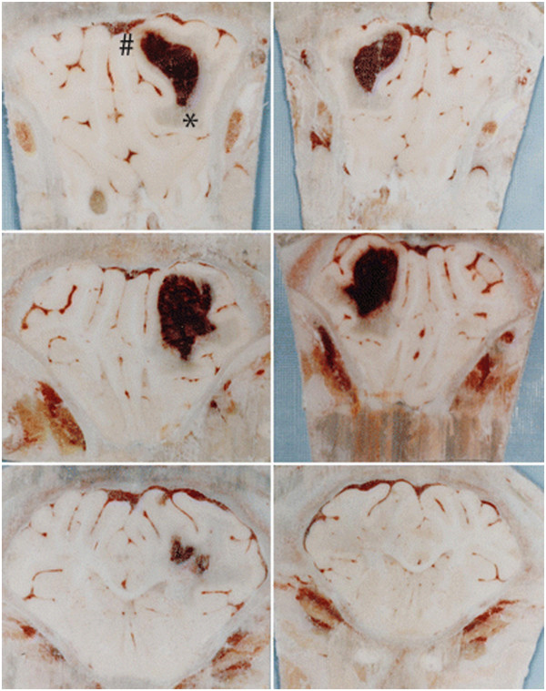
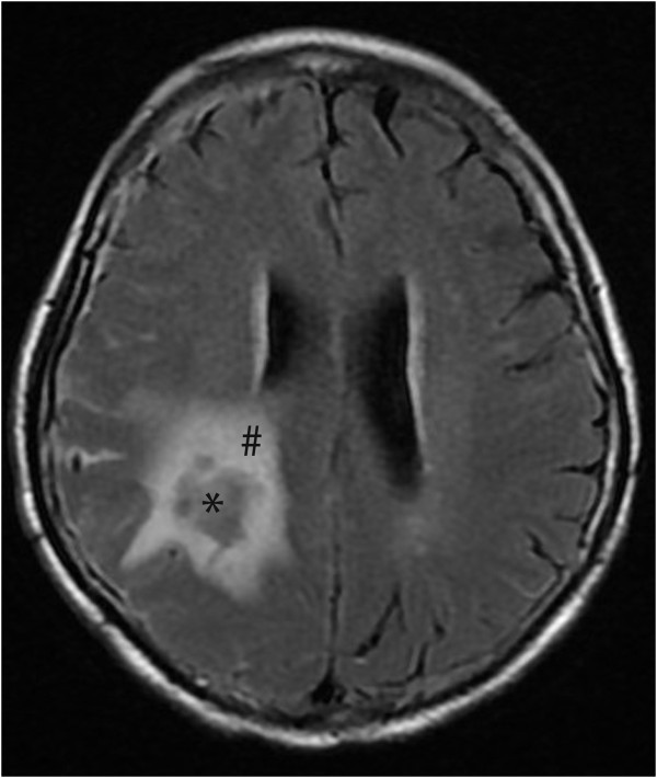
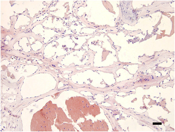
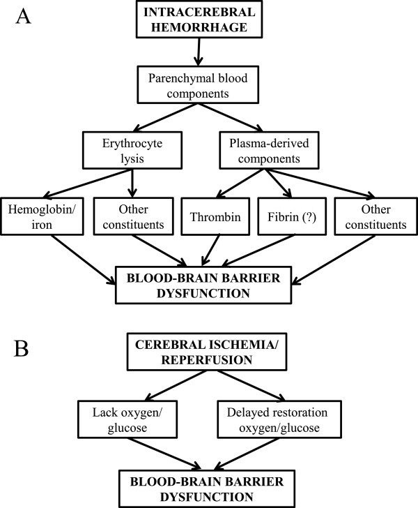
Verification
Claims the paper makes
The claims and statistics of this paper haven’t been checked yet.
Run the check — free, no account needed. It reads the open-access text and checks the sources it cites.
Claims other papers make about this one
Each is checked against this paper’s own text: does it say what the citing paper says it does?
The source backs part of the claim, not all of it — or backs it for a different population, condition or comparison.
In the citing paper
“During and after ischemic stroke, BBB disruption facilitates injury progression and increases the risk of hemorrhage, predicting poor patient outcome and limiting the use of thrombolytic treatment with recombinant tissue-type plasminogen activator (tPA) (308, 370).”
From Blood-Brain Barrier: From Physiology to Disease and Back (2019) · cites this paper as [308]
Experimental — accuracy not yet measuredIn the citing paper
“Further, the virus can access the brain through the blood stream and infect BBB cells, as observed for other viruses [131].”
From Hyperactivation of P2X7 receptors as a culprit of COVID-19 neuropathology (2021) · cites this paper as [131]
Self-check
Percentages, sums and reported differences re-computed from the paper’s own numbers.
The self-check needs the paper’s full text.
Citations
Cites
Tissue Plasminogen Activator for Acute Ischemic Stroke
(1995) · New England Journal of Medicine · doi:10.1056/nejm199512143332401 · Cited by 11,654
Astrocyte-endothelial interactions at the blood-brain barrier
Abbott, N. J., Rönnbäck, L., & Hansson, E. (2006) · Nature Reviews Neuroscience · doi:10.1038/nrn1824 · Cited by 5,595
Harold, D., Abraham, R., Hollingworth, P., Sims, R., Gerrish, A., Hamshere, M. L., Pahwa, J. S., Moskvina, V., Dowzell, K., Williams, A., et al. (2009) · Nature Genetics · doi:10.1038/ng.440 · Cited by 3,326
Genome-wide association study identifies variants at CLU and CR1 associated with Alzheimer's disease
the European Alzheimer's Disease Initiative Investigators, Lambert, J.-C., Heath, S., Even, G., Campion, D., Sleegers, K., Hiltunen, M., Combarros, O., Zelenika, D., Bullido, M. J., et al. (2009) · Nature Genetics · doi:10.1038/ng.439 · Cited by 2,234
Rapid Blood-Pressure Lowering in Patients with Acute Intracerebral Hemorrhage
Anderson, C. S., Heeley, E. L., Huang, Y., Wang, J., Stapf, C., Delcourt, C., Lindley, R. I., Robinson, T. G., Lavados, P. M., Neal, B. C., et al. (2013) · New England Journal of Medicine · doi:10.1056/nejmoa1214609 · Cited by 1,638
Mechanisms of brain injury after intracerebral haemorrhage
Xi, G., Keep, R. F., & Hoff, J. T. (2005) · The Lancet Neurology · doi:10.1016/s1474-4422(05)70283-0 · Cited by 1,366
Self-citationIntracerebral haemorrhage: mechanisms of injury and therapeutic targets
Keep, R. F., Hua, Y., & Xi, G. (2012) · The Lancet Neurology · doi:10.1016/s1474-4422(12)70104-7 · Cited by 1,308
Self-citationEfficacy and Safety of Recombinant Activated Factor VII for Acute Intracerebral Hemorrhage
Mayer, S. A., Brun, N. C., Begtrup, K., Broderick, J., Davis, S., Diringer, M. N., Skolnick, B. E., & Steiner, T. (2008) · New England Journal of Medicine · doi:10.1056/nejmoa0707534 · Cited by 1,255
Yang, Y., Estrada, E. Y., Thompson, J. F., Liu, W., & Rosenberg, G. A. (2007) · Journal of Cerebral Blood Flow & Metabolism · doi:10.1038/sj.jcbfm.9600375 · Cited by 1,060
Molecular Pathophysiology of Cerebral Hemorrhage
Aronowski, J. A., & Zhao, X. (2011) · Stroke · doi:10.1161/strokeaha.110.596718 · Cited by 855
Cited by
Sorted by citations
Blood-Brain Barrier: From Physiology to Disease and Back
Sweeney, M. D., Zhao, Z., Montagne, A., Nelson, A. R., & Zlokovic, B. V. (2019) · Physiological Reviews · doi:10.1152/physrev.00050.2017 · Cited by 2,312
Blood-brain barrier dysfunction and recovery after ischemic stroke
Jiang, X., Andjelkovic, A. V., Zhu, L., Yang, T., Bennett, M. V. L., Chen, J., Keep, R. F., & Shi, Y. (2017) · Progress in Neurobiology · doi:10.1016/j.pneurobio.2017.10.001 · Cited by 967
Self-citationNeuroinflammation after intracerebral hemorrhage
Mracsko, E. Z., & Veltkamp, R. C. (2014) · Frontiers in Cellular Neuroscience · doi:10.3389/fncel.2014.00388 · Cited by 348
Pathogenesis of Brain Edema and Investigation into Anti-Edema Drugs
Michinaga, S., & Koyama, Y. (2015) · International Journal of Molecular Sciences · doi:10.3390/ijms16059949 · Cited by 339
Saunders, N. R., Dzięgielewska, K. M., Møllgård, K., & Habgood, M. D. (2015) · Frontiers in Neuroscience · doi:10.3389/fnins.2015.00385 · Cited by 308
Role of Interleukin-10 in Acute Brain Injuries
Garcia, J. M., Stillings, S. A., Leclerc, J. L., Phillips, H., Edwards, N. J., Robicsek, S. A., Hoh, B. L., Blackburn, S., & Doré, S. (2017) · Frontiers in Neurology · doi:10.3389/fneur.2017.00244 · Cited by 247
The Mitogen-Activated Protein Kinase (MAPK) Signaling Pathway as a Discovery Target in Stroke
Sun, J., & Nan, G. (2016) · Journal of Molecular Neuroscience · doi:10.1007/s12031-016-0717-8 · Cited by 225
Keep, R. F., Andjelkovic, A. V., Xiang, J., Stamatovic, S. M., Antonetti, D. A., Hua, Y., & Xi, G. (2018) · Journal of Cerebral Blood Flow & Metabolism · doi:10.1177/0271678x18774666 · Cited by 188
Self-citationBlood-Brain Barrier Protection as a Therapeutic Strategy for Acute Ischemic Stroke
Sifat, A. E., Vaidya, B., & Abbruscato, T. J. (2017) · The AAPS Journal · doi:10.1208/s12248-017-0091-7 · Cited by 173
Hyperactivation of P2X7 receptors as a culprit of COVID-19 neuropathology
Ribeiro, D. E., Oliveira-Giacomelli, Á., Glaser, T., Arnaud-Sampaio, V. F., Andrejew, R., Dieckmann, L., Baranova, J., Lameu, C., Ratajczak, M. Z., & Ulrich, H. (2021) · Molecular Psychiatry · doi:10.1038/s41380-020-00965-3 · Cited by 145
Zhang, Y., Khan, S., Liu, Y., Wu, G., Yong, V. W., & Xue, M. (2022) · Frontiers in Immunology · doi:10.3389/fimmu.2022.847246 · Cited by 137
Chen, S., Tang, C., Ding, H., Wang, Z., Liu, X., Chai, Y., Jiang, W., Han, Y., & Zeng, H. (2020) · Frontiers in Immunology · doi:10.3389/fimmu.2020.594071 · Cited by 132
Strategies to Extend Thrombolytic Time Window for Ischemic Stroke Treatment: An Unmet Clinical Need
Peña, I. D., Borlongan, C. V., Shen, G., & Davis, W. L. (2017) · Journal of Stroke · doi:10.5853/jos.2016.01515 · Cited by 135
Okada, T., Suzuki, H., Travis, Z. D., & Zhang, J. H. (2020) · Current Neuropharmacology · doi:10.2174/1570159x18666200528143301 · Cited by 141
Wei, C., Kong, Y., Li, G., Guan, Y., Wang, P., & Miao, C. (2017) · Scientific Reports · doi:10.1038/s41598-017-00851-z · Cited by 126
Fang, Y., Gao, S., Wang, X., Cao, Y., Lü, J., Chen, S., Lenahan, C., Zhang, J. H., Shao, A., & Zhang, J. (2020) · Frontiers in Cellular Neuroscience · doi:10.3389/fncel.2020.00068 · Cited by 114
Wang, D., Liu, K., Wake, H., Teshigawara, K., Mori, S., & Nishibori, M. (2017) · Scientific Reports · doi:10.1038/srep46243 · Cited by 101
Imai, T., Iwata, S., Hirayama, T., Nagasawa, H., Nakamura, S., Shimazawa, M., & Hara, H. (2019) · Scientific Reports · doi:10.1038/s41598-019-42370-z · Cited by 94
Chen, Q., Tang, J., Tan, L., Guo, J., Tao, Y., Li, L., Chen, Y., Liu, X. B., Zhang, J. H., Chen, Z., et al. (2015) · Stroke · doi:10.1161/strokeaha.115.009713 · Cited by 94
Yang, Y., Xiong, X., Liu, J., Wu, L., Zhong, Q., Zhou, K., Meng, Z., Liu, L., Wang, F., Gong, Q., et al. (2017) · Journal of the American Heart Association · doi:10.1161/jaha.117.005811 · Cited by 87
Endothelial progenitor cells: Potential novel therapeutics for ischaemic stroke
Bayraktutan, U. (2019) · Pharmacological Research · doi:10.1016/j.phrs.2019.04.017 · Cited by 85
Wu, C., Shyue, S., Hung, T., Wen, S., Lin, C.-C., Chang, C.-F., & Chen, S. (2017) · Journal of Neuroinflammation · doi:10.1186/s12974-017-1005-4 · Cited by 77
Aerobic Training and Mobilization Early Post-stroke: Cautions and Considerations
Marzolini, S., Robertson, A. D., OH, P. I., Goodman, J. M., Corbett, D., Du, X., & MacIntosh, B. J. (2019) · Frontiers in Neurology · doi:10.3389/fneur.2019.01187 · Cited by 83
Carmona-Mora, P., Ander, B. P., Jickling, G. C., Dykstra-Aiello, C., Zhan, X., Ferino, E., Hamade, F., Amini, H., Hull, H., Sharp, F. R., et al. (2020) · Journal of Cerebral Blood Flow & Metabolism · doi:10.1177/0271678x20953912 · Cited by 68
Translational Stroke Research on Blood-Brain Barrier Damage: Challenges, Perspectives, and Goals
Shi, Y., Leak, R. K., Keep, R. F., & Chen, J. (2016) · Translational Stroke Research · doi:10.1007/s12975-016-0447-9 · Cited by 67
Self-citationStamova, B. S., Ander, B. P., Jickling, G. C., Hamade, F., Durocher, M., Zhan, X., Liu, D., Cheng, X., Hull, H., Yee, A., et al. (2018) · Journal of Cerebral Blood Flow & Metabolism · doi:10.1177/0271678x18769513 · Cited by 66
Carnosine Attenuates Brain Oxidative Stress and Apoptosis After Intracerebral Hemorrhage in Rats
Xie, R., Li, D., Liu, X.-C., Yang, M., Fang, J., Sun, B., Zhang, Z., & Yang, X. (2016) · Neurochemical Research · doi:10.1007/s11064-016-2104-9 · Cited by 67
Intracerebral haemorrhage: from clinical settings to animal models
Bai, Q., Sheng, Z., Liu, Y., Zhang, R., Yong, V. W., & Xue, M. (2020) · Stroke and Vascular Neurology · doi:10.1136/svn-2020-000334 · Cited by 68
Xu, F., Shen, G., Su, Z., He, Z., & Yuan, L. (2019) · Brain and Behavior · doi:10.1002/brb3.1254 · Cited by 66
Wang, P., Ren, Q., Shi, M., Liu, Y., Bai, H., & Chang, Y. (2022) · Antioxidants · doi:10.3390/antiox11071257 · Cited by 64
Andjelkovic, A. V., Stamatovic, S. M., Phillips, C. M., Martínez-Revollar, G., & Keep, R. F. (2020) · Fluids and Barriers of the CNS · doi:10.1186/s12987-020-00202-7 · Cited by 68
Self-citationZhao, C., Ma, J., Wang, Z., Li, H., Shen, H., Li, X., & Chen, G. (2020) · Translational Stroke Research · doi:10.1007/s12975-019-00775-y · Cited by 61
Yu, L., Lu, Z., Burchell, S. R., Nowrangi, D., Manaenko, A., Li, X., Xu, Y., Xu, N., Tang, J., Dai, H., et al. (2017) · Journal of Neurochemistry · doi:10.1111/jnc.14238 · Cited by 59
Remodeling of the Neurovascular Unit Following Cerebral Ischemia and Hemorrhage
Satō, Y., Falcone-Juengert, J., Tominaga, T., Su, H., & Liu, J. (2022) · Cells · doi:10.3390/cells11182823 · Cited by 60
Blast-induced temporal alterations in blood–brain barrier properties in a rodent model
Kawoos, U., Abutarboush, R., Gu, M., Chen, Y., Statz, J. K., Goodrich, S. Y., & Ahlers, S. T. (2021) · Scientific Reports · doi:10.1038/s41598-021-84730-8 · Cited by 58
Cofilin as a Promising Therapeutic Target for Ischemic and Hemorrhagic Stroke
Alhadidi, Q., Sayeed, M. S. B., & Shah, Z. A. (2015) · Translational Stroke Research · doi:10.1007/s12975-015-0438-2 · Cited by 57
Kanamaru, H., & Suzuki, H. (2019) · Neural Regeneration Research · doi:10.4103/1673-5374.251190 · Cited by 56
Peña, I. D., Knecht, T., & Borlongan, C. V. (2018) · Brain Circulation · doi:10.4103/bc.bc_21_18 · Cited by 56
Fang, Y., Tian, Y., Huang, Q., Wan, Y., Xu, L., Wang, W., Pan, D., Zhu, S., & Xie, M. (2019) · Journal of Neuroinflammation · doi:10.1186/s12974-019-1485-5 · Cited by 57
Jiang, C. Q., Zuo, F., Wang, Y., Wan, J., Yang, Z.-J., Lu, H., Chen, W., Zang, W., Yang, Q., & Wang, J. (2016) · Neurobiology of Aging · doi:10.1016/j.neurobiolaging.2016.02.029 · Cited by 54
Li, Y., Wu, P., Bihl, J. C., & Shi, H. (2020) · Current Neuropharmacology · doi:10.2174/1570159x18666200106154203 · Cited by 54
Durocher, M., Knepp, B., Yee, A., Jickling, G. C., Rodríguez, F., Ng, K., Zhan, X., Hamade, F., Ferino, E., Amini, H., et al. (2020) · Translational Stroke Research · doi:10.1007/s12975-020-00869-y · Cited by 54
Fang, J., Yuan, Q., Du, Z., Fei, M., Zhang, Q., Yang, L., Wang, M., Yang, W., Yu, J., Wu, G., et al. (2022) · Biochemical and Biophysical Research Communications · doi:10.1016/j.bbrc.2022.06.040 · Cited by 53
Xiang, J., Routhe, L. G., Wilkinson, D. A., Hua, Y., Moos, T., Xi, G., & Keep, R. F. (2017) · Fluids and Barriers of the CNS · doi:10.1186/s12987-017-0056-3 · Cited by 51
Self-citationWang, C., Fei, Y., Xu, C., Zhao, Y., & Pan, Y. (2015) · PubMed Central · Cited by 50
Zhang, Y., Fang, M., Zhu, J., Li, T., Li, N., Su, B., Guo-dong, S., Li, L., & Zhou, C. (2024) · International Journal of Biological Macromolecules · doi:10.1016/j.ijbiomac.2024.132970 · Cited by 48
Wang, Z., Chen, Z., Yang, J., Yang, Z., Yin, J., Zuo, G., Duan, X., Shen, H., Li, H., & Chen, G. (2016) · Journal of Cerebral Blood Flow & Metabolism · doi:10.1177/0271678x16669513 · Cited by 48
Xing, G., Zhao, T., Zhang, X., He, L., Li, X., Cui, P., Li, M., Li, D., Zhang, N., & Jiang, W. (2020) · Frontiers in Cellular Neuroscience · doi:10.3389/fncel.2020.575690 · Cited by 49
Nadeau, C. A., Dietrich, K., Wilkinson, C. M., Crawford, A. M., George, G. N., Nichol, H. K., & Colbourne, F. (2018) · Translational Stroke Research · doi:10.1007/s12975-018-0636-9 · Cited by 47
Loss of mural cell-derived laminin aggravates hemorrhagic brain injury
Gautam, J., Xu, L., Nirwane, A. M., Nguyen, B., & Yao, Y. (2020) · Journal of Neuroinflammation · doi:10.1186/s12974-020-01788-3 · Cited by 47
Duan, T., Li, L., Yu, Y., Li, T., Han, R., Sun, X., Cui, Y., Liu, T., Wang, X., Wang, Y., et al. (2022) · Pharmacological Research · doi:10.1016/j.phrs.2022.106200 · Cited by 47
Min, H., Choi, B., Jang, Y. H., Cho, I., & Lee, S. J. (2017) · Molecular Brain · doi:10.1186/s13041-017-0305-z · Cited by 46
Liu, Y., Li, Z., Khan, S., Zhang, R., Wei, R., Zhang, Y., Xue, M., & Yong, V. W. (2021) · Neuroscience Letters · doi:10.1016/j.neulet.2021.136297 · Cited by 45
Wang, Y., Fan, P., XIE, G., Chen, Z.-Q., Li, H., Tang, T., & Luo, J. (2016) · Experimental and Therapeutic Medicine · doi:10.3892/etm.2016.3330 · Cited by 45
Adjunctive Therapy Approaches for Ischemic Stroke: Innovations to Expand Time Window of Treatment
Knecht, T., Story, J., Liu, J., Davis, W. L., Borlongan, C. V., & Peña, I. D. (2017) · International Journal of Molecular Sciences · doi:10.3390/ijms18122756 · Cited by 45
Yan, J., Xu, W., Lenahan, C., Huang, L., Ocak, U., Wen, J., Li, G., He, W., Le, C., Zhang, J. H., et al. (2022) · Fluids and Barriers of the CNS · doi:10.1186/s12987-022-00305-3 · Cited by 43
Fgr contributes to hemorrhage-induced thalamic pain by activating NF-κB/ERK1/2 pathways
Huang, T., Fu, G., Gao, J., Zhang, Y., Cai, W., Wu, S., Jia, S., Xia, S., Bachmann, T., Bekker, A., et al. (2020) · JCI Insight · doi:10.1172/jci.insight.139987 · Cited by 43
Bahadar, G. A., & Shah, Z. A. (2021) · CNS & Neurological Disorders - Drug Targets · doi:10.2174/1871527320666210223145112 · Cited by 42
Innate and adaptive immune response in stroke: Focus on epigenetic regulation
Picascia, A., Grimaldi, V., Iannone, C., Soricelli, A., & Claudio Napoli, A. (2015) · Journal of Neuroimmunology · doi:10.1016/j.jneuroim.2015.10.013 · Cited by 40
Sugiyama, T., Imai, T., Nakamura, S., Yamauchi, K., Sawada, S., Shimazawa, M., & Hara, H. (2018) · Brain Research · doi:10.1016/j.brainres.2018.08.021 · Cited by 38
Chen, J., Wang, L., Xu, H., Wang, Y., & Liang, Q. (2021) · Journal of Leukocyte Biology · doi:10.1002/jlb.5mr0321-632r · Cited by 38
Isoquercetin Improves Inflammatory Response in Rats Following Ischemic Stroke
Shi, Y., Chen, X., Liu, J., Fan, X., Jin, Y., Gu, J., Liang, J., Liang, X., & Wang, C. (2021) · Frontiers in Neuroscience · doi:10.3389/fnins.2021.555543 · Cited by 36
Central Nervous System Tissue Regeneration after Intracerebral Hemorrhage: The Next Frontier
Zhang, R., Xue, M., & Yong, V. W. (2021) · Cells · doi:10.3390/cells10102513 · Cited by 35
Cheng, S., Gao, W., Xu, X. J., Fan, H., Wu, Y., Li, F., Zhang, J., Zhu, X., & Zhang, Y. (2016) · Brain Research Bulletin · doi:10.1016/j.brainresbull.2016.10.007 · Cited by 36
Intercellular cross-talk in intracerebral hemorrhage
Egashira, Y., Hua, Y., Keep, R. F., & Xi, G. (2015) · Brain Research · doi:10.1016/j.brainres.2015.04.003 · Cited by 34
Self-citationProtective effects of the astaxanthin derivative, adonixanthin, on brain hemorrhagic injury
Iwata, S., Imai, T., Shimazawa, M., Ishibashi, T., Hayashi, M., Hara, H., & Nakamura, S. (2018) · Brain Research · doi:10.1016/j.brainres.2018.08.009 · Cited by 34
Okada, T., & Suzuki, H. (2020) · PubMed · doi:10.14670/hh-18-208 · Cited by 34
Cell-specific activation of RIPK1 and MLKL after intracerebral hemorrhage in mice
Lüle, S., Wu, L., Sarro-Schwartz, A., Edmiston, W. J., Izzy, S., Songtachalert, T., Ahn, S. H., Fernandes, N. D., Jin, G., Chung, J. Y., et al. (2020) · Journal of Cerebral Blood Flow & Metabolism · doi:10.1177/0271678x20973609 · Cited by 34
Song, D., Ji, Y., Huang, X., Ma, Y., Fang, C., Qiu, L., Tan, X., Chen, Y.-L., Wang, S., Chang, J., et al. (2022) · CNS Neuroscience & Therapeutics · doi:10.1111/cns.13832 · Cited by 33
Reyes‐Esteves, S., Nong, J., Glassman, P. M., Omo‐Lamai, S., Ohashi, S. N., Myerson, J. W., Zamora, M. E., Ma, X., Kasner, S. E., Sansing, L. H., et al. (2023) · Journal of Controlled Release · doi:10.1016/j.jconrel.2023.02.037 · Cited by 35
Li, M., Ke, J., Deng, Y., Chen, C., Huang, Y., Bian, Y., Guo, S., Wu, Y., Zhang, H., Liu, M., et al. (2021) · Frontiers in Pharmacology · doi:10.3389/fphar.2021.671783 · Cited by 33
Predictive Value of CTA Spot Sign on Hematoma Expansion in Intracerebral Hemorrhage Patients
Peng, W.-J., Reis, C., Reis, H., Zhang, J. H., & Yang, J. (2017) · BioMed Research International · doi:10.1155/2017/4137210 · Cited by 32
Including Patients With Stroke in Cardiac Rehabilitation
Marzolini, S. (2020) · Journal of Cardiopulmonary Rehabilitation and Prevention · doi:10.1097/hcr.0000000000000540 · Cited by 33
Block, N., Nauclér, P., Wagner, P., Morfeldt, E., & Henriques‐Normark, B. (2022) · Journal of Internal Medicine · doi:10.1111/joim.13488 · Cited by 33
Effects of deferoxamine on blood-brain barrier disruption after subarachnoid hemorrhage
Li, Y., Yang, H., Ni, W., & Gu, Y. (2017) · PLoS ONE · doi:10.1371/journal.pone.0172784 · Cited by 31
Al-Ahmady, Z. S., Dickie, B., Aldred, I., Jasim, D. A., Barrington, J., Haley, M., Lemarchand, E., Coutts, G., Kaur, S., Bates, J., et al. (2022) · Theranostics · doi:10.7150/thno.72167 · Cited by 31
Tobacco Use: A Major Risk Factor of Intracerebral Hemorrhage
Cho, S., Rehni, A. K., & Dave, K. R. (2021) · Journal of Stroke · doi:10.5853/jos.2020.04770 · Cited by 31
Fan, S., Du, H., Yao, Y., Wang, H., Zhang, M., Shi, X.-L., Yan, J., Li, P., Xiao, G., He, S., et al. (2025) · Phytomedicine · doi:10.1016/j.phymed.2025.157162 · Cited by 29
Lu, T., Wang, Z., Sherchan, P., Xu, Y., Wang, T., Zhang, Y., Yu, L., Xu, N., Tang, J., You, W., et al. (2018) · Journal of Neurochemistry · doi:10.1111/jnc.14622 · Cited by 28
Drug Repurposing: Promises of Edaravone Target Drug in Traumatic Brain Injury
Shakkour, Z., Issa, H., Ismail, H., Ashekyan, O., Habashy, K. J., Nasrallah, L., Jourdi, H., Hamade, E., Mondello, S., Sabra, M., et al. (2020) · Current Medicinal Chemistry · doi:10.2174/0929867327666200812221022 · Cited by 34
Caspase-1: A Promising Target for Preserving Blood–Brain Barrier Integrity in Acute Stroke
Ye, X., Song, G., Huang, S., Liang, Q., Fang, Y., Lian, L., & Zhu, S. (2022) · Frontiers in Molecular Neuroscience · doi:10.3389/fnmol.2022.856372 · Cited by 30
The Important Double-Edged Role of Astrocytes in Neurovascular Unit After Ischemic Stroke
Han, G., Song, L., Ding, Z., Wang, Q., Yan, Y., Huang, J., & Ma, C. (2022) · Frontiers in Aging Neuroscience · doi:10.3389/fnagi.2022.833431 · Cited by 32
Alzheimer’s Disease: From Amyloid to Autoimmune Hypothesis
Arshavsky, Y. I. (2020) · The Neuroscientist · doi:10.1177/1073858420908189 · Cited by 28
Zhou, Z.-Y., Zhao, W.-R., Xiao, Y., Zhang, J., Tang, J., & Lee, S. M. (2020) · Frontiers in Pharmacology · doi:10.3389/fphar.2020.551745 · Cited by 27
Girard, R., Li, Y., Stadnik, A., Shenkar, R., Hobson, N., Romanos, S. G., Srinath, A., Moore, T., Lightle, R., Shkoukani, A., et al. (2020) · Neurosurgery · doi:10.1093/neuros/nyaa478 · Cited by 27
Lüle, S., Wu, L., McAllister, L. M., Edmiston, W. J., Chung, J. Y., Levy, E. S., Zheng, Y., Gough, P. J., Bertin, J., Degterev, A., et al. (2017) · Stroke · doi:10.1161/strokeaha.117.017702 · Cited by 26
Li, X., Zhang, Y., Chang, J., Zhang, C., Li, L., Dai, Y., Yang, H., & Wang, Y. (2023) · Cellular and Molecular Life Sciences · doi:10.1007/s00018-023-04716-9 · Cited by 26
Teo, K. C., Lau, K. K., Mak, R. H. Y., Leung, H.-Y., Chang, R. S., Tse, M. Y., Lee, R., Leung, G., Ho, S.-L., Cheung, R. T. F., et al. (2017) · World Neurosurgery · doi:10.1016/j.wneu.2017.06.015 · Cited by 26
Lekic, T., Klebe, D. W., McBride, D. W., Manaenko, A., Rolland, W. B., Flores, J. J., Altay, O., Tang, J., & Zhang, J. H. (2015) · Stroke · doi:10.1161/strokeaha.114.007889 · Cited by 25
Erdei, J., Tóth, A., Nagy, A., Nyakundi, B. B., Fejes, Z., Nagy, B., Novák, L., Bognár, L., Balogh, E., Paragh, G., et al. (2020) · Frontiers in Immunology · doi:10.3389/fimmu.2020.00228 · Cited by 25
Li, Z., Chen, X., Zhang, X., Ren, X., Chen, X., Cao, J., Zang, W., Liu, X., & Guo, F. (2017) · Journal of Molecular Neuroscience · doi:10.1007/s12031-017-0883-3 · Cited by 24
Shi, J., Wu, G., Zou, X., & Jiang, K. (2017) · Pharmacological Reports · doi:10.1016/j.pharep.2017.05.004 · Cited by 24
Failure of bumetanide to improve outcome after intracerebral hemorrhage in rat
Wilkinson, C. M., Fedor, B. A., Aziz, J. R., Nadeau, C. A., Brar, P. S., Clark, J. J. A., & Colbourne, F. (2019) · PLoS ONE · doi:10.1371/journal.pone.0210660 · Cited by 24
Guo, H., Zhang, W., Wang, Z., Li, Z., Zhou, J., & Yang, Z. (2022) · Frontiers in Molecular Neuroscience · doi:10.3389/fnmol.2022.977941 · Cited by 23
Xie, Y., He, W., Ma, L., Ren, R., Yang, S., & Lu, Q. (2023) · CNS Neuroscience & Therapeutics · doi:10.1111/cns.14255 · Cited by 23
Kocsis, A. E., Kucsápszky, N., Santa-Maria, A. R., Hunyadi, A., Deli, M. A., & Walter, F. R. (2025) · Nutrients · doi:10.3390/nu17050766 · Cited by 21
Microglial Mechanisms and Therapeutic Potential in Brain Injury Post-Intracerebral Hemorrhage
Gong, Y., Li, H., Cui, H., & Gong, Y. (2025) · Journal of Inflammation Research · doi:10.2147/jir.s498809 · Cited by 23
Wang, G., Sun, Y., Yang, Q., Dai, D., Zhang, L., Fan, H., Zhang, W., Dong, J., & Zhao, P. (2023) · Food and Chemical Toxicology · doi:10.1016/j.fct.2023.113813 · Cited by 21
Prognostic impact of leukocytosis in intracerebral hemorrhage
Yu, Z., Zheng, J., Guo, R., Ma, L., You, C., & Li, H. (2019) · Medicine · doi:10.1097/md.0000000000016281 · Cited by 21
Navigating Hemorrhagic Shock: Biomarkers, Therapies, and Challenges in Clinical Care
Monge, K. M., Rosa, C., Sublette, C. M., Pratap, A., Kovacs, E. J., & Idrovo, J. (2024) · Biomedicines · doi:10.3390/biomedicines12122864 · Cited by 22
Metadata
- Authors
- Keep, R. F., Zhou, N., Xiang, J., Andjelkovic, A. V., Hua, Y., & Xi, G.
- Affiliations
- University of Michigan (US)
- Corresponding author
- Keep, Richard F
- Venue
- Fluids and Barriers of the CNS
- Published
- 2014
- Volume
- 11
- Issue
- 1
- Pages
- 18
- ISSN
- 2045-8118 (ISSN-L)
- PMC
- PMC4130123
- OpenAlex
- W2103156349
- Cited by
- 220
- References
- 144
- Citations by year
- 2026: 22 · 2025: 20 · 2024: 17 · 2023: 19 · 2022: 23 · 2021: 18 · 2020: 29 · 2019: 17 · 2018: 9 · 2017: 24 · 2016: 12 · 2015: 9 · 2014: 1
- FWCI
- 2.10
- Type
- journal article
- Language
- en
- Open access
- gold
- doi.org
- Licence
- cc-by
- Locations
- Fluids and Barriers of the CNS (publishedVersion, cc-by)
- PubMed Central (publishedVersion, cc-by)
- PubMed (publishedVersion)
- Deep Blue (University of Michigan) (submittedVersion)
- Funders
- National Institutes of Health (NS073959, NS079157)
- National Institute of Neurological Disorders and Stroke (R21NS075757, R01NS034709, R21NS084049, R01NS079157)
- Topics
- Health Sciences › Medicine › Clinical Neurology › Intracerebral and Subarachnoid Hemorrhage Research
- Life Sciences › Neuroscience › Neurology › Barrier Structure and Function Studies
- Health Sciences › Medicine › Clinical Neurology › Traumatic Brain Injury and Neurovascular Disturbances
- Keywords
- intracerebral hemorrhage, blood-brain barrier dysfunction, hematoma expansion, vascular disruption, thrombin, hemoglobin, iron, neuroinflammation, iron chelation, hematoma evacuation, hypertension, vascular malformations, coagulopathy, Alzheimer's disease, secondary hemorrhage
About these checks
Registry, retraction, Cites and statistics results are computed, not judged, with one exception: a printed reference that no index lists is read once by a model before it counts as a citation error. Claims are extracted by a model and kept only when their quote appears verbatim in the text; each cited source is then checked, with a model’s help, for whether it says what the claim relies on. Those verdicts can be wrong — read the claim’s quote and the cited source before relying on one.
Statistics and arithmetic are recomputed, not judged.
Confidence bands are shown only for fields calibrated on our annotated test set. None is calibrated yet, so no band is shown.
What counts as a claim ›How we measure confidence ›How we check this ›
Are you an author, or do you see a problem with this page? Request a review
