Cited by 221Wnt/β-catenin signaling in development and cancerDevelopmental Biology and Gene RegulationSignaling Pathways in Disease

The DOI matches its Crossref record; 61 of the 61 references it cites were found in a registry or an index.

Nuclear localization is required for Dishevelled function in Wnt/β-catenin signaling

Open access

Abstract

Background: Dishevelled (Dsh) is a key component of multiple signaling pathways that are initiated by Wnt secreted ligands and Frizzled receptors during embryonic development. Although Dsh has been detected in a number of cellular compartments, the importance of its subcellular distribution for signaling remains to be determined. Results: We report that Dsh protein accumulates in cell nuclei when Xenopus embryonic explants or mammalian cells are incubated with inhibitors of nuclear export or when a specific nuclear-export signal (NES) in Dsh is disrupted by mutagenesis. Dsh protein with a mutated NES, while predominantly nuclear, remains fully active in its ability to stimulate canonical Wnt signaling. Conversely, point mutations in conserved amino-acid residues that are essential for the nuclear localization of Dsh impair the ability of Dsh to activate downstream targets of Wnt signaling. When these conserved residues of Dsh are replaced with an unrelated SV40 nuclear localization signal, full Dsh activity is restored. Consistent with a signaling function for Dsh in the nucleus, treatment of cultured mammalian cells with medium containing Wnt3a results in nuclear accumulation of endogenous Dsh protein. Conclusions: These findings suggest that nuclear localization of Dsh is required for its function in the canonical Wnt/β-catenin signaling pathway. We discuss the relevance of these findings to existing models of Wnt signal transduction to the nucleus.

Overview

The claims and statistics of this paper haven’t been checked yet.

Run the check — free, no account needed. It reads the open-access text and checks the sources it cites.

A few free checks a day · papers already verified open instantly · results are public

Integrity signals

Registry

Crossref · 10.1186/jbiol20

Cites

61 of 61 references verified0 citation errors · 0 not checkable

Statistics

Figures

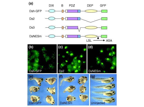
Figure 1 Nuclear export of Dsh is not critical for its activity. (a) The Dsh constructs used to analyze nuclear export. (b-d) RNAs encoding Dsh-GFP, Ds2 and DsNESm (0.5 ng each) were injected into two animal blastomeres of 4–8-cell embryos. Animal-cap explants were excised at stage 10, fixed and examined for GFP fluorescence. (b) Wild type Dsh-GFP localized in punctate structures of the cytoplasm, whereas (c) Ds2 and (d) DsNESm accumulated in the nucleus of animal pole cells. (e,f) One ventral vegetal blastomere of 8-cell embryos was injected with 1 ng Dsh-GFP or DsNESm RNA as indicated. Complete secondary axes were induced in both cases. (g) Uninjected sibling embryos.
Figure 1 Nuclear export of Dsh is not critical for its activity. (a) The Dsh constructs used to analyze nuclear export. (b-d) RNAs encoding Dsh-GFP, Ds2 and DsNESm (0.5 ng each) were injected into two animal blastomeres of 4–8-cell embryos. Animal-cap explants were excised at stage 10, fixed and examined for GFP fluorescence. (b) Wild type Dsh-GFP localized in punctate structures of the cytoplasm, whereas (c) Ds2 and (d) DsNESm accumulated in the nucleus of animal pole cells. (e,f) One ventral vegetal blastomere of 8-cell embryos was injected with 1 ng Dsh-GFP or DsNESm RNA as indicated. Complete secondary axes were induced in both cases. (g) Uninjected sibling embryos.CC BY · PubMed Central
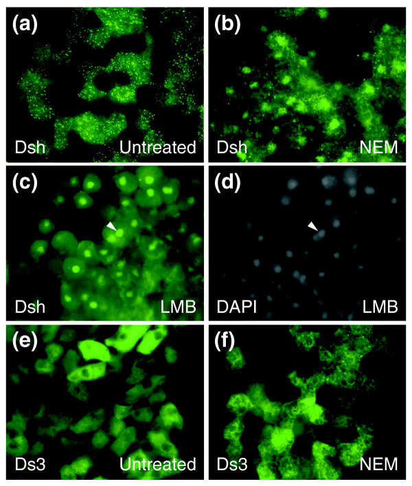
Figure 2 Accumulation of Dsh in the nucleus in the absence of nuclear export. (a-d) Dsh-GFP RNA (0.7 ng) was injected into two animal blastomeres of 4–8 cell embryos. Animal caps were excised at stage 10 and then left (a) untreated or (b) treated with 10 mM NEM or (c,d) 50 ng/ml leptomycin B (LMB), fixed and examined for GFP fluorescence. (a) Dsh-GFP is mainly localized to vesicular structures in the cytoplasm. In the presence of (b) NEM or (c) LMB, Dsh-GFP accumulates in the nucleus, as supported by (d) DAPI staining of nuclei in the same field as in (c). Nuclear staining is marked by arrowheads (c,d). (e,f) The Ds3 construct, lacking amino acids 334–381, remained in the cytoplasm in the (e) absence or (f) presence of NEM.
Figure 2 Accumulation of Dsh in the nucleus in the absence of nuclear export. (a-d) Dsh-GFP RNA (0.7 ng) was injected into two animal blastomeres of 4–8 cell embryos. Animal caps were excised at stage 10 and then left (a) untreated or (b) treated with 10 mM NEM or (c,d) 50 ng/ml leptomycin B (LMB), fixed and examined for GFP fluorescence. (a) Dsh-GFP is mainly localized to vesicular structures in the cytoplasm. In the presence of (b) NEM or (c) LMB, Dsh-GFP accumulates in the nucleus, as supported by (d) DAPI staining of nuclei in the same field as in (c). Nuclear staining is marked by arrowheads (c,d). (e,f) The Ds3 construct, lacking amino acids 334–381, remained in the cytoplasm in the (e) absence or (f) presence of NEM.CC BY · PubMed Central
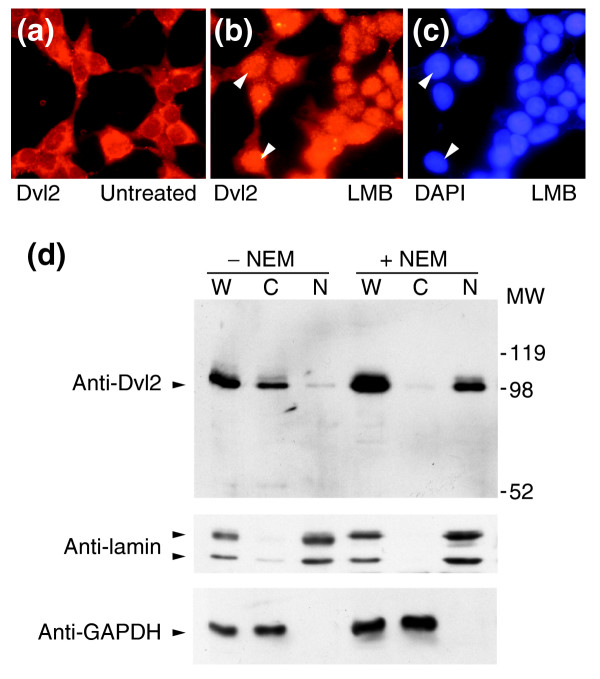
Figure 3 Endogenous Dsh shuttles between the cytoplasm and nucleus. Immunofluorescent staining of HEK293 cells with anti-Dvl2 antibodies reveals different subcellular localization of Dvl2 (a) without or (b) with LMB treatment. (c) DAPI staining shows the location of nuclei in the same field as (b); the arrowheads indicate corresponding nuclei in (b) and (c). (d) Distribution of endogenous Dvl2 recognized by anti-Dvl2 antibodies in the nuclear and the cytoplasmic fractions of Rat-1 fibroblasts. In the absence of NEM, Dvl2 is localized mainly in the cytoplasm (C), while after NEM treatment Dvl2 is exclusively localized in the nuclei (N). W, whole cell lysate. Antibodies to lamin and GAPDH show the separation of the nuclear and cytoplasmic fractions.
Figure 3 Endogenous Dsh shuttles between the cytoplasm and nucleus. Immunofluorescent staining of HEK293 cells with anti-Dvl2 antibodies reveals different subcellular localization of Dvl2 (a) without or (b) with LMB treatment. (c) DAPI staining shows the location of nuclei in the same field as (b); the arrowheads indicate corresponding nuclei in (b) and (c). (d) Distribution of endogenous Dvl2 recognized by anti-Dvl2 antibodies in the nuclear and the cytoplasmic fractions of Rat-1 fibroblasts. In the absence of NEM, Dvl2 is localized mainly in the cytoplasm (C), while after NEM treatment Dvl2 is exclusively localized in the nuclei (N). W, whole cell lysate. Antibodies to lamin and GAPDH show the separation of the nuclear and cytoplasmic fractions.CC BY · PubMed Central
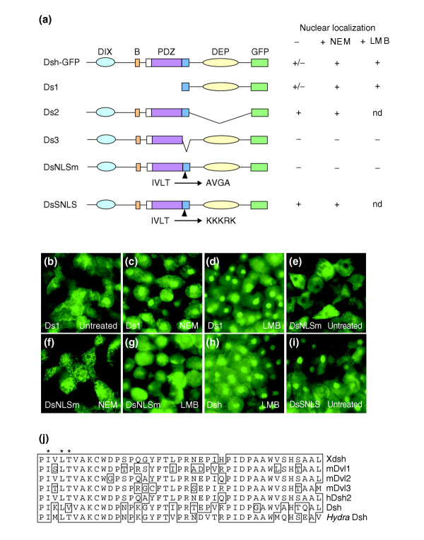
Figure 4 Mapping nuclear localization signals in Dsh. (a) The Dsh constructs used to study nuclear transport and their localization to the nucleus after NEM or LMB treatment; the DIX, PDZ and DEP domains are shown as in Figure 1a; B is the basic region and nd denotes not done. (b-i) Subcellular localization of Dsh-GFP constructs in the absence or presence of NEM or LMB. Embryos were injected with 0.5 ng of each mRNA, and GFP analysis was carried out as in Figure 1b-d. (b-d) Ds1, (e-g) DsNLSm, (h) Dsh, (i) DsSNLS. (b,e,i) no NEM treatment; (c,f) after NEM treatment; (d,g,h) after LMB treatment. (j) Comparison of conserved amino-acid sequences that are required for Dsh nuclear localization; X denotes the Xenopus protein, m the mouse and h the human. Amino-acid residues mutated in DsNLSm are indicated by asterisks.
Figure 4 Mapping nuclear localization signals in Dsh. (a) The Dsh constructs used to study nuclear transport and their localization to the nucleus after NEM or LMB treatment; the DIX, PDZ and DEP domains are shown as in Figure 1a; B is the basic region and nd denotes not done. (b-i) Subcellular localization of Dsh-GFP constructs in the absence or presence of NEM or LMB. Embryos were injected with 0.5 ng of each mRNA, and GFP analysis was carried out as in Figure 1b-d. (b-d) Ds1, (e-g) DsNLSm, (h) Dsh, (i) DsSNLS. (b,e,i) no NEM treatment; (c,f) after NEM treatment; (d,g,h) after LMB treatment. (j) Comparison of conserved amino-acid sequences that are required for Dsh nuclear localization; X denotes the Xenopus protein, m the mouse and h the human. Amino-acid residues mutated in DsNLSm are indicated by asterisks.CC BY · PubMed Central
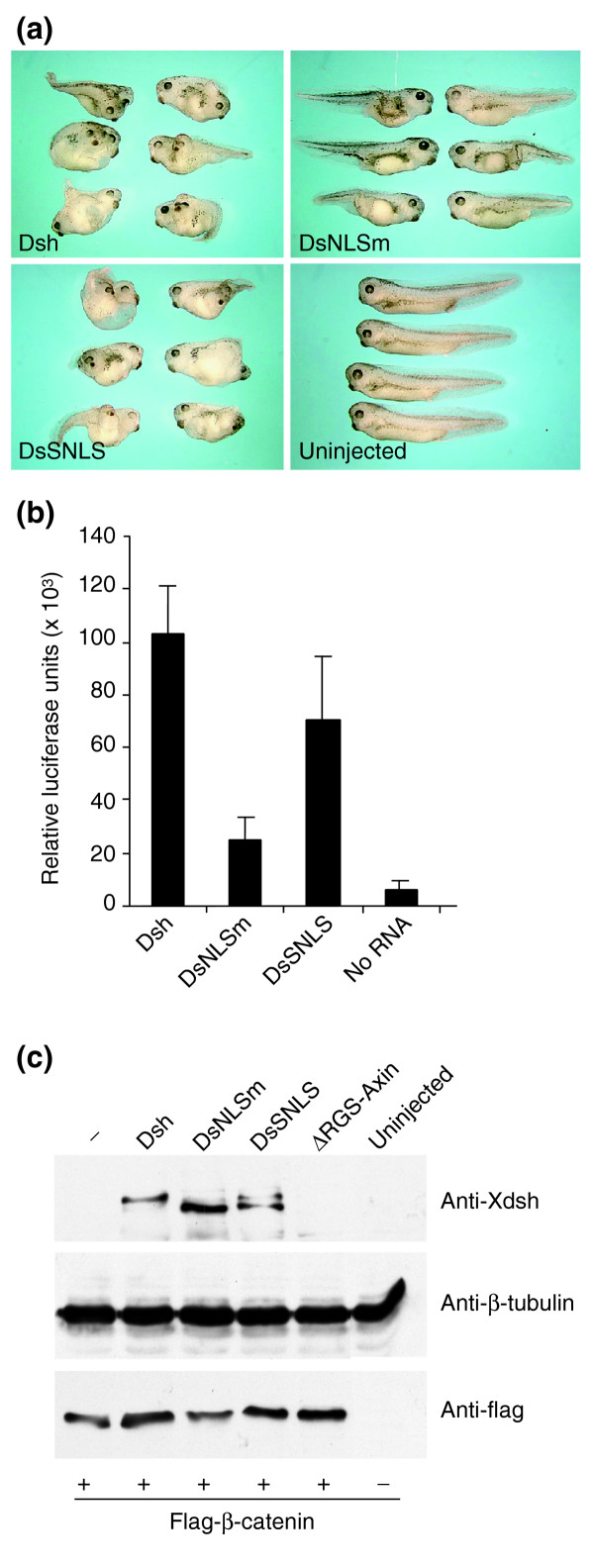
Figure 5 Activation of the Wnt/β-catenin pathway requires nuclear localization of Dsh. (a) Axis-inducing activity of Dsh constructs. One ventral vegetal blastomere of 8-cell embryos was injected with 1 ng Dsh-GFP, DsNLSm, or DsSNLS mRNA as indicated. Uninjected sibling embryos are also shown. (b) Activation of the Siamois reporter gene. The reporter -833pSia-Luc plasmid (20 pg) was coinjected with Dsh-GFP, DsNLSm or DsSNLS mRNA (0.5 ng each) into a single animal ventral blastomere of 8-cell embryos. Injected embryos were lysed at stage 10+ for luciferase activity determination. Results are shown in relative light units as the mean +/- standard deviation from triplicate samples. (c) Requirement for Dsh NLS for the stabilization of β-catenin. Flag-β-catenin mRNA (0.4 ng) was coinjected with Dsh, DsNLSm, DsSNLS or ΔRGS-Axin mRNA (2 ng each) into four animal blastomeres of 4–8-cell embryos. Levels of β-catenin and Dsh constructs were assessed in stage 10 embryo lysates with anti-Flag antibodies and anti-Xdsh antibodies; β-tubulin serves as a loading control. Dsh and DsSNLS, but not DsNLSm, are able to stabilize β-catenin. ΔRGS-Axin was used as a control for an activator of the Wnt pathway.
Figure 5 Activation of the Wnt/β-catenin pathway requires nuclear localization of Dsh. (a) Axis-inducing activity of Dsh constructs. One ventral vegetal blastomere of 8-cell embryos was injected with 1 ng Dsh-GFP, DsNLSm, or DsSNLS mRNA as indicated. Uninjected sibling embryos are also shown. (b) Activation of the Siamois reporter gene. The reporter -833pSia-Luc plasmid (20 pg) was coinjected with Dsh-GFP, DsNLSm or DsSNLS mRNA (0.5 ng each) into a single animal ventral blastomere of 8-cell embryos. Injected embryos were lysed at stage 10+ for luciferase activity determination. Results are shown in relative light units as the mean +/- standard deviation from triplicate samples. (c) Requirement for Dsh NLS for the stabilization of β-catenin. Flag-β-catenin mRNA (0.4 ng) was coinjected with Dsh, DsNLSm, DsSNLS or ΔRGS-Axin mRNA (2 ng each) into four animal blastomeres of 4–8-cell embryos. Levels of β-catenin and Dsh constructs were assessed in stage 10 embryo lysates with anti-Flag antibodies and anti-Xdsh antibodies; β-tubulin serves as a loading control. Dsh and DsSNLS, but not DsNLSm, are able to stabilize β-catenin. ΔRGS-Axin was used as a control for an activator of the Wnt pathway.CC BY · PubMed Central
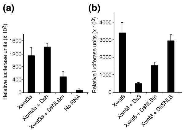
Figure 6 Dominant inhibition of Wnt-dependent transcription by Dsh mutants. Eight-cell embryos were injected (a) in one animal ventral blastomere or (b) in one vegetal ventral blastomere with -833pSia-Luc DNA (20 pg), mRNAs encoding Xwnt3a (5 pg) or Xwnt8 (2 pg), and Dsh-GFP, DsNLSm, Ds3 or DsSNLS mRNA (0.5 ng) as indicated. Luciferase activity was measured as described in Figure 5b.
Figure 6 Dominant inhibition of Wnt-dependent transcription by Dsh mutants. Eight-cell embryos were injected (a) in one animal ventral blastomere or (b) in one vegetal ventral blastomere with -833pSia-Luc DNA (20 pg), mRNAs encoding Xwnt3a (5 pg) or Xwnt8 (2 pg), and Dsh-GFP, DsNLSm, Ds3 or DsSNLS mRNA (0.5 ng) as indicated. Luciferase activity was measured as described in Figure 5b.CC BY · PubMed Central
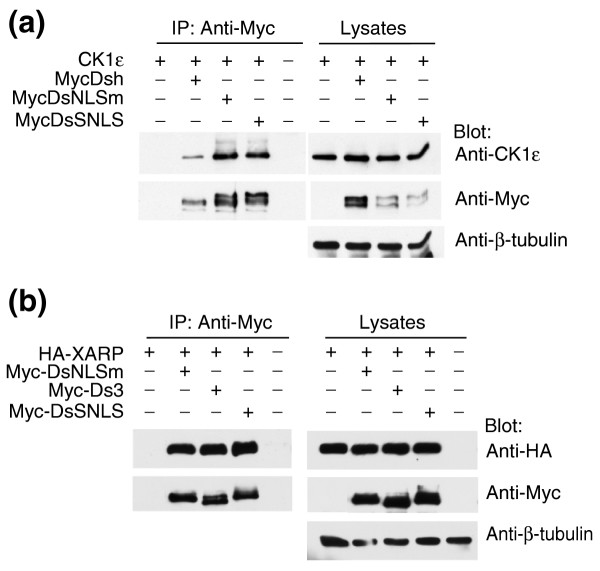
Figure 7 Dsh mutants retain the ability to bind CK1ε and XARP. Four-cell embryos were injected in four sites in the animal hemisphere with CK1ε, HA-XARP, Myc-tagged Dsh, DsNLSm, Ds3 or DsSNLS RNA alone (2 ng each) or in combinations as indicated. The embryonic lysates were collected at stage 10.5 for immunoprecipitation with anti-Myc antibodies. Co-immunoprecipitated (a) CK1ε or (b) HA-XARP was probed with anti-CK1ε or anti-HA antibodies; β-tubulin served as a loading control.
Figure 7 Dsh mutants retain the ability to bind CK1ε and XARP. Four-cell embryos were injected in four sites in the animal hemisphere with CK1ε, HA-XARP, Myc-tagged Dsh, DsNLSm, Ds3 or DsSNLS RNA alone (2 ng each) or in combinations as indicated. The embryonic lysates were collected at stage 10.5 for immunoprecipitation with anti-Myc antibodies. Co-immunoprecipitated (a) CK1ε or (b) HA-XARP was probed with anti-CK1ε or anti-HA antibodies; β-tubulin served as a loading control.CC BY · PubMed Central
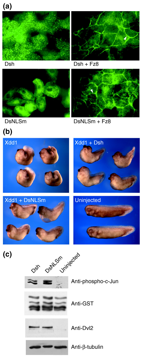
Figure 8 DsNLSm, defective in the β-catenin pathway, is active in noncanonical signaling. (a) Fz8-dependent recruitment of Dsh-GFP constructs to the cell membrane. Dsh-GFP or DsNLSm RNA (0.5 ng) was injected alone or with Fz8 RNA (1 ng) into two animal blastomeres at the 4–8-cell stage. GFP fluorescence was assessed in animal cap explants as in Figure 1b-d. Both Dsh and DsNLSm are efficiently recruited to the cell membrane by Fz8. Arrowheads point to cell membranes. (b) DsNLSm can rescue convergent extension defects caused by Xdd1. Four-cell embryos were injected with 0.6 ng Xdd1 RNA alone or together with 2 ng Dsh-GFP or DsNLSm RNA into two vegetal dorsal blastomeres. The injected embryos were allowed to develop until the sibling embryos reached stage 32. (c) Activation of JNK by the Dsh nuclear import mutant. Four animal blastomeres of four-cell embryos were each injected with 1 ng of RNAs encoding Dsh-GFP or DsNLSm. Embryonic lysates were collected at stage 10.5 for in vitro JNK activity assay using anti-phospho-specific c-Jun antibodies. Total GST-c-Jun levels were assessed with anti-GST antibodies. Dsh-GFP and DsNLSm were equally expressed, as monitored with anti-Dvl2 antibodies; β-tubulin served as a loading control.
Figure 8 DsNLSm, defective in the β-catenin pathway, is active in noncanonical signaling. (a) Fz8-dependent recruitment of Dsh-GFP constructs to the cell membrane. Dsh-GFP or DsNLSm RNA (0.5 ng) was injected alone or with Fz8 RNA (1 ng) into two animal blastomeres at the 4–8-cell stage. GFP fluorescence was assessed in animal cap explants as in Figure 1b-d. Both Dsh and DsNLSm are efficiently recruited to the cell membrane by Fz8. Arrowheads point to cell membranes. (b) DsNLSm can rescue convergent extension defects caused by Xdd1. Four-cell embryos were injected with 0.6 ng Xdd1 RNA alone or together with 2 ng Dsh-GFP or DsNLSm RNA into two vegetal dorsal blastomeres. The injected embryos were allowed to develop until the sibling embryos reached stage 32. (c) Activation of JNK by the Dsh nuclear import mutant. Four animal blastomeres of four-cell embryos were each injected with 1 ng of RNAs encoding Dsh-GFP or DsNLSm. Embryonic lysates were collected at stage 10.5 for in vitro JNK activity assay using anti-phospho-specific c-Jun antibodies. Total GST-c-Jun levels were assessed with anti-GST antibodies. Dsh-GFP and DsNLSm were equally expressed, as monitored with anti-Dvl2 antibodies; β-tubulin served as a loading control.CC BY · PubMed Central
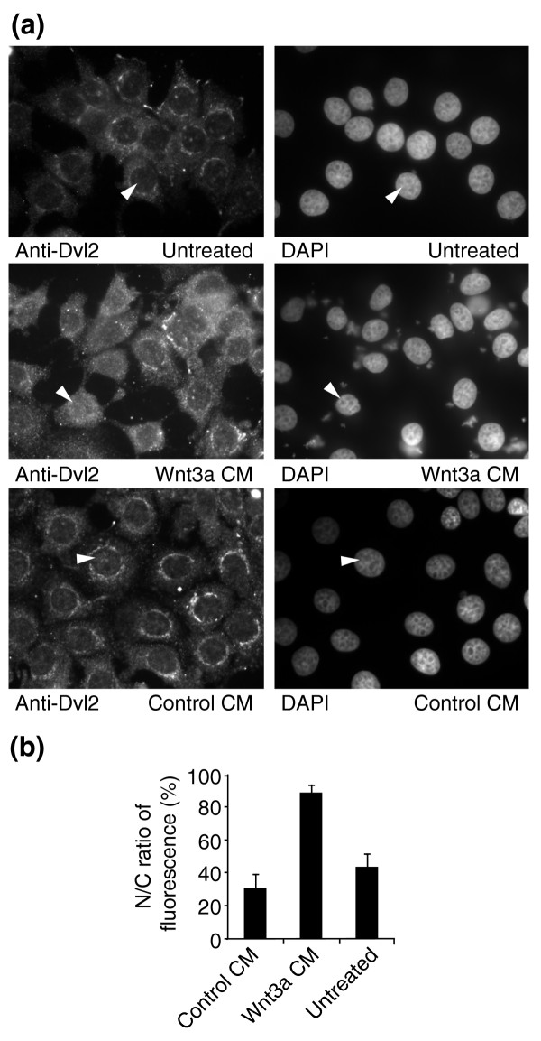
Figure 9 Nuclear translocation of Dvl2 upon Wnt3a treatment. (a) MCF7 cells were treated either with Wnt3a-conditioned or control medium for 6 h, fixed and immunostained with anti-Dvl2 antibodies. In control cells, cytoplasmic and perinuclear staining is visible. Wnt3a-conditioned, but not control, medium enhanced nuclear translocation of Dvl2. DAPI staining indicates the position of cell nuclei. Corresponding cells are shown by arrowheads. (b) Nuclear/cytoplasmic (N/C) ratios of fluorescence were calculated for each panel in (a) as the mean +/- standard deviation.
Figure 9 Nuclear translocation of Dvl2 upon Wnt3a treatment. (a) MCF7 cells were treated either with Wnt3a-conditioned or control medium for 6 h, fixed and immunostained with anti-Dvl2 antibodies. In control cells, cytoplasmic and perinuclear staining is visible. Wnt3a-conditioned, but not control, medium enhanced nuclear translocation of Dvl2. DAPI staining indicates the position of cell nuclei. Corresponding cells are shown by arrowheads. (b) Nuclear/cytoplasmic (N/C) ratios of fluorescence were calculated for each panel in (a) as the mean +/- standard deviation.CC BY · PubMed Central

Verification

Claims the paper makes

The claims and statistics of this paper haven’t been checked yet.

Run the check — free, no account needed. It reads the open-access text and checks the sources it cites.

A few free checks a day · papers already verified open instantly · results are public

Claims other papers make about this one

Each is checked against this paper’s own text: does it say what the citing paper says it does?

Self-check

Percentages, sums and reported differences re-computed from the paper’s own numbers.

The self-check needs the paper’s full text.

Citations

Cites

Show all 61 references

Cited by

Sorted by citations

See all 221

Metadata

Authors
Itoh, K., Brott, B. K., Bae, G.-U., Ratcliffe, M. J., & Sokol, S. Y.
Affiliations
Beth Israel Deaconess Medical Center (US)
Corresponding author
Sokol, Sergei Y
Venue
Journal of Biology
Published
2005-02-15
Volume
4
Issue
1
Pages
3-3
ISSN
1475-4924 (ISSN-L)
OpenAlex
W2122299286
Cited by
221
References
61
Citations by year
2026: 3 · 2025: 6 · 2024: 7 · 2023: 3 · 2022: 9 · 2021: 17 · 2020: 7 · 2019: 10 · 2018: 3 · 2017: 14 · 2016: 5 · 2015: 8 · 2014: 9 · 2013: 13 · 2012: 12
FWCI
8.03
Type
journal article
Language
en
Open access
Locations
  • Journal of Biology (publishedVersion, cc-by)
  • Europe PMC (PubMed Central) (publishedVersion)
  • PubMed Central (publishedVersion)
  • PubMed (publishedVersion)
  • DOAJ (DOAJ: Directory of Open Access Journals) (submittedVersion, cc-by-sa)
Funders
National Institutes of Health
Topics
  • Life Sciences › Biochemistry, Genetics and Molecular Biology › Molecular Biology › Wnt/β-catenin signaling in development and cancer
  • Life Sciences › Biochemistry, Genetics and Molecular Biology › Molecular Biology › Developmental Biology and Gene Regulation
  • Life Sciences › Biochemistry, Genetics and Molecular Biology › Molecular Biology › Signaling Pathways in Disease
Keywords
Dishevelled, nuclear localization, Wnt/β-catenin signaling, nuclear export signal, nuclear localization signal, nuclear accumulation, Wnt3a, Frizzled receptors
MeSH terms
Dishevelled Proteins, Amino Acid Sequence, Animals, Blastomeres, Cell Nucleus, Cells, Cultured, Cytoplasm, Gene Expression Regulation, Humans, Molecular Sequence Data, Mutation, Oligopeptides, Phosphoproteins, Xenopus, Sequence Alignment, Gene Order, Xenopus Proteins, Adaptor Proteins, Signal Transducing, Green Fluorescent Proteins, Nuclear Export Signals, Wnt Proteins, Frizzled Receptors, beta Catenin, Rats

About these checks

Registry, retraction, Cites and statistics results are computed, not judged, with one exception: a printed reference that no index lists is read once by a model before it counts as a citation error. Claims are extracted by a model and kept only when their quote appears verbatim in the text; each cited source is then checked, with a model’s help, for whether it says what the claim relies on. Those verdicts can be wrong — read the claim’s quote and the cited source before relying on one.

Statistics and arithmetic are recomputed, not judged.

Confidence bands are shown only for fields calibrated on our annotated test set. None is calibrated yet, so no band is shown.

What counts as a claim ›How we measure confidence ›How we check this ›

Are you an author, or do you see a problem with this page? Request a review