

The DOI matches its Crossref record; 1 of the 1 references it cites were found in a registry or an index.
Nanoparticles: A New Approach to Upgrade Cancer Diagnosis and Treatment
Open accessAbstract
Traditional cancer therapeutics have been criticized due to various adverse effects and insufficient damage to targeted tumors. The breakthrough of nanoparticles provides a novel approach for upgrading traditional treatments and diagnosis. Actually, nanoparticles can not only solve the shortcomings of traditional cancer diagnosis and treatment, but also create brand-new perspectives and cutting-edge devices for tumor diagnosis and treatment. However, most of the research about nanoparticles stays in vivo and in vitro stage, and only few clinical researches about nanoparticles have been reported. In this review, we first summarize the current applications of nanoparticles in cancer diagnosis and treatment. After that, we propose the challenges that hinder the clinical applications of NPs and provide feasible solutions in combination with the updated literature in the last two years. At the end, we will provide our opinions on the future developments of NPs in tumor diagnosis and treatment.
Overview
The claims and statistics of this paper haven’t been checked yet.
Run the check — free, no account needed. It reads the open-access text and checks the sources it cites.
Integrity signals
Registry
Cites
Statistics
Figures
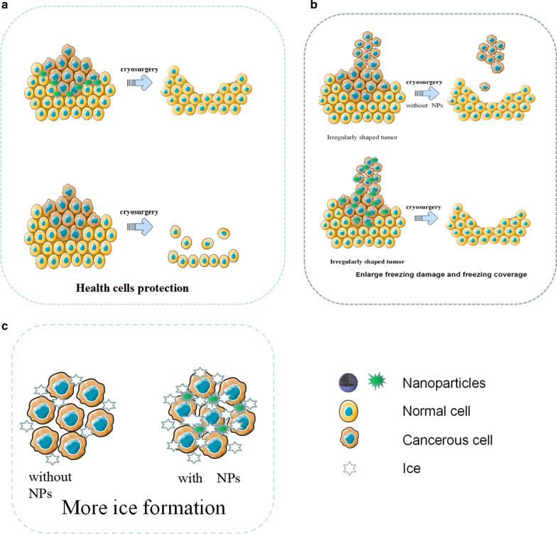
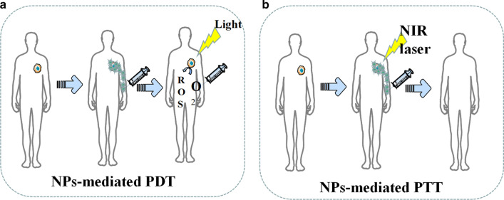
Verification
Claims the paper makes
The claims and statistics of this paper haven’t been checked yet.
Run the check — free, no account needed. It reads the open-access text and checks the sources it cites.
Self-check
Percentages, sums and reported differences re-computed from the paper’s own numbers.
The self-check needs the paper’s full text.
Citations
Cites
Gold nanoparticles for applications in cancer radiotherapy: Mechanisms and recent advancements
Her, S., Jaffray, D. A., & Allen, C. (2017) · Advanced Drug Delivery Reviews · doi:10.1016/j.addr.2015.12.012 · Cited by 865
Research works printed but not linked: 135
- Liu Y, Bhattarai P, Dai Z, Chen X (2019) Photothermal therapy and photoacoustic imaging via nanotheranostics in fighting cancer. Chem Soc Rev 48:2053–2108 · DOI
- Herma R et al (2019) Carbosilane dendrimers with phosphonium terminal groups are low toxic non-viral transfection vectors for sirna cell delivery. Int J Pharmaceut 562:51–65 · DOI
- Woodman C, Vundu G, George A, Wilson CM (2021) Applications and strategies in nanodiagnosis and nanotherapy in lung cancer. Semin Cancer Biol 69:349–364 · DOI
- Israel LL, Galstyan A, Holler E, Ljubimova JY (2020) Magnetic iron oxide nanoparticles for imaging, targeting and treatment of primary and metastatic tumors of the brain. J Control Release 320:45–62 · DOI
- Singh H, Du J, Singh P, Mavlonov GT, Yi T (2018) Development of superparamagnetic iron oxide nanoparticles via direct conjugation with ginsenosides and its in-vitro study. J Photochem Photobiol B 185:100–110 · DOI
- Tao Y, Li M, Ren J, Qu X (2015) Metal nanoclusters: novel probes for diagnostic and therapeutic applications. Chem Soc Rev 44:8636–8663 · DOI
- Sajid M, Potka-Wasylka J (2020) Nanoparticles: synthesis, characteristics, and applications in analytical and other sciences. Microchem J 154:104623 · DOI
- Priyanka KP, Sukirtha TH, Balakrishna KM, Varghese T (2016) Microbicidal activity of TiO2 nanoparticles synthesised by sol–gel method. IET Nanobiotechnol 10(2):81–86 · DOI
- Qi X, Xiong L, Peng J, Tang D (2017) Near infrared laser-controlled drug release of thermoresponsive microgel encapsulated with Fe3O4 nanoparticles. RSC Adv 7:19604–19610 · DOI
- Liu F et al (2015a) Controllable synthesis of polydopamine nanoparticles in microemulsions with pH-activatable properties for cancer detection and treatment. J Mater Chem B 3:6731–6739 · DOI
- Daou TJ et al (2006) Hydrothermal synthesis of monodisperse magnetite nanoparticles. Chem Mater 18:4399–4404 · DOI
- McGilvray KL, Decan MR, Wang D, Scaiano JC (2006) Facile photochemical synthesis of unprotected aqueous gold nanoparticles. J Am Chem Soc 128:15980–15981 · DOI
- Shahriyari F, Yaarali D, Ahmadi R, et al. (2020) Synthesis and characterization of Cu-Sn oxides nanoparticles via wire explosion method with surfactants, evaluation of in-vitro cytotoxic and antibacterial properties. Adv Powder Technol 31(6):2337–2347 · DOI
- de Carvalho JF, de Medeiros SN, Morales MA, Dantas AL, Carriço AS (2013) Synthesis of magnetite nanoparticles by high energy ball milling. Appl Surf Sci 275:84–87 · DOI
- Mascolo MC, Pei Y, Ring TA (2013) Room temperature co-precipitation synthesis of magnetite nanoparticles in a large pH window with different bases. Materials (Basel, Switzerland) 6:5549–5567 · DOI
- Seidi F, Jenjob R, Phakkeeree T, Crespy D (2018) Saccharides, oligosaccharides, and polysaccharides nanoparticles for biomedical applications. J Control Release 284:188–212 · DOI
- Chandler M, Afonin K (2019) Smart-responsive nucleic acid nanoparticles (NANPs) with the potential to modulate immune behavior. Nanomaterials 9:611 · DOI
- Asadian E, Ghalkhani M, Shahrokhian S (2019) Electrochemical sensing based on carbon nanoparticles: a review. Sens Actuators B Chem 293(C):183–209 · DOI
- Wang J, Shah ZH, Zhang S, Lu R (2014) Silica-based nanocomposites via reverse microemulsions: classifications, preparations, and applications. Nanoscale 6:4418–4437 · DOI
- Xie C, Yang Z, Sun Y (2009) Synthesis and characterization of monodispersed SiO2@Y3Al5O12:Er3+ core–shell particles. J Fluoresc 19:623–629 · DOI
- Yu J, Yang L, Zhang L (2018) Pattern generation and motion control of a vortex-like paramagnetic nanoparticle swarm. Int J Robot Res 37(8):912–930 · DOI
- Wei Q, Ji J, Shen J (2008) Synthesis of near-infrared responsive gold nanorod/PNIPAAm core/shell nanohybrids via surface initiated ATRP for smart drug delivery. Macromol Rapid Commun 29:645–650 · DOI
- Nallathamby PD, Hopf J, Irimata LE, McGinnity TL, Roeder RK (2016) Preparation of fluorescent Au–SiO2 core–shell nanoparticles and nanorods with tunable silica shell thickness and surface modification for immunotargeting. J Mater Chem B 4(32):5418–5428 · DOI
- Gour A, Jain NK (2019) Advances in green synthesis of nanoparticles. Artif Cells Nanomed Biotechnol 47:844–851 · DOI · Paper page
- Alijani HQ, Pourseyedi S, Torkzadeh Mahani M, Khatami M (2019) Green synthesis of zinc sulfide (ZnS) nanoparticles using Stevia rebaudiana Bertoni and evaluation of its cytotoxic properties. J Mol Struct 1175:214–218 · DOI
- Miri A, Sarani M (2018) Biosynthesis, characterization and cytotoxic activity of CeO2 nanoparticles. Ceram Int 44:12642–12647 · DOI · Paper page
- Smith B, Gambhir SS (2017) Nanomaterials for in vivo imaging. Chem Rev 117:901–986 · DOI
- Hou Y, Sun Z, Rao W, Liu J (2018) Nanoparticle-mediated cryosurgery for tumor therapy. Nanomed Nanotechnol Biol Med 14:493–506 · DOI
- Lai J et al (2018) MnO nanoparticles with unique excitation-dependent fluorescence for multicolor cellular imaging and MR imaging of brain glioma. Mikrochim Acta 185:244 · DOI
- Huang H et al (2019) GSH-sensitive Pt(IV) prodrug-loaded phase-transitional nanoparticles with a hybrid lipid-polymer shell for precise theranostics against ovarian cancer. Theranostics 9:1047–1065 · DOI
- Wang D et al (2018a) Effective pH-activated theranostic platform for synchronous magnetic resonance imaging diagnosis and chemotherapy. ACS Appl Mater Interfaces 10:31114–31123 · DOI
- Liu Y et al (2019) Folate-Targeted and oxygen/indocyanine green-loaded lipid nanoparticles for dual-mode imaging and photo-sonodynamic/photothermal therapy of ovarian cancer in vitro and in vivo. Mol Pharm 16:4104–4120 · DOI
- Jiang W et al (2019) PEG-coated and Gd-loaded fluorescent silica nanoparticles for targeted prostate cancer magnetic resonance imaging and fluorescence imaging. Int J Nanomed 14:5611–5622 · DOI
- Makela AV, Gaudet JM, Schott MA, Sehl OC, Contag CH, Foster PJ (2020) Magnetic particle imaging of macrophages associated with cancer: filling the voids left by iron-based magnetic resonance imaging. Mol Imaging Biol 22(4):958–968 · DOI
- Huang D et al (1991) Optical coherence tomography. Science 254:1178–1181 · DOI
- Odonnell M (2018) Magnetic nanoparticles as contrast agents for molecular imaging in medicine. Phys C 548:103–106 · DOI
- Gao Z et al (2016) Small is smarter: nano MRI contrast agents—advantages and recent achievements. Small 12:556–576 · DOI
- Estelrich J, Sanchezmartin MJ, Busquets MA (2015) Nanoparticles in magnetic resonance imaging: from simple to dual contrast agents. Int J Nanomed 10:1727–1741
- Ito A, Shinkai M, Honda H, Kobayashi T (2005) Medical application of functionalized magnetic nanoparticles. J Biosci Bioeng 100:1–11 · DOI
- Albanese A, Tang PS, Chan WCW (2012) The effect of nanoparticle size, shape, and surface chemistry on biological systems. Annu Rev Biomed Eng 14:1–16 · DOI
- Oldenburg AL, Toublan FJJ, Suslick KS, Wei A, Boppart SA (2005) Magnetomotive contrast for in vivo optical coherence tomography. Opt Express 13:6597–6614 · DOI
- Laconte LEW, Nitin N, Bao G (2005) Magnetic nanoparticle probes. Mater Today 8:32–38 · DOI
- Dixit S et al (2015) Transferrin receptor-targeted theranostic gold nanoparticles for photosensitizer delivery in brain tumors. Nanoscale 7:1782–1790 · DOI
- Liu C et al (2013) Magnetic/upconversion fluorescent NaGdF4:Yb, Er nanoparticle-based dual-modal molecular probes for imaging tiny tumors in vivo. ACS Nano 7:7227–7240 · DOI
- Bahrami B et al (2017) Nanoparticles and targeted drug delivery in cancer therapy. Immunol Lett 190:64–83 · DOI
- Maiyo F, Singh M (2017) Selenium nanoparticles: potential in cancer gene and drug delivery. Nanomedicine-UK 12:1075–1089 · DOI
- Zhang J, Sun H, Ma PX (2010) Host−guest interaction mediated polymeric assemblies: multifunctional nanoparticles for drug and gene delivery. ACS Nano 4:1049–1059 · DOI
- Surendran SP, Moon MJ, Park R, Jeong YY (2018) Bioactive nanoparticles for cancer immunotherapy. Int J Mol Sci 19:3877 · DOI
- Prabhu RH, Patravale VB, Joshi MD (2015) Polymeric nanoparticles for targeted treatment in oncology: current insights. Int J Nanomed 10:1001–1018
- Smith AM, Duan H, Mohs AM, Nie S (2008) Bioconjugated quantum dots for in vivo molecular and cellular imaging. Adv Drug Deliv Rev 60:1226–1240 · DOI
- Ferreira DDS, Lopes SCDA, Franco MS, De Oliveira MC (2013) PH-sensitive liposomes for drug delivery in cancer treatment. Ther Deliv 4:1099–1123 · DOI
- Jiang L, Vader P, Schiffelers RM (2017) Extracellular vesicles for nucleic acid delivery: progress and prospects for safe RNA-based gene therapy. Gene Ther 24:157–166 · DOI
- Amini MA et al (2019) Combining tumor microenvironment modulating nanoparticles with doxorubicin to enhance chemotherapeutic efficacy and boost antitumor immunity. J Natl Cancer Inst 111:399–408 · DOI
- Salem DS, Sliem MA, Elsesy MS, Shouman SA, Badr Y (2018) Improved chemo-photothermal therapy of hepatocellular carcinoma using chitosan-coated gold nanoparticles. J Photochem Photobiol B 182:92–99 · DOI
- Zhang L, Xie X, Liu D, Xu ZP, Liu R (2018) Efficient co-delivery of neo-epitopes using dispersion-stable layered double hydroxide nanoparticles for enhanced melanoma immunotherapy. Biomaterials 174:54–66 · DOI
- Gu L et al (2017) Folate-modified, indocyanine green-loaded lipid-polymer hybrid nanoparticles for targeted delivery of cisplatin. J Biomater Sci Polym E 28:690–702 · DOI
- Zhang Y, Li N, Suh H, Irvine DJ (2018) Nanoparticle anchoring targets immune agonists to tumors enabling anti-cancer immunity without systemic toxicity. Nat Commun 9:6 · DOI
- Avedian N, Zaaeri F, Daryasari MP, Javar HA, Khoobi M (2018) PH-sensitive biocompatible mesoporous magnetic nanoparticles labeled with folic acid as an efficient carrier for controlled anticancer drug delivery. J Drug Deliv Sci Technol 44:323–332 · DOI
- Zheng X et al (2018) The theranostic efficiency of tumor-specific, ph-responsive, peptide-modified, liposome-containing paclitaxel and superparamagnetic iron oxide nanoparticles. Int J Nanomed 13:1495–1504 · DOI
- Xia Y et al (2020a) Functionalized selenium nanoparticles for targeted sirna delivery silence Derlin1 and promote antitumor efficacy against cervical cancer. Drug Deliv 27:15–25 · DOI
- Bae YH, Park K (2011) Targeted drug delivery to tumors: myths, reality and possibility. J Control Release 153:198–205 · DOI
- Farokhzad OC, Langer R (2009) Impact of nanotechnology on drug delivery. ACS Nano 3:16–20 · DOI
- Sandoval MA et al. (2012) EGFR-targeted stearoyl gemcitabine nanoparticles show enhanced anti-tumor activity. J Control Release 157:287–296 · DOI
- Pandey S et al (2013) Biogenic gold nanoparticles as fotillas to fire berberine hydrochloride using folic acid as molecular road map. Mater Sci Eng C 33:3716–3722 · DOI
- Li L et al (2019) Codelivery of DOX and siRNA by folate-biotin-quaternized starch nanoparticles for promoting synergistic suppression of human lung cancer cells. Drug Deliv 26:499–508 · DOI
- Zhou Y et al (2016) Improving the anticancer efficacy of laminin receptor-specific therapeutic ruthenium nanoparticles (RuBB-loaded EGCG-RuNPs) via ROS-dependent apoptosis in SMMC-7721 cells. ACS Appl Mater Interfaces 8:15000–15012 · DOI
- Chen Q et al (2015) Multifunctional selenium nanoparticles: chiral selectivity of delivering MDR-siRNA for reversal of multidrug resistance and real-time biofluorescence imaging. Nanomed Nanotechnol Biol Med 11:1773–1784 · DOI
- Xia Y et al (2017) Novel functionalized nanoparticles for tumor-targeting co-delivery of doxorubicin and sirna to enhance cancer therapy. Int J Nanomed 13:143–159 · DOI
- Fu X et al (2016) RGD peptide-conjugated selenium nanoparticles: antiangiogenesis by suppressing VEGF-VEGFR2-ERK/AKT pathway. Nanomed Nanotechnol Biol Med 12:1627–1639 · DOI
- Xia Y et al (2020b) Silencing KLK12 expression via RGDfC-decorated selenium nanoparticles for the treatment of colorectal cancer in vitro and in vivo. Mater Sci Eng C 110:110594 · DOI
- Xia Y et al (2020c) Doxorubicin-loaded functionalized selenium nanoparticles for enhanced antitumor efficacy in cervical carcinoma therapy. Mater Sci Eng C 106:110100 · DOI
- Muldrew K et al (2001) Flounder antifreeze peptides increase the efficacy of cryosurgery. Cryobiology 42:182–189 · DOI
- Liu J, Deng Z (2009) Nano-cryosurgery: advances and challenges. J Nanosci Nanotechnol 9:4521–4542 · DOI
- Di D, He Z, Sun Z, Liu J (2012) A new nano-cryosurgical modality for tumor treatment using biodegradable MgO nanoparticles. Nanomed Nanotechnol Biol Med 8:1233–1241 · DOI
- Chua KJ, Chou SK, Ho JC (2007) An analytical study on the thermal effects of cryosurgery on selective cell destruction. J Biomech 40:100–116 · DOI
- Lv Y, Zou Y, Yang L (2012) Uncertainty and sensitivity analysis of properties of phase change micro/nanoparticles for thermal protection during cryosurgery. Forsch Ingenieurwes 76:41–50 · DOI
- Ye P et al (2017) Fe3O4 nanoparticles and cryoablation enhance ice crystal formation to improve the efficiency of killing breast cancer cells. Oncotarget 8:11389–11399 · DOI
- Jelveh S, Chithrani DB (2011) Gold nanostructures as a platform for combinational therapy in future cancer therapeutics. Cancers 3:1081–1110 · DOI
- Wang H et al (2018b) Enhanced cancer therapy with cold-controlled drug release and photothermal warming enabled by one nanoplatform. Biomaterials 180:265–278 · DOI
- Slovak R, Ludwig JM, Gettinger SN, Herbst RS, Kim HS (2017) Immuno-thermal ablations-boosting the anticancer immune response. J ImmunoTherapy Cancer 5:78 · DOI
- Hou X et al (2018) Nanoparticle-based photothermal and photodynamic immunotherapy for tumor treatment. Int J Cancer 143:3050–3060 · DOI
- Zhu X et al (2017a) Folic acid-modified and functionalized CuS nanocrystal-based nanoparticles for combined tumor chemo- and photothermal therapy. J Drug Target 25:425–435 · DOI
- Zhu H et al (2017b) Targeted delivery of siRNA with pH-responsive hybrid gold nanostars for cancer treatment. Int J Mol Sci 18:2029 · DOI
- Ju Y et al (2017) Monodisperse Au-Fe2C Janus nanoparticles: an attractive multifunctional material for triple-modal imaging-guided tumor photothermal therapy. ACS Nano 11:9239–9248 · DOI
- Song S et al (2017) Indocyanine green loaded magnetic carbon nanoparticles for near infrared fluorescence/magnetic resonance dual-modal imaging and photothermal therapy of tumor. ACS Appl Mater Interfaces 9:9484–9495 · DOI
- Pan L, Liu J, Shi J (2017) Nuclear-targeting gold nanorods for extremely low nir activated photothermal therapy. ACS Appl Mater Interfaces 9:15952–15961 · DOI
- Yang G et al (2017) Hollow MnO2 as a tumor-microenvironment-responsive biodegradable nano-platform for combination therapy favoring antitumor immune responses. Nat Commun 8:902 · DOI
- Singh SP et al (2017) NIR triggered liposome gold nanoparticles entrapping curcumin as in situ adjuvant for photothermal treatment of skin cancer. Int J Biol Macromol 110:375–382 · DOI
- Gschwend PM, Conti S, Kaech A, Maake C, Pratsinis SE (2019) Silica-coated TiN particles for killing cancer cells. ACS Appl Mater Interfaces 11:22550–22560 · DOI
- Rizzi M et al (2017) Verteporfin based silica nanoparticle for in vitro selective inhibition of human highly invasive melanoma cell proliferation. J Photochem Photobiol B 167:1–6 · DOI
- Jin J et al (2018) Graphdiyne nanosheet-based drug delivery platform for photothermal/chemotherapy combination treatment of cancer. ACS Appl Mater Interfaces 10:8436–8442 · DOI
- Sun S et al (2019) Ce6-modified carbon dots for multimodal-imaging-guided and single-NIR-laser-triggered photothermal/photodynamic synergistic cancer therapy by reduced irradiation power. ACS Appl Mater Interfaces 11:5791–5803 · DOI
- Chunling H, Zhixiang Z, Sainan L, Xiangjian L, Maolin P (2019) Monodispersed CuSe sensitized covalent organic framework photosensitizer with an enhanced photodynamic and photothermal effect for cancer therapy. ACS Appl Mater Interfaces 11:23072–23082 · DOI
- Ji C et al (2019) Activatable photodynamic therapy for prostate cancer by NIR dye/photosensitizer loaded albumin nanoparticles. J Biomed Nanotechnol 15:311–318 · DOI
- Fletcher JI, Williams R, Henderson MJ, Norris MD, Haber M (2016) ABC transporters as mediators of drug resistance and contributors to cancer cell biology. Drug Resist Update 26:1–9 · DOI · Paper page
- Li Y et al (2015) Multipronged design of light-triggered nanoparticles to overcome cisplatin resistance for efficient ablation of resistant tumor. ACS Nano 9:9626–9637 · DOI
- Wang L et al (2014) Novel insights into combating cancer chemotherapy resistance using a plasmonic nanocarrier: enhancing drug sensitiveness and accumulation simultaneously with localized mild photothermal stimulus of femtosecond pulsed laser. Adv Funct Mater 24:4229–4239 · DOI
- Wang L et al (2015) Using hollow carbon nanospheres as a light-induced free radical generator to overcome chemotherapy resistance. J Am Chem Soc 137:1947–1955 · DOI
- Fay BL, Melamed JR, Day ES (2015) Nanoshell-mediated photothermal therapy can enhance chemotherapy in inflammatory breast cancer cells. Int J Nanomed 10:6931–6941
- Moghissi K, Dixon K (2003) Is Bronchoscopic photodynamic therapy a therapeutic option in lung cancer. Eur Respir J 22:535–541 · DOI
- Gu T et al (2018) Upconversion Composite nanoparticles for tumor hypoxia modulation and enhanced near-infrared-triggered photodynamic therapy. ACS Appl Mater Interfaces 10:15494–15503 · DOI
- Li Y et al (2016) A versatile imaging and therapeutic platform based on dual-band luminescent lanthanide nanoparticles toward tumor metastasis inhibition. ACS Nano 10:2766–2773 · DOI
- Jin J, Zhao Q (2020) Engineering nanoparticles to reprogram radiotherapy and immunotherapy: recent advances and future challenges. J Nanobiotechnol 18:17–75 · DOI
- Baumann M et al (2016) Radiation oncology in the era of precision medicine. Nat Rev Cancer 16:234–249 · DOI
- Kwatra D, Venugopal A, Anant S (2013) Nanoparticles in radiation therapy: a summary of various approaches to enhance radiosensitization in cancer. Transl Cancer Res 2:330–342
- Liu Y et al (2018) Metal-based nanoenhancers for future radiotherapy: radiosensitizing and synergistic effects on tumor cells. Theranostics 8:1824–1849 · DOI
- Yang X, Yang M, Pang B, Vara M, Xia Y (2015) Gold nanomaterials at work in biomedicine. Chem Rev 115:10410–10488 · DOI
- Herold DM, Das IJ, Stobbe CC, Iyer RV, Chapman JD (2000) Gold microspheres: a selective technique for producing biologically effective dose enhancement. Int J Radiat Biol 76:1357–1364 · DOI
- Liu Y et al (2015b) The dependence of radiation enhancement effect on the concentration of gold nanoparticles exposed to low- and high-LET radiations. Phys Med 31:210–218 · DOI
- Charest G, Paquette B, Fortin D, Mathieu D, Sanche L (2010) Concomitant treatment of F98 glioma cells with new liposomal platinum compounds and ionizing radiation. J Neuro-Oncol 97:187–193 · DOI
- Jawaid P et al (2014) Effects of SOD/catalase mimetic platinum nanoparticles on radiation-induced apoptosis in human lymphoma U937 cells. Apoptosis 19:1006–1016 · DOI
- Ma P et al (2017) Enhanced cisplatin chemotherapy by iron oxide nanocarrier-mediated generation of highly toxic reactive oxygen species. Nano Lett 17:928–937 · DOI
- Hauser AK et al (2016) Targeted iron oxide nanoparticles for the enhancement of radiation therapy. Biomaterials 105:127–135 · DOI
- Titus D, Samuel EJJ, Roopan SM (2016) Current scenario of biomedical aspect of metal-based nanoparticles on gel dosimetry. Appl Microbiol Biotechnol 100:4803–4816 · DOI
- Khoei S, Mahdavi SR, Fakhimikabir H, Shakerizadeh A, Hashemian A (2014) The role of iron oxide nanoparticles in the radiosensitization of human prostate carcinoma cell line DU145 at megavoltage radiation energies. Int J Radiat Biol 90:351–356 · DOI
- Huang F et al (2010) Enhancement of irradiation effects on cancer cells by cross-linked dextran-coated iron oxide (CLIO) nanoparticles. Phys Med Biol 55:469–482 · DOI
- Revia RA, Zhang M (2016) Magnetite nanoparticles for cancer diagnosis, treatment, and treatment monitoring: recent advances. Mater Today 19:157–168 · DOI
- Zhang H, Zhang W, Zhou Y, Jiang Y, Li S (2017) Dual functional mesoporous silicon nanoparticles enhance the radiosensitivity of VPA in glioblastoma. Transl Oncol 10:229–240 · DOI
- He L, Lai H, Chen T (2015) Dual-function nanosystem for synergetic cancer chemo-/radiotherapy through ros-mediated signaling pathways. Biomaterials 51:30–42 · DOI
- Luo L et al (2018) Laser immunotherapy in combination with perdurable PD-1 blocking for the treatment of metastatic tumors. ACS Nano 12:7647–7662 · DOI
- Wang X et al (2019) Facile fabrication of magnetic microrobots based on spirulina templates for targeted delivery and synergistic chemo-photothermal therapy. ACS Appl Mater Interfaces 11:4745–4756 · DOI
- Jia G et al (2018) NRP-1 targeted and cargo-loaded exosomes facilitate simultaneous imaging and therapy of glioma in vitro and in vivo. Biomaterials 178:302–316 · DOI · Paper page
- Hennig TL, Unterweger H, Lyer S, Alexiou C, Cicha I (2019) Magnetic accumulation of SPIONs under arterial flow conditions: effect of serum and red blood cells. Molecules (Basel, Switzerland) 24:2588 · DOI
- Manshadi MKD et al (2018) Delivery of magnetic micro/nanoparticles and magnetic-based drug/cargo into arterial flow for targeted therapy. Drug Deliv 25:1963–1973 · DOI
- Li L et al (2018) Small near-infrared photochromic protein for photoacoustic multi-contrast imaging and detection of protein interactions in vivo. Nat Commun 9:2734 · DOI
- Wu Z et al (2019) A microrobotic system guided by photoacoustic computed tomography for targeted navigation in intestines in vivo. Sci Robot 4:x613 · DOI
- Cosco D et al (2016) Rutin-loaded chitosan microspheres: characterization and evaluation of the anti-inflammatory activity. Carbohydr Polym 152:583–591 · DOI
- Tavano R et al (2018) C1q-mediated complement activation and C3 opsonization trigger recognition of stealth poly(2-methyl-2-oxazoline)-coated silica nanoparticles by human phagocytes. ACS Nano 12:5834–5847 · DOI
- Zou Y et al (2020) Polyglycerol grafting shields nanoparticles from protein corona formation to avoid macrophage uptake. ACS Nano 14:7216–7226 · DOI
- Xia Y et al (2020d) Engineering macrophages for cancer immunotherapy and drug delivery. Adv Mater 32:2002054 · DOI · Paper page
- Rong L et al (2019) Iron chelated melanin-like nanoparticles for tumor-associated macrophage repolarization and cancer therapy. Biomaterials 225:119515 · DOI
- Coluccio ML et al (2020) Emerging designs of electronic devices in biomedicine. Micromachines-Basel 11:123 · DOI
- Malara N et al (2018) Superhydrophobic lab-on-chip measures secretome protonation state and provides a personalized risk assessment of sporadic tumour. NPJ Precis Oncol 2:26 · DOI
- Xu X et al (2016) Detection EGFR exon 19 status of lung cancer patients by DNA electrochemical biosensor. Biosens Bioelectron 80:411–417 · DOI
- Amouzadeh Tabrizi M, Shamsipur M, Farzin L (2015) A high sensitive electrochemical aptasensor for the determination of VEGF165 in serum of lung cancer patient. Biosens Bioelectron 74:764–769 · DOI
Cited by
Sorted by citations
Thalmayer, A. S., Götz, K., Lübke, M., & Fischer, G. (2024) · Current Directions in Biomedical Engineering · doi:10.1515/cdbme-2024-2159
Metadata
- Authors
- Yu, Z., Gao, L., Chen, K., Zhang, W., Zhang, Q., Li, Q., & Hu, K.
- Affiliations
- Beijing University of Chinese Medicine (CN)
- China Agricultural University (CN)
- University of New Brunswick (CA)
- Corresponding author
- Hu, Kaiwen
- Venue
- Nanoscale Research Letters
- Published
- 2021-05-20
- Volume
- 16
- Issue
- 1
- Pages
- 88-88
- ISSN
- 1556-276X (ISSN-L)
- PMC
- PMC8137776
- OpenAlex
- W3162697468
- Cited by
- 138
- References
- 1
- Citations by year
- 2026: 6 · 2025: 21 · 2024: 29 · 2023: 35 · 2022: 36 · 2021: 11
- FWCI
- 2.35
- Type
- journal article
- Language
- en
- Open access
- gold
- doi.org
- Locations
- Nanoscale Research Letters (publishedVersion, cc-by)
- PubMed Central (publishedVersion, cc-by)
- PubMed (publishedVersion)
- DOAJ (DOAJ: Directory of Open Access Journals) (submittedVersion, cc-by-sa)
- Topics
- Physical Sciences › Engineering › Biomedical Engineering › Nanoplatforms for cancer theranostics
- Physical Sciences › Materials Science › Biomaterials › Nanoparticle-Based Drug Delivery
- Physical Sciences › Materials Science › General Materials Science › Nanotechnology research and applications
- Keywords
- nanoparticles, cancer diagnosis, cancer therapy, tumor targeting, clinical translation, drug delivery, nanomedicine
About these checks
Registry, retraction, Cites and statistics results are computed, not judged, with one exception: a printed reference that no index lists is read once by a model before it counts as a citation error. Claims are extracted by a model and kept only when their quote appears verbatim in the text; each cited source is then checked, with a model’s help, for whether it says what the claim relies on. Those verdicts can be wrong — read the claim’s quote and the cited source before relying on one.
Statistics and arithmetic are recomputed, not judged.
Confidence bands are shown only for fields calibrated on our annotated test set. None is calibrated yet, so no band is shown.
What counts as a claim ›How we measure confidence ›How we check this ›
Are you an author, or do you see a problem with this page? Request a review
