

The DOI matches its Crossref record; 110 of the 111 references it cites were found in a registry or an index.
Neurovascular Dysfunction in Vascular Dementia, Alzheimer's and Atherosclerosis
Open accessAbstract
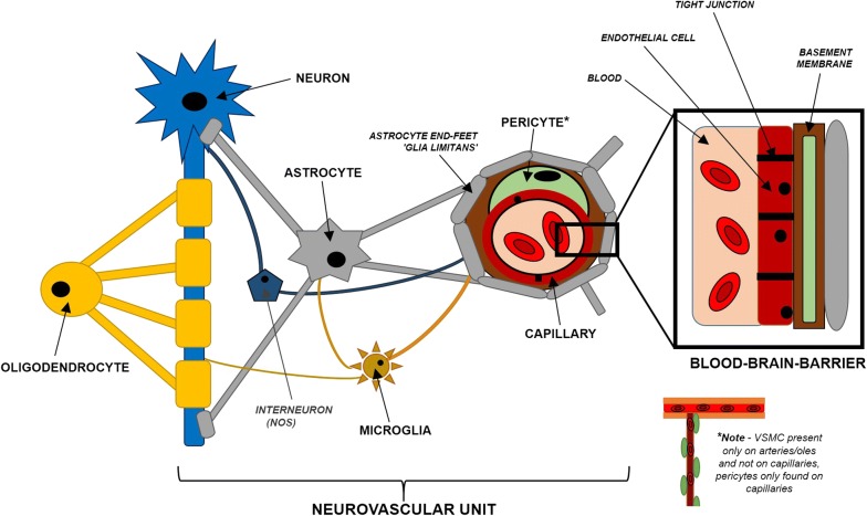
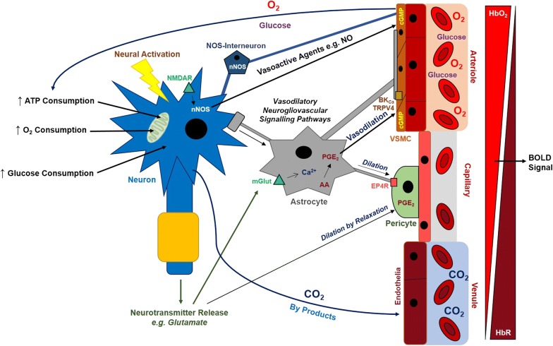
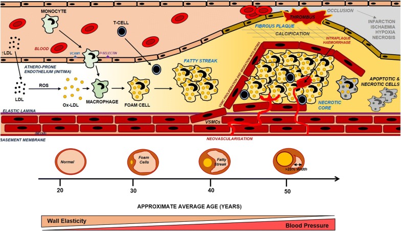
Efficient blood supply to the brain is of paramount importance to its normal functioning and improper blood flow can result in potentially devastating neurological consequences. Cerebral blood flow in response to neural activity is intrinsically regulated by a complex interplay between various cell types within the brain in a relationship termed neurovascular coupling. The breakdown of neurovascular coupling is evident across a wide variety of both neurological and psychiatric disorders including Alzheimer's disease. Atherosclerosis is a chronic syndrome affecting the integrity and function of major blood vessels including those that supply the brain, and it is therefore hypothesised that atherosclerosis impairs cerebral blood flow and neurovascular coupling leading to cerebrovascular dysfunction. This review will discuss the mechanisms of neurovascular coupling in health and disease and how atherosclerosis can potentially cause cerebrovascular dysfunction that may lead to cognitive decline as well as stroke. Understanding the mechanisms of neurovascular coupling in health and disease may enable us to develop potential therapies to prevent the breakdown of neurovascular coupling in the treatment of vascular brain diseases including vascular dementia, Alzheimer's disease and stroke.
Overview
The claims and statistics of this paper haven’t been checked yet.
Run the check — free, no account needed. It reads the open-access text and checks the sources it cites.
Integrity signals
Registry
Cites
Statistics
Figures
Verification
Claims the paper makes
The claims and statistics of this paper haven’t been checked yet.
Run the check — free, no account needed. It reads the open-access text and checks the sources it cites.
Claims other papers make about this one
Each is checked against this paper’s own text: does it say what the citing paper says it does?
In the citing paper
“Unsurprisingly, such a condition, known as cerebral amyloid angiopathy exacerbates the AD pathology (Shabir et al., 2018).”
From Exploring the Therapeutic Potential of Phytochemicals in Alzheimer’s Disease: Focus on Polyphenols and Monoterpenes (2022) · cites this paper as Shabir et al., 2018
Self-check
Percentages, sums and reported differences re-computed from the paper’s own numbers.
The self-check needs the paper’s full text.
Citations
Cites
1 retracted
Montagne, A., Nikolakopoulou, A. M., Zhao, Z., Sagare, A. P., Si, G., Lazic, D., Barnes, S. R., Daianu, M., Ramanathan, A., Go, A., et al. (2018) · Nature Medicine · doi:10.1038/nm.4482 · Cited by 344
RetractedCalcium's Role in Mechanotransduction during Muscle Development
Damm, T. B., & Egli, M. (2014) · Cellular Physiology and Biochemistry · doi:10.1159/000356667 · Cited by 14,552
Antiinflammatory Therapy with Canakinumab for Atherosclerotic Disease
Ridker, P. M., Everett, B. M., Thuren, T., MacFadyen, J. G., Chang, W. H., Ballantyne, C., Fonseca, F., Nicolau, J., Koenig, W., Anker, S. D., et al. (2017) · New England Journal of Medicine · doi:10.1056/nejmoa1707914 · Cited by 9,482
Brain magnetic resonance imaging with contrast dependent on blood oxygenation.
Ogawa, S., Lee, T. M., Kay, A. R., & Tank, D. W. (1990) · Proceedings of the National Academy of Sciences · doi:10.1073/pnas.87.24.9868 · Cited by 6,483
Heart Disease and Stroke Statistics—2015 Update: A Report From the American Heart Association
Mozaffarian, D., Benjamin, E. J., Go, A. S., Arnett, D. K., Blaha, M. J., Cushman, M., de Ferranti, S., Després, J.-P., Fullerton, H. J., Howard, V. J., et al. (2015) · Circulation · doi:10.1161/cir.0000000000000152 · Cited by 5,797
Calcium signalling: dynamics, homeostasis and remodelling
Berridge, M. J., Bootman, M. D., & Roderick, H. L. (2003) · Nature Reviews Molecular Cell Biology · doi:10.1038/nrm1155 · Cited by 5,719
Lusis, A. J. (2000) · Nature · doi:10.1038/35025203 · Cited by 4,880
Gorelick, P. B., Scuteri, A., Black, S. E., DeCarli, C., Greenberg, S. M., Iadecola, C., Launer, L. J., Laurent, S., Lopez, O. L., Nyenhuis, D., et al. (2011) · Stroke · doi:10.1161/str.0b013e3182299496 · Cited by 3,811
Association of apolipoprotein E allele ϵ4 with late‐onset familial and sporadic Alzheimer's disease
Saunders, A. M., Strittmatter, W. J., Schmechel, D., St. George-Hyslop, P. H., Pericak-Vance, M. A., Joo, S. H., Rosi, B. L., Gusella, J. F., Crapper-MacLachlan, D. R., Alberts, M. J., et al. (1993) · Neurology · doi:10.1212/wnl.43.8.1467 · Cited by 3,786
Compensatory Enlargement of Human Atherosclerotic Coronary Arteries
Glagov, S., Weisenberg, E. S., Zarins, C. K., Stankunavicius, R., & Kolettis, G. (1987) · New England Journal of Medicine · doi:10.1056/nejm198705283162204 · Cited by 3,614
Cited by
Sorted by citations
Metal Ions in Alzheimer’s Disease: A Key Role or Not?
Liu, Y., Nguyen, M., Robert, A., & Meunier, B. (2019) · Accounts of Chemical Research · doi:10.1021/acs.accounts.9b00248 · Cited by 353
Neuroprotective effect of SGLT2 inhibitors.
Pawlos, A., Broncel, M., Woźniak, E., & Gorzelak-Pabiś, P. (2021) · Molecules · doi:10.3390/molecules26237213 · Cited by 304
Rajeev, V., Fann, D. Y., Dinh, Q. N., Kim, H. A., Silva, T. M. D., Lai, M. K. P., Chen, C., Drummond, G. R., Sobey, C. G., & Arumugam, T. V. (2022) · Theranostics · doi:10.7150/thno.68304 · Cited by 320
Oxidative Stress and Beta Amyloid in Alzheimer’s Disease. Which Comes First: The Chicken or the Egg?
Tamagno, E., Guglielmotto, M., Vasciaveo, V., & Tabaton, M. (2021) · Antioxidants · doi:10.3390/antiox10091479 · Cited by 282
Rajeev, V., Chai, Y. L., Poh, L., Selvaraji, S., Fann, D. Y., Jo, D., Silva, T. M. D., Drummond, G. R., Sobey, C. G., Arumugam, T. V., et al. (2023) · Acta Neuropathologica Communications · doi:10.1186/s40478-023-01590-1 · Cited by 241
Neutrophil counts and cardiovascular disease
Luo, J. M., Thomassen, J. Q., Nordestgaard, B. G., Tybjærg‐Hansen, A., & Frikke‐Schmidt, R. (2023) · European Heart Journal · doi:10.1093/eurheartj/ehad649 · Cited by 183
Fekete, M., Varga, P., Ungvari, Z. I., Fekete, J. T., Buda, A., Szappanos, Á., Lehoczki, A., Mózes, N., Grosso, G., Godos, J., et al. (2025) · GeroScience · doi:10.1007/s11357-024-01488-3 · Cited by 162
The Neurovascular Unit Dysfunction in Alzheimer’s Disease
Soto-Rojas, L. O., Pacheco‐Herrero, M., Martínez-Gómez, P. A., Campa-Córdoba, B. B., Apátiga-Pérez, R., Villegas-Rojas, M. M., Harrington, C. R., de la Cruz, F., Garcés‐Ramírez, L., & Luna‐Muñoz, J. (2021) · International Journal of Molecular Sciences · doi:10.3390/ijms22042022 · Cited by 146
Canine Cognitive Dysfunction and Alzheimer’s Disease – Two Facets of the Same Disease?
Mihevc, S. P., & Majdič, G. (2019) · Frontiers in Neuroscience · doi:10.3389/fnins.2019.00604 · Cited by 152
Vascular Dementia and Underlying Sex Differences
Akhter, F., Persaud, A., Zaokari, Y., Zhao, Z., & Zhu, D. (2021) · Frontiers in Aging Neuroscience · doi:10.3389/fnagi.2021.720715 · Cited by 119
The Role of Neurovascular System in Neurodegenerative Diseases
Ahmad, A., Patel, V., Xiao, J., & Khan, M. M. (2020) · Molecular Neurobiology · doi:10.1007/s12035-020-02023-z · Cited by 97
Brain injury-induced dysfunction of the blood brain barrier as a risk for dementia
Abrahamson, E. E., & Ikonomović, M. D. (2020) · Experimental Neurology · doi:10.1016/j.expneurol.2020.113257 · Cited by 91
Statins Use in Alzheimer Disease: Bane or Boon from Frantic Search and Narrative Review
Alsubaie, N., Al-Kuraishy, H. M., Al-Gareeb, A. I., Alharbi, B., WAARD, M. M. D., Sabatier, J., Saad, . M., & Batiha, G. E. (2022) · Brain Sciences · doi:10.3390/brainsci12101290 · Cited by 87
Interactions between glial cells and the blood-brain barrier and their role in Alzheimer's disease
Zhao, M., Jiang, X., Zhang, H., Sun, J., Pei, H., Ma, L., Cao, Y., & Li, H. (2021) · Ageing Research Reviews · doi:10.1016/j.arr.2021.101483 · Cited by 86
Neurovascular Alterations in Vascular Dementia: Emphasis on Risk Factors
Lecordier, S., Manrique‐Castano, D., Moghrabi, Y. E., & ElAli, A. (2021) · Frontiers in Aging Neuroscience · doi:10.3389/fnagi.2021.727590 · Cited by 78
Diet’s Role in Modifying Risk of Alzheimer’s Disease: History and Present Understanding
Grant, W. B., & Blake, S. M. (2023) · Journal of Alzheimer s Disease · doi:10.3233/jad-230418 · Cited by 82
Piccialli, I., Tedeschi, V., Caputo, L., D’Errico, S., Ciccone, R., De Feo, V., Secondo, A., & Pannaccione, A. (2022) · Frontiers in Pharmacology · doi:10.3389/fphar.2022.876614 · Cited by 73
Retinal Neurovascular Coupling in Diabetes
Garhöfer, G., Chua, J., Tan, B., Wong, D. W. K., Schmidl, D., & Schmetterer, L. F. (2020) · Journal of Clinical Medicine · doi:10.3390/jcm9092829 · Cited by 72
Schulz, M., Malherbe, C., Cheng, B., Thomalla, G., & Schlemm, E. (2021) · BMC Medicine · doi:10.1186/s12916-021-01962-1 · Cited by 71
Associations between coronary heart disease and risk of cognitive impairment: A meta‐analysis
Liang, X., Huang, Y., & Han, X. (2021) · Brain and Behavior · doi:10.1002/brb3.2108 · Cited by 70
Pascale, J. V., Park, E. J., Adebesin, A. M., Falck, J. R., Schwartzman, M. L., & Garcia, V. G. (2021) · British Journal of Pharmacology · doi:10.1111/bph.15525 · Cited by 59
Li, P., Mu, J., Ma, X., Ding, D., Ma, S., Zhang, H., Liu, J., & Zhang, M. (2021) · Journal of Cerebral Blood Flow & Metabolism · doi:10.1177/0271678x211007960 · Cited by 51
Zhang, M., Zhang, Z., Li, H., Xia, Y., Xing, M., Xiao, C., Cai, W., Bu, L.-L., Li, Y., Park, T., et al. (2024) · Translational Neurodegeneration · doi:10.1186/s40035-023-00388-4 · Cited by 50
The epigenetic modification of DNA methylation in neurological diseases
Li, L., Chen, R., Zhang, H., Li, J., Huang, H., Weng, J., Tan, H., Guo, T., Wang, M., & Xie, J. (2024) · Frontiers in Immunology · doi:10.3389/fimmu.2024.1401962 · Cited by 54
Yang, S., & Webb, A. J. S. (2023) · European Stroke Journal · doi:10.1177/23969873231196981 · Cited by 45
Lv, X., Zhang, M., Cheng, Z., Wang, Q., Wang, P., Xie, Q., Ni, M., Shen, Y., Tang, Q., & Gao, F. (2023) · Alzheimer s Research & Therapy · doi:10.1186/s13195-023-01199-5 · Cited by 44
Comparing dynamic causal models of neurovascular coupling with fMRI and EEG/MEG
Jafarian, A., Vladimir Litvak, A., Cagnan, H., Friston, K. J., & Zeidman, P. (2020) · NeuroImage · doi:10.1016/j.neuroimage.2020.116734 · Cited by 44
Wan, J., Zhou, S., Mea, H. J., Guo, Y., Ku, H., & Urbina, B. (2022) · Chemical Reviews · doi:10.1021/acs.chemrev.1c00480 · Cited by 45
Liu, L.-Y., Lu, Y., Shen, L., Li, C., Yu, J., Chua, R. Y., Ye, K. X., Chao, Y., Shen, Q.-F., Mahendran, R., et al. (2021) · Journal of Psychiatric Research · doi:10.1016/j.jpsychires.2021.11.041 · Cited by 40
Shabir, O., Pendry, B., Lee, L., Eyre, B., Sharp, P. S., Rebollar, M. A., Drew, D., Howarth, C., Heath, P. R., Wharton, S. B., et al. (2022) · eLife · doi:10.7554/elife.68242 · Cited by 40
Soto-Rojas, L. O., Campa-Córdoba, B. B., Harrington, C. R., Salas‐Casas, A., Hernándes‐Alejandro, M., Villanueva‐Fierro, I., Bravo-Muñoz, M., Garcés‐Ramírez, L., de la Cruz‐López, F., Ontiveros‐Torres, M. Á., et al. (2021) · International Journal of Molecular Sciences · doi:10.3390/ijms22073654 · Cited by 41
Rhizoma Coptidis for Alzheimer’s Disease and Vascular Dementia: A Literature Review
Wang, Z., Yang, Y., Liu, M., Wei, Y., Liu, J., Pei, H., & Li, H. (2019) · Current Vascular Pharmacology · doi:10.2174/1570161117666190710151545 · Cited by 38
Karamanova, N., Truran, S., Serrano, G. E., Beach, T. G., Madine, J., Weissig, V., Davies, H. A., Veldhuizen, J., Nikkhah, M., Hansen, M., et al. (2020) · Journal of the American Heart Association · doi:10.1161/jaha.119.014810 · Cited by 40
de Montgolfier, O., Thorin‐Trescases, N., & Thorin, É. (2020) · American Journal of Hypertension · doi:10.1093/ajh/hpaa001 · Cited by 45
Altered Neurovascular Coupling in Subcortical Ischemic Vascular Disease
Liu, X., Cheng, R., Chen, L., Gong, J., Luo, T., & Lv, F. (2021) · Frontiers in Aging Neuroscience · doi:10.3389/fnagi.2021.598365 · Cited by 38
Atrial Fibrillation and Cognitive Impairment: An Associated Burden or Burden by Association?
Manolis, T. A., Manolis, A. S., Apostolopoulos, E. J., Melita, H., & Manolis, A. S. (2020) · Angiology · doi:10.1177/0003319720910669 · Cited by 40
Late-onset epilepsy and subsequent increased risk of dementia
Tsai, Z.-R., Zhang, H., Tseng, C., Peng, H.-C., Kok, V. C., Li, G. P., Hsiung, C. A., & Hsu, C. (2021) · Aging · doi:10.18632/aging.202299 · Cited by 36
Atherosclerosis: The Culprit and Co-victim of Vascular Dementia
Huang, Y., Hong, F., & Yang, S. (2021) · Frontiers in Neuroscience · doi:10.3389/fnins.2021.673440 · Cited by 34
Potential of PET/CT in assessing dementias with emphasis on cerebrovascular disorders
Borja, A. J., Hancin, E. C., Zhang, V., Revheim, M.-E. R., & Alavi, A. A. (2020) · European Journal of Nuclear Medicine and Molecular Imaging · doi:10.1007/s00259-020-04697-y · Cited by 30
Ewees, M. G., Elmahdy, M., Hannawi, Y., & Zweier, J. L. (2024) · Journal of Cerebral Blood Flow & Metabolism · doi:10.1177/0271678x241270415 · Cited by 29
Butters, E., Srinivasan, S., O'Brien, J. T., Su, L., & Bale, G. M. (2023) · Ageing Research Reviews · doi:10.1016/j.arr.2023.101992 · Cited by 29
Gulej, R., Patai, R., Ungvari, A., Kállai, A., Tarantini, S., Yabluchanskiy, A., Huffman, D. M., Conboy, M. J., Conboy, I. M., Mika Kivimaki, B., et al. (2025) · GeroScience · doi:10.1007/s11357-025-01657-y · Cited by 29
Wan, C., Zong, R.-Y., & Chen, X. (2022) · Frontiers in Cardiovascular Medicine · doi:10.3389/fcvm.2022.928701 · Cited by 28
St‐Pierre, M., Carrier, M., Ibáñez, F. G., Khakpour, M., Wallman, M., Parent, M., & Tremblay, M. (2023) · Journal of Neuroinflammation · doi:10.1186/s12974-023-02752-7 · Cited by 28
NAD+ and Vascular Dysfunction: From Mechanisms to Therapeutic Opportunities
Abdellatif, M., Bugger, H., Kroemer, G., & Sedej, S. (2022) · Journal of Lipid and Atherosclerosis · doi:10.12997/jla.2022.11.2.111 · Cited by 26
He, R., He, F., Hu, Z., He, Y., Zeng, X., Liu, Y., Tang, L., Xiang, J., Li, J., He, B., et al. (2023) · BioMed Research International · doi:10.1155/2023/1235552 · Cited by 26
Heutz, R. A. M., Claassen, J. A. H. R., Feiner, S., Davies, A., Gurung, D., Panerai, R. B., de Heus, R. A. A., & Beishon, L. (2023) · Journal of Cerebral Blood Flow & Metabolism · doi:10.1177/0271678x231173449 · Cited by 26
Ruan, Z., Sun, D., Zhou, X., Yu, M., Li, S., Sun, W., Li, Y., Gao, L., & Xu, H. (2023) · Frontiers in Aging Neuroscience · doi:10.3389/fnagi.2023.1224525 · Cited by 26
Contrasting Metabolic Insufficiency in Aging and Dementia
Turner, D. A. (2021) · Aging and Disease · doi:10.14336/ad.2021.0104 · Cited by 25
Cerebral small vessel disease pathology in COVID-19 patients: A systematic review
Owens, C. D., Pinto, C. B., Detwiler, S., Mukli, P., Péterfi, A., Szarvas, Z., Hoffmeister, J. R., Galindo, J., Noori, J., Kirkpatrick, A. C., et al. (2023) · Ageing Research Reviews · doi:10.1016/j.arr.2023.101962 · Cited by 30
Bilirubin and Redox Stress in Age-Related Brain Diseases
Llido, J. P., Jayanti, S. D., Tiribelli, C., & Gazzin, S. (2023) · Antioxidants · doi:10.3390/antiox12081525 · Cited by 27
Neuromechanobiology: An Expanding Field Driven by the Force of Greater Focus
Motz, C. T., Kabat, V., Saxena, T. K., Bellamkonda, R. V., & Zhu, C. (2021) · Advanced Healthcare Materials · doi:10.1002/adhm.202100102 · Cited by 25
Association of coffee, green tea, and caffeine with the risk of dementia in older Japanese people
Matsushita, N., Nakanishi, Y. Y., Watanabe, Y., Kitamura, K., Kabasawa, K., Takahashi, A., Saito, T., Kobayashi, R., Takachi, R., Oshiki, R., et al. (2021) · Journal of the American Geriatrics Society · doi:10.1111/jgs.17407 · Cited by 24
Researches on cognitive sequelae of burn injury: Current status and advances
Xie, C., Hu, J., Cheng, Y., & Yao, Z. (2022) · Frontiers in Neuroscience · doi:10.3389/fnins.2022.1026152 · Cited by 27
Wang, Y., Zhang, H., Liu, L., Li, Z., Zhou, Y., Wei, J., Xu, Y., Zhou, Y., & Tang, Y. (2023) · Frontiers in Aging Neuroscience · doi:10.3389/fnagi.2023.1229559 · Cited by 27
Trigiani, L. J., Royea, J., Tong, X., & Hamel, É. (2019) · The FASEB Journal · doi:10.1096/fj.201901002r · Cited by 24
GPR39 Knockout Worsens Microcirculatory Response to Experimental Stroke in a Sex-Dependent Manner
Xu, Y., Zhang, W. H., Allen, E. M., Fedorov, L. M., Barnes, A. P., Qian, Z. Y., Bah, T. M., Li, Y., Wang, R. K., Shangraw, R. E., et al. (2022) · Translational Stroke Research · doi:10.1007/s12975-022-01093-6 · Cited by 21
Alzheimer’s disease is an inherent, natural part of human brain aging: an integrated perspective
Ferrer, I. (2022) · Free Neuropathology · doi:10.17879/freeneuropathology-2022-3806 · Cited by 25
Does sauna bathing protect against dementia?
Knekt, P., Järvinen, R., Rissanen, H. A., Heliövaara, M., & Aromaa, A. (2020) · Preventive Medicine Reports · doi:10.1016/j.pmedr.2020.101221 · Cited by 21
He, Y., He, T., Li, H., Chen, W., Zhong, B., Wu, Y., Chen, R., Hu, Y., Ma, H., Wu, B., et al. (2024) · Biomedicine & Pharmacotherapy · doi:10.1016/j.biopha.2024.116428 · Cited by 22
Ciftciler, R., & Haznedaroglu, I. C. (2020) · Frontiers in Endocrinology · doi:10.3389/fendo.2020.00425 · Cited by 20
Saiyasit, N., Butlig, E.-A. R., Chaney, S. D., Traylor, M. K., Hawley, N. A., Randall, R. B., Bobinger, H. V., Frizell, C. A., Trimm, F., Crook, E. D., et al. (2022) · Frontiers in Neuroscience · doi:10.3389/fnins.2022.915405 · Cited by 22
Arjmand, S., Ilaghi, M., Sisakht, A. K., Guldager, M. B., Wegener, G., Landau, A. M., & GJEDDE, A. H. (2024) · Basic & Clinical Pharmacology & Toxicology · doi:10.1111/bcpt.14035 · Cited by 23
Wang, X., Padawer-Curry, J. A., Bice, A. R., Kim, B., Rosenthal, Z. P., Lee, J., Goyal, M. S., Macauley, S. L., & Bauer, A. Q. (2024) · Cell Reports · doi:10.1016/j.celrep.2024.114723 · Cited by 23
Biophysics of Blood Circulation in Vascular Dementia and Alzheimer’s Disease
Volobuev, A. N., & Иванович, Р. П. (2019) · Bulletin of Science and Practice · doi:10.33619/2414-2948/41/08 · Cited by 18
Does protracted radon exposure play a role in the development of dementia?
Zhang, Y., Lü, L., Chen, C., Field, R. W., D’Alton, M. E., & He, K. (2022) · Environmental Research · doi:10.1016/j.envres.2022.112980 · Cited by 19
Immune Response at the Crossroads of Atherosclerosis and Alzheimer's Disease
Stahr, N. A., & Galkina, E. (2022) · Frontiers in Cardiovascular Medicine · doi:10.3389/fcvm.2022.870144 · Cited by 18
Vollhardt, A., Frölich, L., Stockbauer, A., Danek, A., Schmitz, C., & Wahl, A.-S. (2025) · Neurobiology of Disease · doi:10.1016/j.nbd.2025.106845 · Cited by 18
Chen, Y., Liu, Q., Liu, J., Wei, P., Li, B., Wang, N., Liu, Z., & Wang, Z. (2021) · NeuroMolecular Medicine · doi:10.1007/s12017-021-08670-2 · Cited by 17
Preclinical models of disease and multimorbidity with focus upon cardiovascular disease and dementia
Shabir, O., Moll, T., Matuszyk, M. M., Eyre, B., Dake, M. D., Berwick, J., & Francis, S. (2020) · Mechanisms of Ageing and Development · doi:10.1016/j.mad.2020.111361 · Cited by 17
Key brain cell interactions and contributions to the pathogenesis of Alzheimer’s disease
Saavedra, J., Nascimento, M., Liz, M. A., & Cardoso, I. (2022) · Frontiers in Cell and Developmental Biology · doi:10.3389/fcell.2022.1036123 · Cited by 16
Oral anticoagulants: A plausible new treatment for Alzheimer's disease?
Toribio‐Fernández, R., Cerón, C., Tristão‐Pereira, C., Fernandez‐Nueda, I., Pérez‐Castillo, A., Fernández‐Ferro, J., Moro, M. Á., Ibáñez, B., Fuster, V., & Cortés‐Canteli, M. (2023) · British Journal of Pharmacology · doi:10.1111/bph.16032 · Cited by 16
Tasaki, S., Kim, N., Truty, T., Zhang, A., Buchman, A. S., Lamar, M., & Bennett, D. A. (2023) · npj Digital Medicine · doi:10.1038/s41746-023-00904-w · Cited by 17
Role of bone marrow-derived macrophages (BMDMs) in neurovascular interactions during stroke
Srivastava, A., Srivastava, P., & Verma, R. (2019) · Neurochemistry International · doi:10.1016/j.neuint.2019.104480 · Cited by 16
Maliszewska‐Cyna, E., Vecchio, L. M., Thomason, L. A. M., Oore, J., Steinman, J., Joo, I. L., Dorr, A. E., McLaurin, J., Sled, J. G., Stefanovic, B., et al. (2020) · NeuroImage · doi:10.1016/j.neuroimage.2020.117269 · Cited by 17
Monomeric C‐reactive protein induces the cellular pathology of Alzheimer's disease
Gan, Q., Wong, A., Zhang, Z., Na, H., Tian, H., Tao, Q., Rajab, I. M., Potempa, L. A., & Qiu, W. Q. (2022) · Alzheimer s & Dementia Translational Research & Clinical Interventions · doi:10.1002/trc2.12319 · Cited by 17
Daniel, J. M., Lindsey, S. H., Mostany, R., Schrader, L. A., & Zsombok, A. (2023) · Frontiers in Neuroendocrinology · doi:10.1016/j.yfrne.2023.101068 · Cited by 19
Rosa, M. D., D’Alia, S., Guarasci, F., Soraci, L., Pierpaoli, E., Lenci, F., Ricci, M., Onder, G., Volpato, S., Ruggiero, C., et al. (2020) · Journal of Clinical Medicine · doi:10.3390/jcm9072202 · Cited by 15
Cerebrovascular morphology: Insights into normal variations, aging effects and disease implications
Deshpande, A., Zhang, L. Q., Balu, R., Yahyavi‐Firouz‐Abadi, N., Badjatia, N., Laksari, K., & Tahsili‐Fahadan, P. (2025) · Journal of Cerebral Blood Flow & Metabolism · doi:10.1177/0271678x251328537 · Cited by 16
Yang, S., & Webb, A. J. S. (2024) · Journal of Cerebral Blood Flow & Metabolism · doi:10.1177/0271678x241302172 · Cited by 13
Neurovascular dysfunctions in hypertensive disorders of pregnancy
Ijomone, O. K., Osahon, I. R., Okoh, C. O. A., Akingbade, G. T., & Ijomone, O. M. (2021) · Metabolic Brain Disease · doi:10.1007/s11011-021-00710-x · Cited by 17
The heart-brain crosstalk in age related cardiovascular and neurodegenerative diseases
Fahim, M. A., Yao, Y., Tipparaju, S. M., & Xuan, W. (2025) · Fluids and Barriers of the CNS · doi:10.1186/s12987-025-00700-6 · Cited by 13
Genomic Insights into Dementia: Precision Medicine and the Impact of Gene-Environment Interaction
Tripathi, A., Pandey, V. K., Sharma, G., Sharma, A. R., Taufeeq, A., Jha, A. K., & Kim, J. (2024) · Aging and Disease · doi:10.14336/ad.2024.0322 · Cited by 13
Qiu, B., Zhao, L., Zhang, X., Wang, Y., Wang, Q., Nie, Y., Chen, X., & Cheung, C. Y.-L. (2021) · Frontiers in Medicine · doi:10.3389/fmed.2021.778283 · Cited by 13
Moyamoya disease: A human model for chronic hypoperfusion and intervention in Alzheimer's disease
Zou, X., Yuan, Y., Liao, Y., Jiang, C., Zhao, F., Ding, D., Gu, Y., Chen, L., Chu, Y., Hsu, Y., et al. (2022) · Alzheimer s & Dementia Translational Research & Clinical Interventions · doi:10.1002/trc2.12285 · Cited by 13
Clinical Applications of PET Imaging in Alzheimer’s Disease
Patil, S., Ayubcha, C., Teichner, E. M., Subtirelu, R. C., Cho, J. H., Ghonim, M., Ghonim, M., Werner, T. J., Høilund‐Carlsen, P. F., Alavi, A., et al. (2024) · PET Clinics · doi:10.1016/j.cpet.2024.09.015 · Cited by 13
Li, Y., Shi, H., Chen, T., Xue, J., Wang, C., Peng, M., & Si, G. (2022) · Annals of Translational Medicine · doi:10.21037/atm-21-6762 · Cited by 12
Su, Z., Yan, J., Ji, H., Liu, M., Zhang, X., Xiaoli Li, A., & Yuan, Y. (2022) · Cerebral Cortex · doi:10.1093/cercor/bhac370 · Cited by 12
Arora, Y., & Dutta, A. (2022) · Brain Sciences · doi:10.3390/brainsci12101294 · Cited by 10
Sun, R., Shang, J., Yan, X., Zhao, J., Wang, W., Wang, W., Li, W., Gao, C., Wang, F., Zhang, H., et al. (2023) · Journal of Alzheimer s Disease · doi:10.3233/jad-221059 · Cited by 11
Duval, G. T., Raud, E., Gohier, H., Dramé, M., Tabué-Teguo, M., & Annweiler, C. (2023) · Maturitas · doi:10.1016/j.maturitas.2023.107866 · Cited by 11
Role of orphan G-protein coupled receptors in tissue ischemia: A comprehensive review
Bajestani, A. K., Alavi, M. S., Etemad, L., & Roohbakhsh, A. (2024) · European Journal of Pharmacology · doi:10.1016/j.ejphar.2024.176762 · Cited by 13
Carotid Atherosclerotic Calcification Characteristics Relate to Post-stroke Cognitive Impairment
Wang, Y., Li, C., Ding, M., Lin, L., Li, P., Wang, Y., Dong, Q., Yang, Y., & Cui, M. (2021) · Frontiers in Aging Neuroscience · doi:10.3389/fnagi.2021.682908 · Cited by 11
Effects of far infrared light on Alzheimer’s disease-transgenic mice
Fukui, K., Shunsuke, K., Kato, Y., & Kohno, M. (2021) · PLoS ONE · doi:10.1371/journal.pone.0253320 · Cited by 11
Kim, G. S., Lee, H., & Jeong, Y. (2021) · NeuroImage · doi:10.1016/j.neuroimage.2021.118740 · Cited by 14
Nuthikattu, S., Milenković, D., Norman, J., Rutledge, J. C., & Villablanca, A. C. (2022) · Nutrients · doi:10.3390/nu14173451 · Cited by 11
Human mini-brains for reconstituting central nervous system disorders
Kang, Y. J., Xue, Y., Shin, J. H., & Cho, H. (2023) · Lab on a Chip · doi:10.1039/d2lc00897a · Cited by 11
Xu, P., Cui, D., Jin, M., & Sun, L. (2024) · Journal of Neurorestoratology · doi:10.1016/j.jnrt.2024.100094 · Cited by 12
SDMA as a marker and mediator in cerebrovascular disease
Riddell, A., Flynn, A., Bergugnat, H., Dowsett, L., & Miller, A. A. (2024) · Clinical Science · doi:10.1042/cs20241021 · Cited by 10
Zhang, X., Wu, M., Cheng, L., Cao, W., Liu, Z., Yang, S., & Kim, M. S. (2024) · Molecular Psychiatry · doi:10.1038/s41380-024-02756-6 · Cited by 13
Metadata
- Authors
- Shabir, O., Berwick, J., & Francis, S. E.
- Affiliations
- University of Sheffield (GB)
- Corresponding author
- Shabir, Osman
- Venue
- BMC Neuroscience
- Published
- 2018-10-17
- Volume
- 19
- Issue
- 1
- Pages
- 62-62
- ISSN
- 1471-2202 (ISSN-L)
- PMC
- PMC6192291
- OpenAlex
- W2896976060
- Cited by
- 191
- References
- 111
- Citations by year
- 2026: 12 · 2025: 27 · 2024: 32 · 2023: 32 · 2022: 25 · 2021: 31 · 2020: 23 · 2019: 8
- FWCI
- 1.46
- Type
- journal article
- Language
- en
- Open access
- gold
- doi.org
- Licence
- Locations
- BMC Neuroscience (publishedVersion, cc-by)
- Europe PMC (PubMed Central) (publishedVersion, other-oa)
- PubMed Central (publishedVersion, cc-by)
- PubMed (publishedVersion)
- White Rose Research Online (University of Leeds, The University of Sheffield, University of York) (acceptedVersion)
- DOAJ (DOAJ: Directory of Open Access Journals) (submittedVersion, cc-by-sa)
- Funders
- Medical Research Council (MR/M013553/1)
- Alzheimer’s Research UK (ARUK-IRG2014)
- Topics
- Life Sciences › Neuroscience › Neurology › Neurological Disease Mechanisms and Treatments
- Life Sciences › Neuroscience › Neurology › Barrier Structure and Function Studies
- Health Sciences › Medicine › Clinical Neurology › Traumatic Brain Injury and Neurovascular Disturbances
- Keywords
- neurovascular coupling, vascular dementia, Alzheimer's disease, atherosclerosis, cerebral blood flow, cerebrovascular dysfunction, stroke, cognitive decline, cerebrovascular disease
- MeSH terms
- Neurovascular Coupling, Alzheimer Disease, Animals, Brain, Humans, Dementia, Vascular, Atherosclerosis
About these checks
Registry, retraction, Cites and statistics results are computed, not judged, with one exception: a printed reference that no index lists is read once by a model before it counts as a citation error. Claims are extracted by a model and kept only when their quote appears verbatim in the text; each cited source is then checked, with a model’s help, for whether it says what the claim relies on. Those verdicts can be wrong — read the claim’s quote and the cited source before relying on one.
Statistics and arithmetic are recomputed, not judged.
Confidence bands are shown only for fields calibrated on our annotated test set. None is calibrated yet, so no band is shown.
What counts as a claim ›How we measure confidence ›How we check this ›
Are you an author, or do you see a problem with this page? Request a review
