

The DOI matches its Crossref record; 110 of the 111 references it cites were found in a registry or an index.
Neurovascular Dysfunction in Vascular Dementia, Alzheimer's and Atherosclerosis
Open accessAbstract
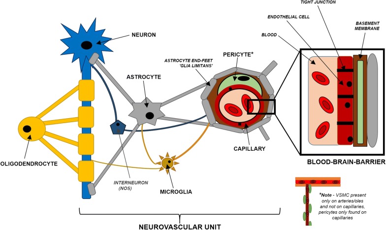
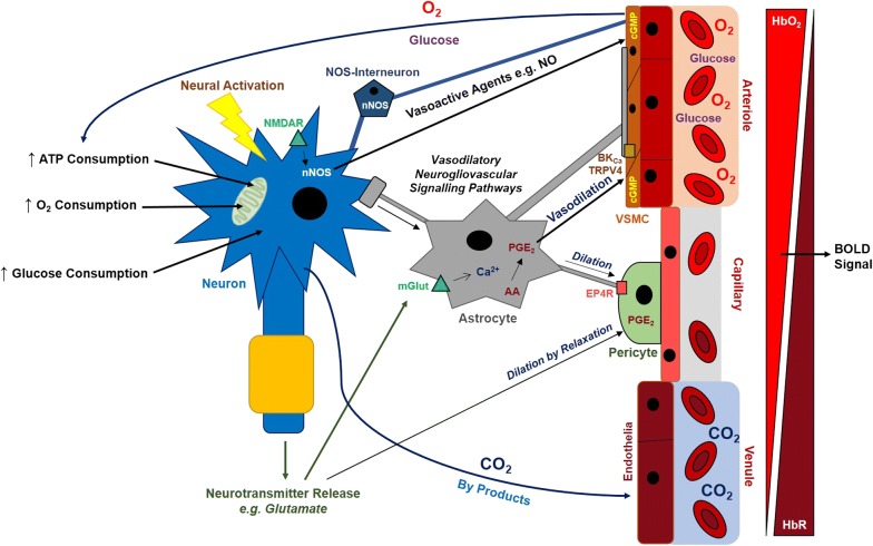
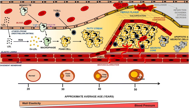
Efficient blood supply to the brain is of paramount importance to its normal functioning and improper blood flow can result in potentially devastating neurological consequences. Cerebral blood flow in response to neural activity is intrinsically regulated by a complex interplay between various cell types within the brain in a relationship termed neurovascular coupling. The breakdown of neurovascular coupling is evident across a wide variety of both neurological and psychiatric disorders including Alzheimer's disease. Atherosclerosis is a chronic syndrome affecting the integrity and function of major blood vessels including those that supply the brain, and it is therefore hypothesised that atherosclerosis impairs cerebral blood flow and neurovascular coupling leading to cerebrovascular dysfunction. This review will discuss the mechanisms of neurovascular coupling in health and disease and how atherosclerosis can potentially cause cerebrovascular dysfunction that may lead to cognitive decline as well as stroke. Understanding the mechanisms of neurovascular coupling in health and disease may enable us to develop potential therapies to prevent the breakdown of neurovascular coupling in the treatment of vascular brain diseases including vascular dementia, Alzheimer's disease and stroke.
Overview
The claims and statistics of this paper haven’t been checked yet.
Run the check — free, no account needed. It reads the open-access text and checks the sources it cites.
Integrity signals
Registry
Cites
Statistics
Figures
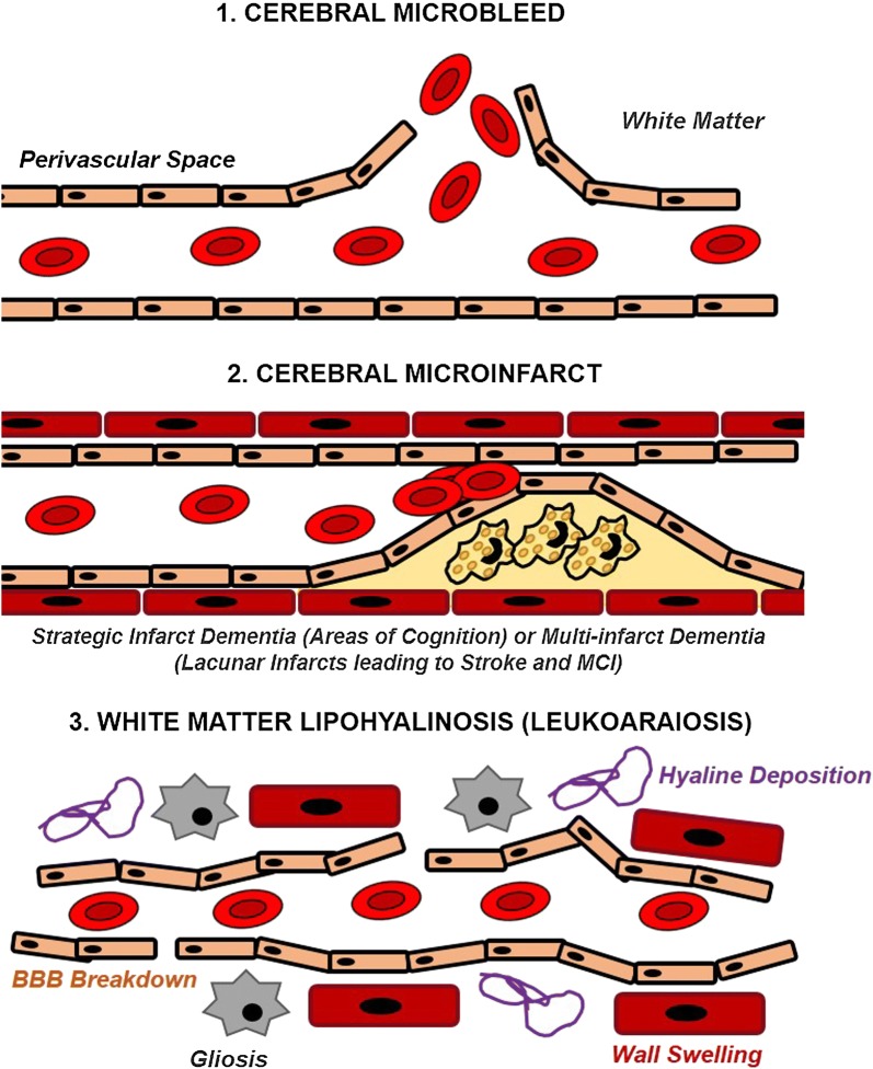
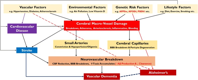
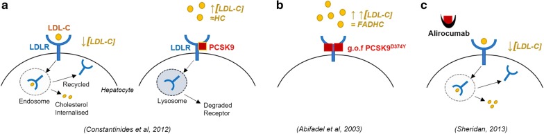
Verification
Claims the paper makes
The claims and statistics of this paper haven’t been checked yet.
Run the check — free, no account needed. It reads the open-access text and checks the sources it cites.
Claims other papers make about this one
Each is checked against this paper’s own text: does it say what the citing paper says it does?
In the citing paper
“Unsurprisingly, such a condition, known as cerebral amyloid angiopathy exacerbates the AD pathology (Shabir et al., 2018).”
From Exploring the Therapeutic Potential of Phytochemicals in Alzheimer’s Disease: Focus on Polyphenols and Monoterpenes (2022) · cites this paper as Shabir et al., 2018
Self-check
Percentages, sums and reported differences re-computed from the paper’s own numbers.
The self-check needs the paper’s full text.
Citations
Cites
1 retracted
Montagne, A., Nikolakopoulou, A. M., Zhao, Z., Sagare, A. P., Si, G., Lazic, D., Barnes, S. R., Daianu, M., Ramanathan, A., Go, A., et al. (2018) · Nature Medicine · doi:10.1038/nm.4482 · Cited by 344
RetractedCalcium's Role in Mechanotransduction during Muscle Development
Damm, T. B., & Egli, M. (2014) · Cellular Physiology and Biochemistry · doi:10.1159/000356667 · Cited by 14,552
Antiinflammatory Therapy with Canakinumab for Atherosclerotic Disease
Ridker, P. M., Everett, B. M., Thuren, T., MacFadyen, J. G., Chang, W. H., Ballantyne, C., Fonseca, F., Nicolau, J., Koenig, W., Anker, S. D., et al. (2017) · New England Journal of Medicine · doi:10.1056/nejmoa1707914 · Cited by 9,482
Brain magnetic resonance imaging with contrast dependent on blood oxygenation.
Ogawa, S., Lee, T. M., Kay, A. R., & Tank, D. W. (1990) · Proceedings of the National Academy of Sciences · doi:10.1073/pnas.87.24.9868 · Cited by 6,483
Heart Disease and Stroke Statistics—2015 Update: A Report From the American Heart Association
Mozaffarian, D., Benjamin, E. J., Go, A. S., Arnett, D. K., Blaha, M. J., Cushman, M., de Ferranti, S., Després, J.-P., Fullerton, H. J., Howard, V. J., et al. (2015) · Circulation · doi:10.1161/cir.0000000000000152 · Cited by 5,797
Calcium signalling: dynamics, homeostasis and remodelling
Berridge, M. J., Bootman, M. D., & Roderick, H. L. (2003) · Nature Reviews Molecular Cell Biology · doi:10.1038/nrm1155 · Cited by 5,719
Lusis, A. J. (2000) · Nature · doi:10.1038/35025203 · Cited by 4,880
Gorelick, P. B., Scuteri, A., Black, S. E., DeCarli, C., Greenberg, S. M., Iadecola, C., Launer, L. J., Laurent, S., Lopez, O. L., Nyenhuis, D., et al. (2011) · Stroke · doi:10.1161/str.0b013e3182299496 · Cited by 3,811
Association of apolipoprotein E allele ϵ4 with late‐onset familial and sporadic Alzheimer's disease
Saunders, A. M., Strittmatter, W. J., Schmechel, D., St. George-Hyslop, P. H., Pericak-Vance, M. A., Joo, S. H., Rosi, B. L., Gusella, J. F., Crapper-MacLachlan, D. R., Alberts, M. J., et al. (1993) · Neurology · doi:10.1212/wnl.43.8.1467 · Cited by 3,786
Compensatory Enlargement of Human Atherosclerotic Coronary Arteries
Glagov, S., Weisenberg, E. S., Zarins, C. K., Stankunavicius, R., & Kolettis, G. (1987) · New England Journal of Medicine · doi:10.1056/nejm198705283162204 · Cited by 3,614
Cited by
Sorted by citations
Items 101–191 of 191
Sheng, L., Zheng, X., Zhao, D., Liu, J., & Song, W. (2025) · Frontiers in Neurology · doi:10.3389/fneur.2025.1522485 · Cited by 11
Low-invasive neural recording in mouse models with diabetes via an ultrasmall needle-electrode
Sanda, R., Yamashita, K., Sawahata, H., Sakamoto, K., Yamagiwa, S., Yokoyama, S., Numano, R., Koida, K., & Kawano, T. (2023) · Biosensors and Bioelectronics · doi:10.1016/j.bios.2023.115605 · Cited by 8
Park, H. K., Song, M., Kim, J., & Han, J. (2020) · Medicine · doi:10.1097/md.0000000000021263 · Cited by 9
The Participation of Hormones in the Processes of Cognitive and Socio-Emotional Aging
Bulgakova, S. V., & Romanchuk, N. (2020) · Bulletin of Science and Practice · doi:10.33619/2414-2948/57/09 · Cited by 9
Understanding Cognitive Deficits in People with Coronary Heart Disease (CHD)
Kang, W., & Malvaso, A. (2023) · Journal of Personalized Medicine · doi:10.3390/jpm13020307 · Cited by 11
Effects of repeated sleep deprivation on brain pericytes in mice
Wu, Y., Li, P., Bhat, N. R., Fan, H., & Liu, M. (2023) · Scientific Reports · doi:10.1038/s41598-023-40138-0 · Cited by 11
Hernandez Torres, L. D., Rezende, F., Peschke, E., Will, O., Hövener, J.-B., Spiecker, F., Özorhan, Ü., Lampe, J., Stölting, I., Aherrahrou, Z., et al. (2024) · Frontiers in Endocrinology · doi:10.3389/fendo.2024.1338458 · Cited by 8
The role of cerebral blood flow in the pathogenesis of Alzheimer's Disease Dementia
Desai, B., Edwards, O., & Beishon, L. (2024) · Aging and Health Research · doi:10.1016/j.ahr.2024.100188 · Cited by 7
Tranexamic Acid Improves Memory and Learning Abilities in Aging Mice
Hiramoto, K., Yamate, Y., Matsuda, K., Sugiyama, D., & Iizuka, Y. (2020) · Journal of Experimental Pharmacology · doi:10.2147/jep.s284532 · Cited by 10
Purroy, F. F., Sánchez, E., Lecube, A., Arqué, G., Pascual, M. V., Mauri, G., Torreguitart, N., Hernández, M., Barbé, F., Fernández, E., et al. (2021) · Frontiers in Neurology · doi:10.3389/fneur.2021.785640 · Cited by 8
Readman, M. R., Wang, Y., Wan, F., Fairman, I., Linkenauger, S. A., Crawford, T. J., & Plack, C. J. (2024) · Parkinsonism & Related Disorders · doi:10.1016/j.parkreldis.2024.107219 · Cited by 7
Shaikh, S. A., & Muthuraman, A. (2023) · Pharmaceuticals · doi:10.3390/ph16060828 · Cited by 9
Toloza–Ramirez, D., & Martella, D. (2019) · Revista médica de Chile · doi:10.4067/s0034-98872019001201594 · Cited by 14
Vascular Dysfunction in the Brain; Implications for Heavy Metal Exposures
Olung, N. F., Aluko, O. M., Jeje, S. O., Adeagbo, A. S. O., & Ijomone, O. M. (2021) · Current Hypertension Reviews · doi:10.2174/1573402117666210225085528 · Cited by 8
Molecular Imaging with PET in the Assessment of Vascular Dementia and Cerebrovascular Disease
Patil, S., Patel, D., Kata, R., Teichner, E. M., Subtirelu, R. C., Ayubcha, C., Werner, T. J., & Alavi, A. A. (2024) · PET Clinics · doi:10.1016/j.cpet.2024.09.001 · Cited by 6
Global brain glucose uptake on 18F-FDG-PET/CT is influenced by chronic cardiovascular risk
Borja, A. J., Hancin, E. C., Zhang, V., Koa, B., Bhattaru, A., Rojulpote, C., Detchou, D. K. E. D., Aly, M., Kaghazchi, F., Gerke, O., et al. (2020) · Nuclear Medicine Communications · doi:10.1097/mnm.0000000000001349 · Cited by 6
Zhang, X., Hollander, C. M., Khan, M. Y., D’silva, M., Ma, H., Yang, X., Bai, R., Keeter, W. C., Galkina, E., Nadler, J. L., et al. (2023) · Communications Biology · doi:10.1038/s42003-023-05304-0 · Cited by 7
Qin, R., Li, T., Li, C., Li, L., Wang, X., & Wang, L. (2023) · PeerJ · doi:10.7717/peerj.16408 · Cited by 6
Applications of Hybrid PET/Magnetic Resonance Imaging in Central Nervous System Disorders
Borja, A. J., Hancin, E. C., Khosravi, M., Ghorpade, R., Koa, B., Miao, X., Werner, T. J., Newberg, A. B., & Alavi, A. A. (2020) · PET Clinics · doi:10.1016/j.cpet.2020.06.004 · Cited by 6
Liu, S., Zhang, C., Zhang, Y., Wu, Z., Wu, P., Tian, S., Zhang, M., Lang, L., Li, L., Wang, R., et al. (2024) · Scientific Reports · doi:10.1038/s41598-024-70446-y · Cited by 6
Lee, H., Lee, H., Choi, J., Hwang, G., Lee, H., Lee, H.-M., Kim, S., Kim, M., Nam, H.-J., Shim, J., et al. (2024) · Healthcare · doi:10.3390/healthcare12050576 · Cited by 7
Higher serum Lp-PLA2 is associated with cognitive impairment in Parkinson’s disease patients
Wu, Z., Shu, D., Wu, S., Cai, P., & Liang, T. (2024) · Frontiers in Neuroscience · doi:10.3389/fnins.2024.1374567 · Cited by 5
Morgan, G. C., Gregory, A., Tang, C., Hwang, S. H., Border, J. J., Xu, J., Liu, Y., Bai, S., Lee, T. J., Cantwell, C., et al. (2026) · GeroScience · doi:10.1007/s11357-026-02140-y · Cited by 4
Huang, S., Nunez, J., Toresco, D. L., Wen, C., Slotabec, L. A., Wang, H., Zhang, H., Rouhi, N., Adenawoola, M. I., & Li, J. (2025) · The FASEB Journal · doi:10.1096/fj.202402725rr · Cited by 4
Pyne, J. D., Morales, C. D., Kraal, A. Z., Alshikho, M. J., Lao, P. J., Turney, I. C., Amarante, E., Lippert, R. V., Chang, J. F., Gutierrez, J., et al. (2025) · Frontiers in Neuroscience · doi:10.3389/fnins.2025.1538956 · Cited by 3
de Liyis, B. G., Sutedja, J. C., Kesuma, P. M. I., Liyis, S., & Widyadharma, I. P. E. (2023) · The Egyptian Journal of Neurology Psychiatry and Neurosurgery · doi:10.1186/s41983-023-00621-x · Cited by 4
Perfusion MRI Techniques: Beyond DSC
Peret, A., Capel, K. W., Jen, M., Chang, W., Kuner, A. D., Manunga, J. M., Samsonov, A. A., Holmes, J. H., Johnson, K. M., & Eisenmenger, L. B. (2023) · Neurographics · doi:10.3174/ng.2100041 · Cited by 4
Khaled, M., Al-Jamal, H., Tajer, L., & El-Mir, R. (2024) · Journal of Alzheimer s Disease · doi:10.3233/jad-231432 · Cited by 4
Le, S., Xu, F., Luo, Z., Shi, W., Lu, S., Zhang, Z., Guo, Z., Xu, W., Yang, M., Li, T., et al. (2025) · BMC Genomics · doi:10.1186/s12864-025-11256-y · Cited by 4
Kumar, B. S., Menon, S. C., Gayathri, S. R., & Chakravarthy, V. S. (2022) · Frontiers in Neuroscience · doi:10.3389/fnins.2022.917196 · Cited by 3
Association of blood pressure variability during acute care hospitalization and incident dementia
Ebinger, J. E., Driver, M. P., Botting, P. G., Wang, M., Cheng, S. C., & Tan, Z. S. (2023) · Frontiers in Neurology · doi:10.3389/fneur.2023.1085885 · Cited by 4
Visual task complexity and eye movement patterns influence measures of human neurovascular coupling
Spence, E. E. M., Hodge, S. V. L., Rosentreter, R. E., Lam, T., Squair, J. W., Fisher, J. P., & Phillips, A. A. (2020) · Physiology & Behavior · doi:10.1016/j.physbeh.2020.113198 · Cited by 4
Kumar, B. S., Mayakkannan, N., Narasimha, S. M., & Chakravarthy, V. S. (2021) · Scientific Reports · doi:10.1038/s41598-021-92661-7 · Cited by 5
Les germanies als Paîsos Catalans
Duran, E. (1982) · Dialnet (Universidad de la Rioja) · Cited by 4
Management of Alzheimer’s Disease with nutraceuticals
Yadav, J. K. (2020) · Elsevier eBooks · doi:10.1016/b978-0-12-820593-8.00028-8 · Cited by 3
Ferreira, J., Ferreira, P., Azevedo, E. I., & Castro, P. (2024) · Ultrasound in Medicine & Biology · doi:10.1016/j.ultrasmedbio.2024.02.003 · Cited by 3
Psychiatric Issues in Older Adults with Gastrointestinal Disorders
Gonzalez-Alonso, R., Gomez, M. F., & O’Dowd, M. A. (2021) · Springer eBooks · doi:10.1007/978-3-030-30192-7_92 · Cited by 3
Reducing excess dietary saturated fat intake to improve cognition in vascular dementia
Blake, S. M., Harding, T., Blake, C. P., & Borman, P. L. (2022) · GSC Advanced Research and Reviews · doi:10.30574/gscarr.2022.12.2.0220 · Cited by 3
Belsheva, M. N., Safonova, L. P., Tatiana, P., Николаевна, М. С., Anastasia, S., & Larisa, D. (2023) · Systems and Technologies of the Digital HealthCare (STDH) · doi:10.1109/stdh59314.2023.10490856 · Cited by 3
Yin, X., Yang, W., Song, L., Liu, J., Li, M., Yang, Z., Liu, W., Wang, Z., Wang, H., & Guo, W. (2024) · Clinical Kidney Journal · doi:10.1093/ckj/sfae353 · Cited by 2
The Glymphatic System, Sleep, and Neurodegeneration
Gorbachevskii, A. V., Kicherova, O. A., & Рейхерт, Л. И. (2024) · Neuroscience and Behavioral Physiology · doi:10.1007/s11055-024-01734-3 · Cited by 2
Wang, H., Zhao, H., Gao, J., Liu, S., Xu, Y., Zhao, X., Yang, S., Gang, B., & Zhang, Z. (2026) · Free Radical Biology and Medicine · doi:10.1016/j.freeradbiomed.2026.03.013 · Cited by 2
The effects of partial sleep restriction and subsequent caffeine ingestion on neurovascular coupling
Lester, A. B., Buckingham, G., & Bond, B. (2024) · Journal of Sleep Research · doi:10.1111/jsr.14145 · Cited by 2
The relationship between atherosclerosis and dementia
Janoutová, J., Machaczka, O., Kovaľová, M., Zatloukalová, A., Ambrož, P., Koutná, V., Mrázková, E., Bar, M., Roubec, M., Bártová, P., et al. (2024) · Central European Journal of Public Health · doi:10.21101/cejph.a7848 · Cited by 2
Sengking, J., Thangwong, P., Jearjaroen, P., Panthiya, L., Buamard, N., Benjakul, S., Tocharus, J., & Tocharus, C. (2025) · Molecular Nutrition & Food Research · doi:10.1002/mnfr.70079 · Cited by 2
Kanso, H., Awde, M. H., Rammal, Z., Mokashar, A. M., Taher, B., Chebli, D., Soloh, N., Ismail, N. A., Salameh, J., Nasrallah, J., et al. (2024) · Nutritional neurosciences · doi:10.1007/978-981-97-4117-5_1 · Cited by 2
Li, C., Liu, Y., Liu, J., & Xu, X. (2024) · Frontiers in Cellular Neuroscience · doi:10.3389/fncel.2024.1353281 · Cited by 2
Morgan, G. C., Gregory, A., Tang, C., Hwang, S. H., Border, J. J., Xu, J., Liu, Y., Bai, S., Lee, T. J., Cantwell, C., et al. (2025) · bioRxiv (Cold Spring Harbor Laboratory) · doi:10.1101/2025.08.21.669945 · Cited by 2
Cerebral blood flow in bipolar disorder
Mio, M., Dimick, M. K., Toma, S., MacIntosh, B. J., & Goldstein, B. I. (2022) · Oxford University Press eBooks · doi:10.1093/med/9780197574522.003.0006 · Cited by 1
Glymphatic system, sleep, neurodegeneration
Gorbachevskii, A. V., Kicherova, O. A., & Рейхерт, Л. И. (2024) · Журнал высшей нервной деятельности им И П Павлова · doi:10.31857/s0044467724030024 · Cited by 1
Liu, Z., Hu, B., Tang, J., Liu, X., Cheng, B., Jia, C., & Zhang, L. (2024) · Journal of Alzheimer s Disease · doi:10.1177/13872877241289381 · Cited by 2
High level expression of OSBPL11 is associated with atherosclerosis and Alzheimer's disease
Zhang, H., Chen, Y., Xu, Q., Wu, X., He, N., Ren, Z., Wang, G., Tang, Z., Xiang, Q., & Liu, L. (2025) · Inflammation · doi:10.1007/s10753-025-02252-1 · Cited by 1
Maparu, K., & Singh, S. (2025) · Brain Disorders · doi:10.1016/j.dscb.2025.100244 · Cited by 2
Zhu, X., Diep, Y. N., Tran, V. T. A., Kang, Y. J., Lee, L. P., & Cho, H. (2025) · BioChip Journal · doi:10.1007/s13206-025-00228-9 · Cited by 1
Pena, G. S., Liu, Y., Silva, M. C. D., Ranadive, S. M., Smith, J. C., & Kuzmiak‐Glancy, S. (2025) · Journal of the American Heart Association · doi:10.1161/jaha.125.041233 · Cited by 1
izydorczak, M., Oumov, M., Udhwani, M. V., Fostok, F., Coronas‐Samano, G., Sanganahalli, B. G., Hermán, P., Hyder, F., & Verhagen, J. V. (2026) · Frontiers in Aging Neuroscience · doi:10.3389/fnagi.2026.1622135 · Cited by 1
Inflammatory and metabolic pathways underlying the association between PM2.5 exposure and dementia
Li, S., Guo, J., Wang, Y., Shi, D., 练新尧, Lu, J., & Li, J. (2026) · Environmental Pollution · doi:10.1016/j.envpol.2026.127961 · Cited by 2
Smart Drug-Delivery Approaches for Enhanced Management of Comorbid Conditions in Alzheimer’s Disease
Stanciu, G. D., Costăchescu, I., Dascalu, C., & Tamba, B.-I. (2026) · Life · doi:10.3390/life16030510 · Cited by 2
Sengking, J., Thangwong, P., Jearjaroen, P., Yawoot, N., Wangtueai, S., Tocharus, J., & Tocharus, C. (2026) · International Journal of Molecular Sciences · doi:10.3390/ijms27062856 · Cited by 1
Yang, W., Song, L., Li, M., Bai, X., Xu, B., min, L., Yang, Z., Ma, T., Gong, Y., Fu, G., et al. (2026) · Brain Imaging and Behavior · doi:10.1007/s11682-026-01161-7 · Cited by 1
Ball, J. D., Panerai, R. B., Williams, C. A. L., & Beishon, L. (2022) · Cerebral Circulation - Cognition and Behavior · doi:10.1016/j.cccb.2022.100039 · Cited by 1
Chen, Z., Liao, S., Chen, G., Li, C., Liu, C., Liu, J., Wu, G., Lyu, Z., Liu, M., Wu, X., et al. (2025) · Translational Vision Science & Technology · doi:10.1167/tvst.14.3.14 · Cited by 1
Psychiatric Issues in Older Adults with Gastrointestinal Disorders
Gonzalez-Alonso, R., Gomez, M. F., & O’Dowd, M. A. (2020) · Springer eBooks · doi:10.1007/978-3-319-90761-1_92-1 · Cited by 1
Cholesterol and Dementia: A Possible Therapeutic Approach
Alavez-Rubio, J. S., & Juárez‐Cedillo, T. (2022) · Springer eBooks · doi:10.1007/978-981-16-7606-2_13 · Cited by 1
Alladi, S., Arshad, F., & Paplikar, A. (2021) · Elsevier eBooks · doi:10.1016/b978-0-12-819641-0.00128-6 · Cited by 1
A systematic review of near-infrared spectroscopy in dementiaPreprint
Butters, E., Srinivasan, S., O'Brien, J. T., Su, L., & Bale, G. M. (2022) · medRxiv · doi:10.1101/2022.11.23.22282361 · Cited by 1
Zhukov, D. Y. (2019) · International Multidisciplinary Scientific GeoConference SGEM ... · doi:10.5593/sgem2019/6.1/s25.105
Neurovascular Function in a Novel Model of Experimental AtherosclerosisPreprint
Shabir, O., Pendry, B., Heath, P. R., Rebollar, M. A., Howarth, C., Wharton, S. B., Berwick, J., & Francis, S. (2020) · bioRxiv (Cold Spring Harbor Laboratory) · doi:10.1101/2020.01.29.924936
Liu, C. S. (2020) · TSpace (University of Toronto)
The profiling and imaging of sterol molecules in the human brain
Griffiths, L. (2023) · Cronfa (Swansea University) · doi:10.23889/suthesis.62587
Pregnancy as a window of opportunity for dementia prevention: a narrative review
Hand, L. K., Taylor, M. K., Sullivan, D. K., Siengsukon, C. F., Morris, J. K., Martin, L. E., & Hull, H. R. (2024) · Nutritional Neuroscience · doi:10.1080/1028415x.2024.2371727
Yahagi‐Estevam, M. (2024) · Digital Library of Theses and Dissertations (Universidade de São Paulo) · doi:10.11606/d.5.2024.tde-28062024-150045
Inoue, T., Nomura, S., Yamakawa, T., Takara, S., Imoto, H., Maruta, Y., Niwayama, M., & Suzuki, M. (2025) · Clinical Neurophysiology · doi:10.1016/j.clinph.2025.02.262
izydorczak, M., Oumov, M., Udhwani, M. V., Fostok, F., Coronas‐Samano, G., Sanganahalli, B. G., Hermán, P., Hyder, F., & Verhagen, J. V. (2025) · bioRxiv (Cold Spring Harbor Laboratory) · doi:10.1101/2025.04.20.649712
Ritson, M., Xia, D., Wheeler-Jones, C. P. D., & Stolp, H. B. (2025) · Journal of Neurochemistry · doi:10.1111/jnc.70130
Kang, S., Lim, C., Lim, S., Kim, K.-M., & Cho, S. (2025) · Genes · doi:10.3390/genes16070746
Gong, P., Shi, J., Kang, Y., & Zhu, Z. (2025) · Dementia and Geriatric Cognitive Disorders · doi:10.1159/000547668
Eu, I., Lu, C. C., Lee, E., & Soni, K. (2025) · Berkeley Pharma Tech Journal of Medicine · doi:10.52243/bptjm.v5i1.67
Neurovascular de‐coupling underlies dual‐task cost across cognitive abilities
Fitzgibbon‐Collins, L. K., Best, S., Noguchi, M., Guest, C., BORRIE, M. J., Shoemaker, J. K., & Bhangu, J. S. (2025) · Alzheimer s & Dementia Translational Research & Clinical Interventions · doi:10.1002/trc2.70156
Bota, P. M., Picón-Pagès, P., Fanlo-Ucar, H., Almabhouh, S., Bagudanch, O., Zeylan, M. E., Senyuz, S., Gohl, P., Molina-Fernández, R., Fernández‐Fuentes, N., et al. (2025) · bioRxiv (Cold Spring Harbor Laboratory) · doi:10.1101/2025.11.05.686746
Early detection of mild cognitive impairment utilizing ocular biomarker-based risk scoring nomogram
Hussen, M. S., Lam, B. Y., Gao, W., Zhou, L., Choi, K. Y., & Chan, H. H. (2025) · Frontiers in Aging Neuroscience · doi:10.3389/fnagi.2025.1669948
Ivanova, L. G., Mkrtchyan, V. R., Khaikin, V. D., Kudukhova, K. I., Gastasheva, M. A., & Akzhigitov, R. G. (2025) · Vestnik nevrologii psihiatrii i nejrohirurgii (Bulletin of Neurology Psychiatry and Neurosurgery) · doi:10.33920/med-01-2511-11
Lv, X., Zhang, M., Cheng, Z., Wang, Q., Wang, P., Xie, Q., Ni, M., Shen, Y., Tang, Q., & Gao, F. (2023) · Figshare · doi:10.6084/m9.figshare.22620493
Schulz, M., Malherbe, C., Cheng, B., Thomalla, G., & Schlemm, E. (2021) · Figshare · doi:10.6084/m9.figshare.14538908
Bota, P. M., Picón-Pagès, P., Fanlo-Ucar, H., Almabhouh, S., Bagudanch, O., Zeylan, M. E., Senyuz, S., Gohl, P., Molina-Fernández, R., Fernandez-Fuentes, N., et al. (2025) · Computational and Structural Biotechnology Journal · doi:10.1016/j.csbj.2025.12.032
Edwards, O., Nicholls, J. K., Desai, B., Panerai, R. B., Ball, J. D., Sensier, Y., Mukaetova‐Ladinska, E. B., & Beishon, L. (2026) · Journal of Neuroscience Methods · doi:10.1016/j.jneumeth.2026.110784
Han, J., Zhuo, W., Chen, B., Wieczorek, J., Zhang, Y., He, Y., & Feng, B. (2026) · Environment & Health · doi:10.1021/envhealth.5c00835 · Cited by 1
Donna, M. G. D., Bagnato, M. R., Bonomi, C. G., Motta, C., Centonze, D., Martorana, A., & Diomedi, M. (2026) · Alzheimer s & Dementia Translational Research & Clinical Interventions · doi:10.1002/trc2.70250
Irisin upregulation as a contributory mechanism for the therapeutic benefits of SGLT-2 inhibitors
Noto, N. M., Martin, K., Fidelia, S., Parmar, M. S., Cubeddu, L. X., Gurusamy, N., & Speth, R. C. (2026) · Pharmacology & Therapeutics · doi:10.1016/j.pharmthera.2026.109090
Yun, Y. O. N. G., Liu, T., Si, M., Yang, L., Bao, X., Luo, H., Chai, C., & Hua, J. (2026) · Experimental Neurology · doi:10.1016/j.expneurol.2026.116065
Metadata
- Authors
- Shabir, O., Berwick, J., & Francis, S. E.
- Affiliations
- University of Sheffield (GB)
- Corresponding author
- Shabir, Osman
- Venue
- BMC Neuroscience
- Published
- 2018-10-17
- Volume
- 19
- Issue
- 1
- Pages
- 62-62
- ISSN
- 1471-2202 (ISSN-L)
- PMC
- PMC6192291
- OpenAlex
- W2896976060
- Cited by
- 191
- References
- 111
- Citations by year
- 2026: 12 · 2025: 27 · 2024: 32 · 2023: 32 · 2022: 25 · 2021: 31 · 2020: 23 · 2019: 8
- FWCI
- 1.46
- Type
- journal article
- Language
- en
- Open access
- gold
- doi.org
- Licence
- Locations
- BMC Neuroscience (publishedVersion, cc-by)
- Europe PMC (PubMed Central) (publishedVersion, other-oa)
- PubMed Central (publishedVersion, cc-by)
- PubMed (publishedVersion)
- White Rose Research Online (University of Leeds, The University of Sheffield, University of York) (acceptedVersion)
- DOAJ (DOAJ: Directory of Open Access Journals) (submittedVersion, cc-by-sa)
- Funders
- Medical Research Council (MR/M013553/1)
- Alzheimer’s Research UK (ARUK-IRG2014)
- Topics
- Life Sciences › Neuroscience › Neurology › Neurological Disease Mechanisms and Treatments
- Life Sciences › Neuroscience › Neurology › Barrier Structure and Function Studies
- Health Sciences › Medicine › Clinical Neurology › Traumatic Brain Injury and Neurovascular Disturbances
- Keywords
- neurovascular coupling, vascular dementia, Alzheimer's disease, atherosclerosis, cerebral blood flow, cerebrovascular dysfunction, stroke, cognitive decline, cerebrovascular disease
- MeSH terms
- Neurovascular Coupling, Alzheimer Disease, Animals, Brain, Humans, Dementia, Vascular, Atherosclerosis
About these checks
Registry, retraction, Cites and statistics results are computed, not judged, with one exception: a printed reference that no index lists is read once by a model before it counts as a citation error. Claims are extracted by a model and kept only when their quote appears verbatim in the text; each cited source is then checked, with a model’s help, for whether it says what the claim relies on. Those verdicts can be wrong — read the claim’s quote and the cited source before relying on one.
Statistics and arithmetic are recomputed, not judged.
Confidence bands are shown only for fields calibrated on our annotated test set. None is calibrated yet, so no band is shown.
What counts as a claim ›How we measure confidence ›How we check this ›
Are you an author, or do you see a problem with this page? Request a review
