

The DOI matches its Crossref record; 8 of the 8 references it cites were found in a registry or an index.
Oligosaccharides increase the genotoxic effect of colibactin produced by pks+ Escherichia coli strains
Published version of a preprint
Abstract
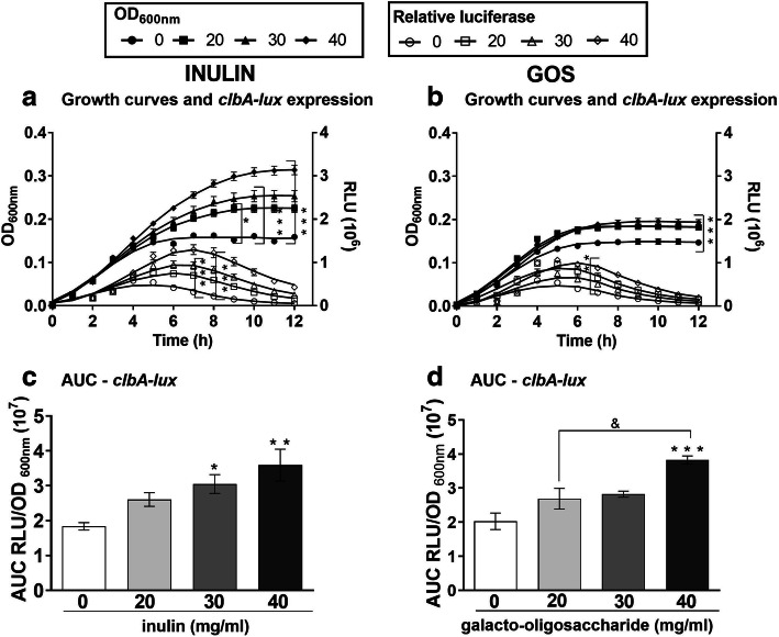
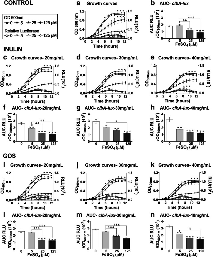
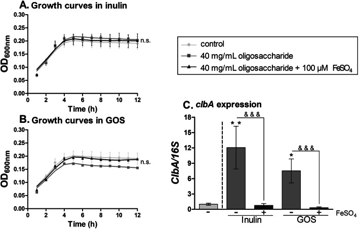
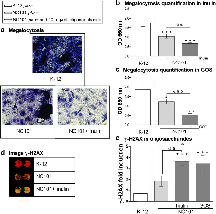
Background: Colibactin is a genotoxin that induces DNA double-strand breaks that may lead to carcinogenesis and is produced by Escherichia coli strains harboring the pks island. Human and animal studies have shown that colibactin-producing gut bacteria promote carcinogenesis and enhance the progression of colorectal cancer through cellular senescence and chromosomal abnormalities. In this study, we investigated the impact of prebiotics on the genotoxicity of colibactin-producing E. coli strains Nissle 1917 and NC101. Methods: Bacteria were grown in medium supplemented with 20, 30 and 40 mg/mL of prebiotics inulin or galacto-oligosaccharide, and with or without 5 μM, 25 μM and 125 μM of ferrous sulfate. Colibactin expression was assessed by luciferase reporter assay for the clbA gene, essential for colibactin production, in E. coli Nissle 1917 and by RT-PCR in E. coli NC101. The human epithelial colorectal adenocarcinoma cell line, Caco-2, was used to assess colibactin-induced megalocytosis by methylene blue binding assay and genotoxicity by γ-H2AX immunofluorescence analysis. Results: Inulin and galacto-oligosaccharide enhanced the expression of clbA in pks+ E. coli . However, the addition of 125 μM of ferrous sulfate inhibited the expression of clbA triggered by oligosaccharides. In the presence of either oligosaccharide, E. coli NC101 increased dysplasia and DNA double-strand breaks in Caco-2 cells compared to untreated cells. Conclusion: Our results suggest that, in vitro, prebiotic oligosaccharides exacerbate DNA damage induced by colibactin-producing bacteria. Further studies are necessary to establish whether oligosaccharide supplementation may lead to increased colorectal tumorigenesis in animal models colonized with pks+ E. coli .
Overview
The claims and statistics of this paper haven’t been checked yet.
Run the check — free, no account needed. It reads the open-access text and checks the sources it cites.
Integrity signals
Registry
Cites
Statistics
Figures
Verification
Claims the paper makes
The claims and statistics of this paper haven’t been checked yet.
Run the check — free, no account needed. It reads the open-access text and checks the sources it cites.
Claims other papers make about this one
Each is checked against this paper’s own text: does it say what the citing paper says it does?
The source backs part of the claim, not all of it — or backs it for a different population, condition or comparison.
In the citing paper
“The apparent difference in colibactin mutagenesis observed between species, or between the cohorts studied, might result from a different prevalence of pks+ E. coli strains36 or a different expression of colibactin by pks+ E. coli across species37.”
From Somatic mutation rates scale with lifespan across mammals (2022) · cites this paper as [37]
Experimental — accuracy not yet measured
Self-check
Percentages, sums and reported differences re-computed from the paper’s own numbers.
The self-check needs the paper’s full text.
Citations
Cites
Intestinal inflammation targets cancer-inducing activity of the microbiota
Arthur, J. C., Perez-Chanona, E., Mühlbauer, M., Tomkovich, S., Uronis, J. M., Fan, T.-J., Campbell, B. J., Abujamel, T., Dogan, B., Rogers, A. B., et al. (2012) · Science · doi:10.1126/science.1224820 · Cited by 2,263
Wirbel, J., Pyl, P. T., Kartal, E., Zych, K., Kashani, A., Milanese, A., Fleck, J. S., Voigt, A. Y., Palleja, A., Ponnudurai, R., et al. (2019) · Nature Medicine · doi:10.1038/s41591-019-0406-6 · Cited by 1,373
Mutational signature in colorectal cancer caused by genotoxic pks+ E. coli
Pleguezuelos-Manzano, C., Puschhof, J., Rosendahl Huber, A., van Hoeck, A., Wood, H. M., Nomburg, J., Gurjao, C., Manders, F., Dalmasso, G., Stege, P. B., et al. (2020) · Nature · doi:10.1038/s41586-020-2080-8 · Cited by 1,208
Escherichia coli Induces DNA Double-Strand Breaks in Eukaryotic Cells
Nougayrède, J.-P., Homburg, S., Taieb, F., Boury, M., Brzuszkiewicz, E., Gottschalk, G., Buchrieser, C., Hacker, J., Dobrindt, U., & Oswald, E. (2006) · Science · doi:10.1126/science.1127059 · Cited by 1,188
Patients with familial adenomatous polyposis harbor colonic biofilms containing tumorigenic bacteria
Dejea, C. M., Fathi, P., Craig, J. M., Boleij, A., Taddese, R., Geis, A. L., Wu, X., DeStefano Shields, C. E., Hechenbleikner, E. M., Huso, D. L., et al. (2018) · Science · doi:10.1126/science.aah3648 · Cited by 1,105 · cited 4×
“coli in murine models showed a direct link between colibactin production and colon carcinogenesis [4, 5].”
BackgroundInfluentialHigh Prevalence of Mucosa-Associated E. coli Producing Cyclomodulin and Genotoxin in Colon Cancer
Buc, E., Dubois, D., Sauvanet, P., Raisch, J., Delmas, J., Darfeuille-Michaud, A., Pezet, D., & Bonnet, R. (2013) · PLoS ONE · doi:10.1371/journal.pone.0056964 · Cited by 535
Poeker, S. A., Geirnaert, A., Berchtold, L., Greppi, A., Krych, L., Steinert, R. E., de Wouters, T., & Lacroix, C. (2018) · Scientific Reports · doi:10.1038/s41598-018-22438-y · Cited by 176
Interplay between Siderophores and Colibactin Genotoxin Biosynthetic Pathways in Escherichia coli
Martin, P., Marcq, I., Magistro, G., Penary, M., Garcie, C., Payros, D., Boury, M., Olier, M., Nougayrède, J.-P., Audebert, M., et al. (2013) · PLoS Pathogens · doi:10.1371/journal.ppat.1003437 · Cited by 138
Research works printed but not linked: 45
- Rawla P, Sunkara T, Barsouk A. Epidemiology of colorectal cancer: incidence, mortality, survival, and risk factors. Prz Gastroenterol. 2019;14(2):89–103. https://doi.org/10.5114/pg.2018.81072. · DOI
- Liu W, Zhang R, Shu R, Yu J, Li H, Long H, Jin S, Li S, Hu Q, Yao F, et al. Study of the relationship between microbiome and colorectal Cancer susceptibility using 16SrRNA sequencing. Biomed Res Int. 2020;2020:7828392. https://doi.org/10.1155/2020/7828392. · DOI
- Zou S, Fang L, Lee MH. Dysbiosis of gut microbiota in promoting the development of colorectal cancer. Gastroenterol Rep (Oxf). 2018;6(1):1–12. https://doi.org/10.1093/gastro/gox031. · DOI
- Homburg S, Oswald E, Hacker J, Dobrindt U. Expression analysis of the colibactin gene cluster coding for a novel polyketide in Escherichia coli. FEMS Microbiol Lett. 2007;275(2):255–62. https://doi.org/10.1111/j.1574-6968.2007.00889.x. · DOI
- Secher T, Samba-Louaka A, Oswald E, Nougayrède J-P. Escherichia coli producing colibactin triggers premature and transmissible senescence in mammalian cells. PLoS One. 2013;8(10):e77157. https://doi.org/10.1371/journal.pone.0077157. · DOI
- Kamada N, Chen GY, Inohara N, Nunez G. Control of pathogens and pathobionts by the gut microbiota. Nat Immunol. 2013;14(7):685–90. https://doi.org/10.1038/ni.2608. · DOI · Paper page
- Massot M, Daubié A-S, Clermont O, Jauréguy F, Couffignal C, Dahbi G, Mora A, Blanco J, Branger C, Mentré F, et al. Phylogenetic, virulence and antibiotic resistance characteristics of commensal strain populations of Escherichia coli from community subjects in the Paris area in 2010 and evolution over 30 years. Microbiology (Reading). 2016;162(4):642–50. https://doi.org/10.1099/mic.0.000242. · DOI
- Cole BK, Ilikj M, McCloskey CB, Chavez-Bueno S. Antibiotic resistance and molecular characterization of bacteremia Escherichia coli isolates from newborns in the United States. PLoS One. 2019;14(7):e0219352. https://doi.org/10.1371/journal.pone.0219352. · DOI
- Gibson GR, Hutkins R, Sanders ME, Prescott SL, Reimer RA, Salminen SJ, Scott K, Stanton C, Swanson KS, Cani PD, et al. Expert consensus document: the international scientific Association for Probiotics and Prebiotics (ISAPP) consensus statement on the definition and scope of prebiotics. Nat Rev Gastroenterol Hepatol. 2017;14(8):491–502. https://doi.org/10.1038/nrgastro.2017.75. · DOI · Paper page
- Pham VT, Seifert N, Richard N, Raederstorff D, Steinert RE, Prudence K, Mohajeri MH. The effects of fermentation products of prebiotic fibres on gut barrier and immune functions in vitro. PeerJ. 2018;6:e5288. https://doi.org/10.7717/peerj.5288. · DOI
- Önal Darilmaz D, Sönmez Ş, Beyatli Y. The effects of inulin as a prebiotic supplement and the synbiotic interactions of probiotics to improve oxalate degrading activity. Int J Food Sci Technology. 2019;54(1):121–31. https://doi.org/10.1111/ijfs.13912. · DOI
- Fooks LJ, Gibson GR. In vitro investigations of the effect of probiotics and prebiotics on selected human intestinal pathogens. FEMS Microbiol Ecol. 2002;39:67–75. · DOI
- Shoaf K, Mulvey GL, Armstrong GD, Hutkins RW. Prebiotic galactooligosaccharides reduce adherence of enteropathogenic Escherichia coli to tissue culture cells. Infect Immun. 2006;74(12):6920–8. https://doi.org/10.1128/IAI.01030-06. · DOI
- Tronnet S, Garcie C, Brachmann AO, Piel J, Oswald E, Martin P: High iron supply inhibits the synthesis of the genotoxin colibactin by pathogenic Escherichia coli through a non-canonical Fur/RyhB-mediated pathway. Pathogens Disease 2017, 75(5). https://doi.org/10.1093/femspd/ftx066. · DOI
- Tronnet S, Garcie C, Rehm N, Dobrindt U, Oswald E, Martin P. Iron homeostasis regulates the Genotoxicity of Escherichia coli that produces Colibactin. Infect Immun. 2016;84(12):3358–68. https://doi.org/10.1128/IAI.00659-16. · DOI
- Makui H, Soares RJ, Jiang W, Constante M, Santos MM. Contribution of Hfe expression in macrophages to the regulation of hepatic hepcidin levels and iron loading. Blood. 2005;106(6):2189–95. https://doi.org/10.1182/blood-2005-02-0629. · DOI
- Bossuet-Greif N, Belloy M, Boury M, Oswald E, Nougayrede J-P: Protocol for HeLa Cells Infection with Escherichia coli Strains Producing Colibactin and Quantification of the Induced DNA-damage. Bio-Protocol 2017, 7(16). https://doi.org/10.21769/BioProtoc.2520. · DOI
- Bruballa AC, Shiromizu CM, Bernal AM, Pineda GE, Sabbione F, Trevani AS, Bentancor LV, Ramos MV, Fernandez-Brando RJ, Munoz MJ et al: Role of Shiga Toxins in Cytotoxicity and Immunomodulatory Effects of Escherichia coli O157:H7 during Host-Bacterial Interactions in vitro. Toxins (Basel) 2020, 12(1). https://doi.org/10.3390/toxins12010048. · DOI
- Tronnet S, Oswald E: Quantification of Colibactin-associated Genotoxicity in HeLa Cells by In Cell Western (ICW) Using γ-H2AX as a Marker. Bio-Protocol 2018, 8(6). https://doi.org/10.21769/BioProtoc.2771. · DOI
- Wallenstein A, Rehm N, Brinkmann M, Selle M, Bossuet-Greif N, Sauer D, Bunk B, Spröer C, Wami HT, Homburg S et al: ClbR Is the Key Transcriptional Activator of Colibactin Gene Expression in Escherichia coli. 2020, 5(4):e00591–00520. https://doi.org/10.1128/mSphere.00591-20%J mSphere. · DOI
- Kim SC, Tonkonogy SL, Albright CA, Tsang J, Balish EJ, Braun J, Huycke MM, Sartor RB. Variable phenotypes of enterocolitis in interleukin 10-deficient mice monoassociated with two different commensal bacteria. Gastroenterology. 2005;128(4):891–906. https://doi.org/10.1053/j.gastro.2005.02.009. · DOI
- Bakthavatchalu V, Wert KJ, Feng Y, Mannion A, Ge Z, Garcia A, Scott KE, Caron TJ, Madden CM, Jacobsen JT, et al. Cytotoxic Escherichia coli strains encoding colibactin isolated from immunocompromised mice with urosepsis and meningitis. PLoS One. 2018;13(3):e0194443. https://doi.org/10.1371/journal.pone.0194443. · DOI
- Song M, Chan AT, Sun J. Influence of the gut microbiome, diet, and environment on risk of colorectal Cancer. Gastroenterology. 2020;158(2):322–40. https://doi.org/10.1053/j.gastro.2019.06.048. · DOI · Paper page
- Raimondi S, Righini L, Candeliere F, Musmeci E, Bonvicini F, Gentilomi G, Starcic Erjavec M, Amaretti A, Rossi M: Antibiotic Resistance, Virulence Factors, Phenotyping, and Genotyping of E. coli Isolated from the Feces of Healthy Subjects. Microorganisms 2019, 7(8). https://doi.org/10.3390/microorganisms7080251. · DOI
- Iyadorai T, Mariappan V, Vellasamy KM, Wanyiri JW, Roslani AC, Lee GK, Sears C, Vadivelu J. Prevalence and association of pks+ Escherichia coli with colorectal cancer in patients at the university Malaya medical Centre, Malaysia. PLoS One. 2020;15(1):e0228217. https://doi.org/10.1371/journal.pone.0228217. · DOI
- Suresh A, Ranjan A, Jadhav S, Hussain A, Shaik S, Alam M, Baddam R, Wieler LH, Ahmed N. Molecular genetic and functional analysis of pks-harboring, Extra-Intestinal Pathogenic Escherichia coli From India. Front Microbiol. 2018;9:2631. https://doi.org/10.3389/fmicb.2018.02631. · DOI
- Yoshikawa Y, Tsunematsu Y, Matsuzaki N, Hirayama Y, Higashiguchi F, Sato M, Iwashita Y, Miyoshi N, Mutoh M, Ishikawa H, et al. Characterization of colibactin-producing Escherichia coli isolated from Japanese patients with colorectal cancer. Jpn J Infect Dis. 2020. https://doi.org/10.7883/yoken.JJID.2020.066. · DOI
- Olier M, Marcq I, Salvador-Cartier C, Secher T, Dobrindt U, Boury M, Bacquie V, Penary M, Gaultier E, Nougayrede JP, et al. Genotoxicity of Escherichia coli Nissle 1917 strain cannot be dissociated from its probiotic activity. Gut Microbes. 2012;3(6):501–9. https://doi.org/10.4161/gmic.21737. · DOI
- Bossuet-Greif N, Vignard J, Taieb F, Mirey G, Dubois D, Petit C, Oswald E, Nougayrede JP: The Colibactin Genotoxin Generates DNA Interstrand Cross-Links in Infected Cells. mBio 2018, 9(2). https://doi.org/10.1128/mBio.02393-17. · DOI
- Busti F, Marchi G, Ugolini S, Castagna A, Girelli D: Anemia and Iron Deficiency in Cancer Patients: Role of Iron Replacement Therapy. Pharmaceuticals (Basel) 2018, 11(4). https://doi.org/10.3390/ph11040094. · DOI
- Constante M, Fragoso G, Lupien-Meilleur J, Calve A, Santos MM. Iron supplements modulate Colon microbiota composition and potentiate the protective effects of probiotics in dextran sodium sulfate-induced colitis. Inflamm Bowel Dis. 2017;23(5):753–66. https://doi.org/10.1097/MIB.0000000000001089. · DOI
- Dziubanska-Kusibab PJ, Berger H, Battistini F, Bouwman BAM, Iftekhar A, Katainen R, Cajuso T, Crosetto N, Orozco M, Aaltonen LA, et al. Colibactin DNA-damage signature indicates mutational impact in colorectal cancer. Nat Med. 2020;26(7):1063–9. https://doi.org/10.1038/s41591-020-0908-2. · DOI
- Wolff RK, Hoffman MD, Wolff EC, Herrick JS, Sakoda LC, Samowitz WS, Slattery ML. Mutation analysis of adenomas and carcinomas of the colon: early and late drivers. Genes Chromosom Cancer. 2018;57(7):366–76. https://doi.org/10.1002/gcc.22539. · DOI
- Raisch J, Buc E, Bonnet M, Sauvanet P, Vazeille E, de Vallee A, Dechelotte P, Darcha C, Pezet D, Bonnet R, et al. Colon cancer-associated B2 Escherichia coli colonize gut mucosa and promote cell proliferation. World J Gastroenterol. 2014;20(21):6560–72. https://doi.org/10.3748/wjg.v20.i21.6560. · DOI
- Kosari F, Taheri M, Moradi A, Hakimi Alni R, Alikhani MY. Evaluation of cinnamon extract effects on clbB gene expression and biofilm formation in Escherichia coli strains isolated from colon cancer patients. BMC Cancer. 2020;20(1):267. https://doi.org/10.1186/s12885-020-06736-1. · DOI
- Kaewkod T, Tobe R, Tragoolpua Y, Mihara H. Medicinal plant extracts protect epithelial cells from infection and DNA damage caused by colibactin-producing Escherichia coli, and inhibit the growth of bacteria. J Appl Microbiol. 2020. https://doi.org/10.1111/jam.14817. · DOI
- Wu F, Groopman JD, Pestka JJ. Public health impacts of foodborne Mycotoxins. Annu Rev Food Sci Technol. 2014;5(1):351–72. https://doi.org/10.1146/annurev-food-030713-092431. · DOI
- Payros D, Dobrindt U, Martin P, Secher T, Bracarense AP, Boury M, Laffitte J, Pinton P, Oswald E, Oswald IP: The Food Contaminant Deoxynivalenol Exacerbates the Genotoxicity of Gut Microbiota. mBio 2017, 8(2). https://doi.org/10.1128/mBio.00007-17. · DOI
- Jung TH, Jeon WM, Han KS. In vitro effects of dietary inulin on human fecal microbiota and butyrate production. J Microbiol Biotechnol. 2015;25(9):1555–8. https://doi.org/10.4014/jmb.1505.05078. · DOI · Paper page
- Singh V, Vijay-Kumar M. Beneficial and detrimental effects of processed dietary fibers on intestinal and liver health: health benefits of refined dietary fibers need to be redefined! Gastroenterol Rep (Oxf). 2020;8(2):85–9. https://doi.org/10.1093/gastro/goz072. · DOI
- Pajari AM, Rajakangas J, Paivarinta E, Kosma VM, Rafter J, Mutanen M. Promotion of intestinal tumor formation by inulin is associated with an accumulation of cytosolic beta-catenin in min mice. Int J Cancer. 2003;106(5):653–60. https://doi.org/10.1002/ijc.11270. · DOI
- Misikangas M, Tanayama H, Rajakangas J, Linden J, Pajari AM, Mutanen M. Inulin results in increased levels of beta-catenin and cyclin D1 as the adenomas increase in size from small to large in the min/+ mouse. Br J Nutr. 2008;99(5):963–70. https://doi.org/10.1017/S0007114507853414. · DOI
- Li Y, Elmen L, Segota I, Xian Y, Tinoco R, Feng Y, Fujita Y, Segura Munoz RR, Schmaltz R, Bradley LM, et al. Prebiotic-induced anti-tumor immunity attenuates tumor growth. Cell Rep. 2020;30(6):1753–66 e1756. https://doi.org/10.1016/j.celrep.2020.01.035. · DOI · Paper page
- Fernandez J, Moreno FJ, Olano A, Clemente A, Villar CJ, Lombo F. A Galacto-oligosaccharides preparation derived from lactulose protects against colorectal Cancer development in an animal model. Front Microbiol. 2018;9:2004. https://doi.org/10.3389/fmicb.2018.02004. · DOI
- Murphy N, Moreno V, Hughes DJ, Vodicka L, Vodicka P, Aglago EK, Gunter MJ, Jenab M. Lifestyle and dietary environmental factors in colorectal cancer susceptibility. Mol Asp Med. 2019;69:2–9. https://doi.org/10.1016/j.mam.2019.06.005. · DOI
Cited by
Sorted by citations
Somatic mutation rates scale with lifespan across mammals
Cagan, A., Baez-Ortega, A., Brzozowska, N., Abascal, F., Coorens, T. H. H., Sanders, M. A., Lawson, A. R. J., Harvey, L. M. R., Bhosle, S., Jones, D., et al. (2022) · Nature · doi:10.1038/s41586-022-04618-z · Cited by 481
Rosendahl Huber, A., Pleguezuelos-Manzano, C., Puschhof, J., Ubels, J., Boot, C., Saftien, A., Verheul, M., Trabut, L. T., Groenen, N., van Roosmalen, M., et al. (2024) · Cancer Cell · doi:10.1016/j.ccell.2024.02.009 · Cited by 106
Inulin impacts tumorigenesis promotion by colibactin-producing Escherichia coli in ApcMin/+ mice
Oliero, M., Hajjar, R., Cuisiniere, T., Fragoso, G., Calvé, A., & Santos, M. M. (2023) · Frontiers in Microbiology · doi:10.3389/fmicb.2023.1067505 · Cited by 19
Self-citation
Metadata
- Authors
- Oliero, M., Calvé, A., Fragoso, G., Cuisiniere, T., Hajjar, R., Dobrindt, U., & Santos, M. M.
- Venue
- BMC Cancer
- Published
- 2021-02-17
- Volume
- 21
- Issue
- 1
- ISSN
- 1471-2407
- PMC
- PMC7890614
- Cited by
- 47
- References
- 8
- Language
- en
- Funders
- Canadian Institutes of Health Research (PJT-159775)
- Natural Sciences and Engineering Research Council of Canada (RGPIN-2018-06442)
- Institut du Cancer de Montréal (scholarship)
- Université de Montréal (scholarship)
- Fonds de Recherche du Québec - Santé (MSSS Resident Physician Health Research Career Training Program)
- Interdisciplinary Center for Clinical Research of the Medical Faculty Münster (Dob2/013/12)
About these checks
Registry, retraction, Cites and statistics results are computed, not judged, with one exception: a printed reference that no index lists is read once by a model before it counts as a citation error. Claims are extracted by a model and kept only when their quote appears verbatim in the text; each cited source is then checked, with a model’s help, for whether it says what the claim relies on. Those verdicts can be wrong — read the claim’s quote and the cited source before relying on one.
Statistics and arithmetic are recomputed, not judged.
Confidence bands are shown only for fields calibrated on our annotated test set. None is calibrated yet, so no band is shown.
What counts as a claim ›How we measure confidence ›How we check this ›
Are you an author, or do you see a problem with this page? Request a review
How to cite
Cite CoChat for the checks on this page, and the paper for its own findings.
- This page
- CoChat. (2026). Citation check: Oligosaccharides increase the genotoxic effect of colibactin produced by pks+ Escherichia coli strains. https://app.cochat.ai/papers/10.1186/s12885-021-07876-8
- The paper
- Oliero, M., Calvé, A., Fragoso, G., Cuisiniere, T., Hajjar, R., Dobrindt, U., & Santos, M. M. (2021). Oligosaccharides increase the genotoxic effect of colibactin produced by pks+ Escherichia coli strains. BMC Cancer. https://doi.org/10.1186/s12885-021-07876-8
