

The DOI matches its Crossref record; 28 of the 28 references it cites were found in a registry or an index; none of the 10 sources its claims cite is checked for support yet.
Pediatric osmotic demyelination syndrome triggered by post-traumatic pituitary stalk injury: a case report
Open accessAbstract
BACKGROUND: Osmotic demyelination syndrome (ODS) is an acute neurological disorder resulting from demyelination, typically triggered by rapid osmotic shifts. This syndrome is rare in adults and even more uncommon in children.To date, no reported cases have documented ODS secondary to hyponatremia caused by post-traumatic pituitary stalk injury. We report a pediatric patient with traumatic brain injury who developed ODS subsequent to pituitary stalk disruption and hyponatremia, and discuss the corresponding diagnostic and therapeutic strategies. CASE PRESENTATION: We report the case of a 13-year-old Chinese girl who presented with seizures, central diabetes insipidus, dysarthria, dysphagia, and painful hypertonia following traumatic brain injury. Laboratory investigation revealed Severe hyponatremia. Magnetic resonance imaging (MRI)confirmed the diagnosis of ODS. The child was managed accordingly and demonstrated a favorable clinical response, with substantial recovery in physical function and quality of life. CONCLUSION: Early diagnosis of ODS is based on recognizing key risk factors, typical clinical symptoms, and characteristic neuroimaging findings. Contrast-enhanced MRI can be critical in elucidating the underlying etiology. B-complex vitamins and hyperbaric oxygen therapy have the potential to improve disease outcomes.
Overview
CoChat Summary
TL;DR
A 13-year-old Chinese girl with traumatic brain injury developed osmotic demyelination syndrome after severe hyponatremia and post-traumatic pituitary stalk injury, with MRI showing extrapontine lesions and no pontine involvement. She improved after sodium correction, desmopressin, B-complex vitamins, and hyperbaric oxygen therapy, with central diabetes insipidus remitting by 3 months.
Key findings
- The patient’s serum sodium was 113 mmol/L; during correction with intermittent 150 mL 3% hypertonic saline, levels were 117, 120, and 128 mmol/L in the first 24 h and subsequent measurements were 130, 135, and 144 mmol/L. Total NaCl supplementation was 17.2 g in the first 24 h and an additional 19.6 g over the next 24 h.
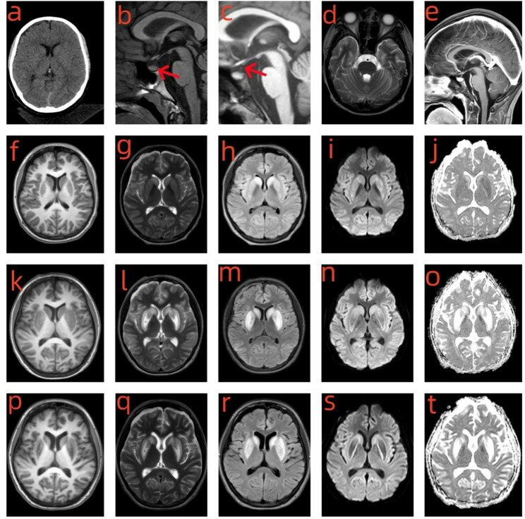
- Initial MRI showed T2/FLAIR-hyperintense and T1-hypointense lesions with focal restricted diffusion in the bilateral caudate nuclei, putamen, and insulae; repeat contrast-enhanced MRI showed progression plus an absent T1-weighted posterior pituitary bright spot and an ill-defined pituitary stalk, without pontine T2-hyperintense lesions or abnormal enhancement.
- After treatment with B-complex vitamins (vitamin B1 10 mg TID, vitamin B2 5 mg TID, vitamin B6 10 mg once daily, and vitamin B12 0.5 mg TID for 1 month), rehabilitation, and hyperbaric oxygen therapy, follow-up MRI on hospital day 18 showed partial resolution; at 3-month telephone follow-up, dysarthria and dysphagia had lessened, left-sided hypertonia had resolved, and central diabetes insipidus had completely remitted.
- The paper states no prior case reports have documented ODS secondary to hyponatremia caused by post-traumatic pituitary stalk injury; pediatric ODS is rare, with 106 pediatric cases noted in a cited review from 1960–2018.
Methods
- Single case report of a 13-year-old Chinese girl with traumatic brain injury transferred after seizures; the clinical course, laboratory tests, hormone measurements, CT, and serial brain MRI were described.
- Diagnostic evaluation included serum sodium and osmolality, hormone levels (T3, fT3, T4, fT4, TSH, cortisol, ACTH), contrast-enhanced pituitary MRI, and multidisciplinary exclusion of hypoglycemic encephalopathy and CO poisoning; ODS was diagnosed from clinical history, hyponatremia, and neuroimaging.
- No controlled comparison or formal statistical analysis was performed; management included hypertonic saline, desmopressin, neuroprotective agents, muscle relaxants, analgesics, B-complex vitamins, and hyperbaric oxygen therapy.
Limitations
- Stated: no standardized treatment protocol for ODS has been established, clinical outcomes remain inconsistent, and the mechanisms—especially those pertaining to B-complex vitamin supplementation—remain to be elucidated.
- Evident: this is a single pediatric case report with no control group; the causal link between pituitary stalk injury, hyponatremia correction, and ODS is inferred from the clinical timeline and imaging rather than experimentally established.
- Evident: follow-up was limited to a 3-month telephone assessment, and some laboratory data (e.g., urine sodium and osmolality on day 1) were not available.
Model-written summary. Check it against the paper before relying on it.
Integrity signals
Registry
Cites
Statistics
Figures
Verification
Claims the paper makes
0 Valid · 0 Not valid · 10 Not checked · 23 Own results and interpretations
A result the paper reports from its own data — it cites no other source for it, so there is none to check. The quote shows it is in the paper.
In the paper
“Her serum sodium was severely low at 113 mmol/L (Table 1).”
§ Pediatric osmotic demyelination syndrome triggered by post-traumatic pituitary stalk injury: a case report > Case report
plainSingle case- Asserted: passes — The authors put it forward as true.
- Checkable: passes — It can be checked against evidence.
- Load-bearing: passes — The paper’s conclusions depend on it.
- Based: passes — The paper gives a reason why it holds.
A result the paper reports from its own data — it cites no other source for it, so there is none to check. The quote shows it is in the paper.
In the paper
“Initial brain MRI demonstrated T2/FLAIR-hyperintense and T1-hypointense lesions with focal restricted diffusion involving the bilateral caudate nuclei, putamen, and insulae.”
§ Pediatric osmotic demyelination syndrome triggered by post-traumatic pituitary stalk injury: a case report > Case report
boostedSingle case- Asserted: passes — The authors put it forward as true.
- Checkable: passes — It can be checked against evidence.
- Load-bearing: passes — The paper’s conclusions depend on it.
- Based: passes — The paper gives a reason why it holds.
A result the paper reports from its own data — it cites no other source for it, so there is none to check. The quote shows it is in the paper.
In the paper
“Repeat contrast-enhanced brain MRI demonstrated marked progression of T2/FLAIR-hyperintense and T1-hypointense lesions with more prominent focal restricted diffusion in the previously involved bilateral basal ganglia and insular cortices.”
§ Pediatric osmotic demyelination syndrome triggered by post-traumatic pituitary stalk injury: a case report > Case report
boostedSingle case- Asserted: passes — The authors put it forward as true.
- Checkable: passes — It can be checked against evidence.
- Load-bearing: passes — The paper’s conclusions depend on it.
- Based: passes — The paper gives a reason why it holds.
A conclusion the paper draws from its own results — it cites no other source for it, so there is none to check. The quote shows it is in the paper.
In the paper
“Contrast-enhanced MRI can be critical in elucidating the underlying etiology.”
§ Pediatric osmotic demyelination syndrome triggered by post-traumatic pituitary stalk injury: a case report > Abstract > Conclusion
plainstates a possible causeSingle case- Asserted: passes — The authors put it forward as true.
- Checkable: passes — It can be checked against evidence.
- Load-bearing: passes — The paper’s conclusions depend on it.
- Based: passes — The paper gives a reason why it holds.
A conclusion the paper draws from its own results — it cites no other source for it, so there is none to check. The quote shows it is in the paper.
In the paper
“Later, the clinical picture evolved into central diabetes insipidus. This progression occurred due to failure of AVP transport to the posterior pituitary, a consequence of the pituitary stalk injury.”
§ Pediatric osmotic demyelination syndrome triggered by post-traumatic pituitary stalk injury: a case report > Discussion
plainstates a causeSingle case- Asserted: passes — The authors put it forward as true.
- Checkable: passes — It can be checked against evidence.
- Load-bearing: passes — The paper’s conclusions depend on it.
- Based: passes — The paper gives a reason why it holds.
7 more claims
Create a free account to see all 33 claims and to verify the 10 references still open.
- It may state two separate claims as one. 16
- The quote only partly states this claim. 1
- Probably not a substantial claim. 8
Self-check
Percentages, sums and reported differences re-computed from the paper’s own numbers.
Each contradiction between two passages listed here was confirmed by an independent check.
Rated minor: it would not touch a result — a value no result rests on, a p-value that stays on the same side of the significance level, or a conflict that is clerical, resolved elsewhere in the paper, or outside its own findings.
In the paper
§ Pediatric osmotic demyelination syndrome triggered by post-traumatic pituitary stalk injury: a case report > Discussion
“In the early phase of this case, the patient’s urine osmolality and urinary sodium level were low, while serum osmolality, serum hormone levels and blood pressure remained within normal limits.”
§ Pediatric osmotic demyelination syndrome triggered by post-traumatic pituitary stalk injury: a case report > Case report
“| serum osmolality(mOsm/L) | 242.2 | 306.8 | 303.6 | 295.7 |”
Citations
Cites
ADAMS, R. D. (1959) · A.M.A. Archives of Neurology & Psychiatry · doi:10.1001/archneurpsyc.1959.02340140020004 · Cited by 621
Kelly, D. F., Gaw Gonzalo, I. T., Cohan, P., Berman, N., Swerdloff, R., & Wang, C. (2000) · Journal of Neurosurgery · doi:10.3171/jns.2000.93.5.0743 · Cited by 455
Hypopituitarism Secondary to Head Trauma
Benvenga, S. (2000) · Journal of Clinical Endocrinology & Metabolism · doi:10.1210/jc.85.4.1353 · Cited by 423
PONTINE AND EXTRAPONTINE MYELINOLYSIS
Wright, D. G., Laureno, R., & Victor, M. (1979) · Brain · doi:10.1093/brain/102.2.361 · Cited by 395
Lampl, C., & Yazdi, K. (2002) · European Neurology · doi:10.1159/000047939 · Cited by 307
Prevalence of Neuroendocrine Dysfunction in Patients Recovering from Traumatic Brain Injury
Lieberman, S. A. (2001) · Journal of Clinical Endocrinology & Metabolism · doi:10.1210/jc.86.6.2752 · Cited by 261
Osmotic demyelination disorders: central pontine and extrapontine myelinolysis
Brown, W. D. (2000) · Current Opinion in Neurology · doi:10.1097/00019052-200012000-00014 · Cited by 246
Diagnosis and Management of Central Diabetes Insipidus in Adults
Tomkins, M., Lawless, S., Martin-Grace, J., Sherlock, M., & Thompson, C. J. (2022) · The Journal of Clinical Endocrinology & Metabolism · doi:10.1210/clinem/dgac381 · Cited by 159
Kallakatta, R. N., Radhakrishnan, A., Fayaz, R. K., Unnikrishnan, J. P., Kesavadas, C., & Sarma, S. P. (2010) · Journal of Neurology Neurosurgery & Psychiatry · doi:10.1136/jnnp.2009.201764 · Cited by 142
Gankam Kengne, F., & Decaux, G. (2018) · Kidney International Reports · doi:10.1016/j.ekir.2017.08.015 · Cited by 122
Berl, T., & Rastegar, A. (2010) · American Journal of Kidney Diseases · doi:10.1053/j.ajkd.2009.12.024 · Cited by 87
Hypernatremia in patients with severe traumatic brain injury: a systematic review
Kolmodin, L., Sekhon, M. S., Henderson, W. R., Turgeon, A. F., & Griesdale, D. E. G. (2013) · Annals of Intensive Care · doi:10.1186/2110-5820-3-35 · Cited by 69
Hannon, M. J., & Thompson, C. J. (2014) · Journal of Clinical Medicine · doi:10.3390/jcm3041084 · Cited by 61
Central pontine myelinolysis complicating treatment of the hyperglycaemic hyperosmolar state
O’Malley, G., Moran, C., Draman, M. S., King, T., Smith, D., Thompson, C. J., & Agha, A. (2008) · Annals of Clinical Biochemistry International Journal of Laboratory Medicine · doi:10.1258/acb.2008.007171 · Cited by 57
Huang, W., Weng, W., Peng, T., Ro, L., Yang, C., & Chen, K. (2007) · Renal Failure · doi:10.1080/08860220701392314 · Cited by 54
de Souza, A., & Desai, P. K. (2012) · Neurological Research · doi:10.1179/1743132812y.0000000009 · Cited by 45
Clinical picture and the treatment of TBI-induced hypopituitarism
Caputo, M., Mele, C., Prodam, F., Marzullo, P., & Aimaretti, G. (2019) · Pituitary · doi:10.1007/s11102-019-00956-w · Cited by 34
Osmotic Demyelination Syndrome in Children
Bansal, L. R., & Zinkus, T. (2019) · Pediatric Neurology · doi:10.1016/j.pediatrneurol.2019.03.018 · Cited by 32
Krahulik, D., Aleksijevic, D., Smolka, V., Klaskova, E., Venhacova, P., Vaverka, M., Mihal, V., & Zapletalova, J. (2017) · Biomedical Papers · doi:10.5507/bp.2016.047 · Cited by 31
Central Pontine Myelinolysis: A Metabolic Disorder of Myelin
Hurley, R. A., Filley, C. M., & Taber, K. H. (2011) · The Journal of Neuropsychiatry and Clinical Neurosciences · doi:10.1176/jnp.23.4.jnp369 · Cited by 30
Osmotic demyelination syndrome associated with hypophosphataemia: 2 cases and a review of literature
Turnbull, J., Lumsden, D. E., Siddiqui, A. U., Lin, J., & Lim, M. J. (2012) · Acta Paediatrica · doi:10.1111/apa.12143 · Cited by 30
Aljboor, G. S., Tulemat, A., Al-Saedi, A. R., Radoi, M. P., Toader, C., & Papacocea, T. M. (2024) · Neurosurgical Review · doi:10.1007/s10143-024-03088-3 · Cited by 30
Osmotic demyelination syndrome: plasmapheresis versus intravenous immunoglobulin?
Atchaneeyasakul, K. C., Tipirneni, A., Gloria, S., Berry, A. C., Shah, K., & Yavagal, D. R. (2016) · Internal and Emergency Medicine · doi:10.1007/s11739-016-1452-4 · Cited by 26
Therapeutic Effect of Steroids in Osmotic Demyelination of Infancy
Bansal, L. R. (2018) · Child Neurology Open · doi:10.1177/2329048x18770576 · Cited by 15
Ocular Motor and Imaging Abnormalities of Midbrain Dysfunction in Osmotic Demyelination Syndrome
Hawthorne, K. M., Compton, C. J., Vaphiades, M. S., Roberson, G. H., & Kline, L. B. (2009) · Journal of Neuro-Ophthalmology · doi:10.1097/wno.0b013e3181c25390 · Cited by 11
An Approach to Traumatic Brain Injury-Related Hypopituitarism: Overcoming the Pediatric Challenges
Vlad, R. M., Albu, A. I., Nicolaescu, I. D., Dobritoiu, R., Carsote, M., Sandru, F., Albu, D., & Păcurar, D. (2023) · Diagnostics · doi:10.3390/diagnostics13020212 · Cited by 7
West, A. N., Diaz-Thomas, A. M., & Shafi, N. I. (2020) · Frontiers in Neurology · doi:10.3389/fneur.2020.551923 · Cited by 6
Central Pontine and Extrapontine Myelinolysis: The Great Masquerader—An Autopsy Case Report
Jacob, S., Gupta, H., Nikolic, D., Gundogdu, B., & Ong, S. (2014) · Case Reports in Neurological Medicine · doi:10.1155/2014/745347 · Cited by 4
Research works printed but not linked: 2
- Bhoi KK, Pandit A, Guha G, et al. Reversible parkinsonism in central pontine and extrapontine myelinolysis: a report of five cases from India and review of the literature. Neurol Asia. 2007;12:101–9.
- Cyran E. Hypophysenschadigung durch Schadelbasisfraktur. Dtsch Med Wochenschr. 1918;44:1261.
Cited by
Still being checked.
Metadata
- Authors
- Sheng, M., Zhang, M., & Lu, W.
- Affiliations
- Second Affiliated Hospital of Nanjing Medical University (CN)
- ORCID
- Zhang, Ming
- Venue
- BMC Pediatrics
- Published
- 2026-05-28
- Volume
- 26
- Issue
- 1
- ISSN
- 1471-2431 (ISSN-L)
- PMC
- PMC13411744
- OpenAlex
- W7162672094
- Cited by
- 0
- References
- 28
- FWCI
- 0.00
- Type
- journal article
- Language
- en
- Open access
- gold
- doi.org
- Licence
- cc-by-nc-nd
- creativecommons.org/licenses/by-nc-nd/4.0 (tdm)
- creativecommons.org/licenses/by-nc-nd/4.0 (vor, 61 d)
- Locations
- BMC Pediatrics (publishedVersion, cc-by-nc-nd)
- PubMed Central (publishedVersion, cc-by-nc-nd)
- PubMed (publishedVersion)
- DOAJ (DOAJ: Directory of Open Access Journals) (submittedVersion)
- Topics
- Health Sciences › Medicine › Nephrology › Electrolyte and hormonal disorders
- Health Sciences › Medicine › Clinical Neurology › Traumatic Brain Injury and Neurovascular Disturbances
- Health Sciences › Medicine › Endocrinology, Diabetes and Metabolism › Pituitary Gland Disorders and Treatments
- Keywords
- central pontine myelinolysis, traumatic brain injury, hyponatremia, central diabetes insipidus, pediatric patients, contrast-enhanced MRI, B vitamins, hyperbaric oxygen therapy, seizures, dysarthria, dysphagia
- MeSH terms
- Brain Injuries, Traumatic, Adolescent, Demyelinating Diseases, Female, Humans, Hyponatremia, Magnetic Resonance Imaging, Pituitary Gland, Syndrome
About these checks
Registry, retraction, Cites and statistics results are computed, not judged, with one exception: a printed reference that no index lists is read once by a model before it counts as a citation error. Claims are extracted by a model and kept only when their quote appears verbatim in the text; each cited source is then checked, with a model’s help, for whether it says what the claim relies on. Those verdicts can be wrong — read the claim’s quote and the cited source before relying on one.
A language model drew the claims from the paper on 2026-11-14, under version 3 of the claim definition (model deepseek-v4.1-flash, prompt extract-v3).
A second model labelled each claim on 2026-11-14 (model jev-1.13-20260917, definition revision gold-guideline-v0.2).
Statistics and arithmetic are recomputed, not judged.
Confidence bands are shown only for fields calibrated on our annotated test set. None is calibrated yet, so no band is shown.
What counts as a claim ›How we measure confidence ›How we check this ›
Are you an author, or do you see a problem with this page? Request a review
Data sources
Bibliographic data: Semantic Scholar, OpenAlex, Crossref.
Contains information from the Semantic Scholar Academic Graph Datasets, made available under the ODC Attribution License (ODC-BY 1.0).
Semantic Scholar’s data is described in: Kinney, R., et al. (2023). The Semantic Scholar Open Data Platform. arXiv:2301.10140.
