

The DOI matches its Crossref record; 3 of the 3 references it cites were found in a registry or an index.
SALL4 promotes angiogenesis in gastric cancer by regulating VEGF expression and targeting SALL4/VEGF pathway inhibits cancer progression
Open accessAbstract
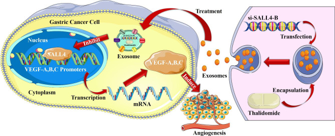
Background: Spalt-like protein 4 (SALL4) is a stemness-related transcription factor whose abnormal re-expression contributes to cancer initiation and progression. However, the role of SALL4 in cancer angiogenesis remains unknown. Methods: Analyses of clinical specimens via TCGA datasets were performed to determine the expression level and clinical significance of SALL4 in STAD (Stomach Adenocarcinoma). SALL4 knockdown, knockout, and overexpression were achieved by siRNA, CRISPR/Cas9, and plasmid transfection. The effects of conditioned medium (CM) from SALL4 knockdown or overexpression of gastric cancer cells on endothelial cell proliferation, migration, and tube formation were investigated by CCK-8 assay, transwell migration assay, and tube formation assay. The regulation of VEGF gene expression by SALL4 was studied by qRT-PCR, western blot, chromatin immunoprecipitation (ChIP) assay, and electrophoretic mobility shift assay (EMSA). Engineered exosomes from 293T cells loaded with si-SALL4-B and thalidomide were produced to test their therapeutic effect on gastric cancer progression. Results: SALL4 expression was increased in STAD and positively correlated with tumor progression and poor prognosis. SALL4-B knockdown or knockout decreased while over-expression increased the promotion of human umbilical vein endothelial cells (HUVEC) cell proliferation, migration, and tube formation by gastric cancer cell-derived CM. Further investigation revealed a widespread association of SALL4 with angiogenic gene transcription through the TCGA datasets. Additionally, SALL4-B knockdown reduced, while over-expression enhanced the expression levels of VEGF-A, B, and C genes. The results of ChIP and EMSA assays indicated that SALL4 could directly bind to the promoters of VEGF-A, B, and C genes and activate their transcription, which may be associated with increased histone H3-K79 and H3-K4 modifications in their promoter regions. Furthermore, si-SALL4-B and thalidomide-loaded exosomes could be efficiently uptaken by gastric cancer cells and significantly reduced SALL4-B and Vascular Endothelial Growth Factor (VEGF) expression levels in gastric cancer cells, thus inhibiting the pro-angiogenic role of their derived CM. Conclusion: These findings suggest that SALL4 plays an important role in angiogenesis by transcriptionally regulating VEGF expression. Co-delivery of the functional siRNA and anticancer drug via exosomes represents a useful approach to inhibiting cancer angiogenesis by targeting SALL4/VEGF pathway.
Overview
The claims and statistics of this paper haven’t been checked yet.
Run the check — free, no account needed. It reads the open-access text and checks the sources it cites.
Integrity signals
Registry
Cites
Statistics
Figures
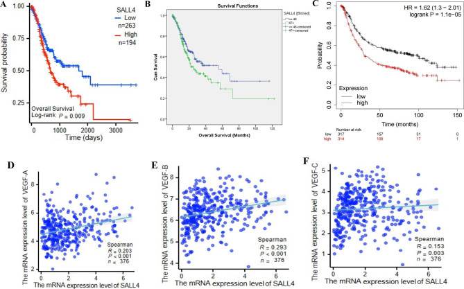
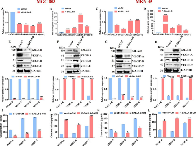
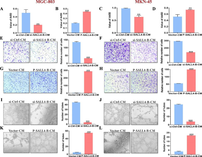
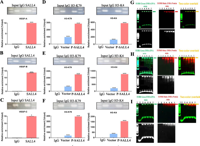
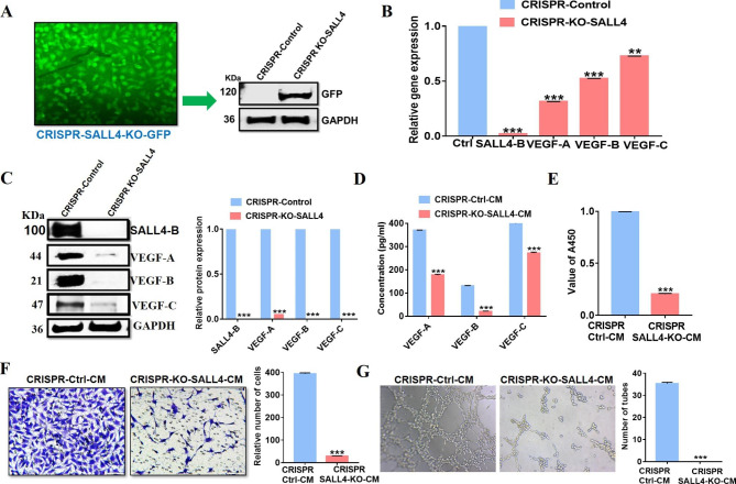
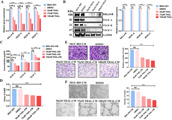
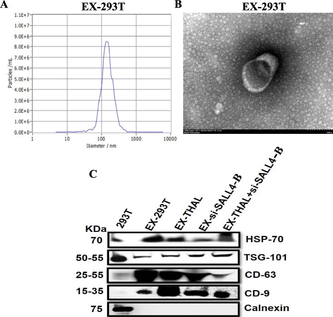
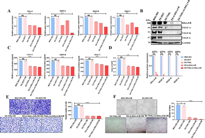
Verification
Claims the paper makes
The claims and statistics of this paper haven’t been checked yet.
Run the check — free, no account needed. It reads the open-access text and checks the sources it cites.
Claims other papers make about this one
Each is checked against this paper’s own text: does it say what the citing paper says it does?
The source backs part of the claim, not all of it — or backs it for a different population, condition or comparison.
In the citing paper
“SALL4 is overexpressed in many cancers with poor prognosis 61-67”
From Tetramerisation governs SALL transcription factor function in development and disease (2025) · cites this paper as [66]
Experimental — accuracy not yet measured
Self-check
Percentages, sums and reported differences re-computed from the paper’s own numbers.
The self-check needs the paper’s full text.
Citations
Cites
Donovan, K. A., An, J., Nowak, R. P., Yuan, J. C., Fink, E. C., Berry, B. C., Ebert, B. L., & Fischer, E. S. (2018) · eLife · doi:10.7554/elife.38430 · Cited by 509 · cited 2×
“angiogenesis Thalidomide has been regarded as a potent inhibitor of SALL4 in numerous studies [21].”
BackgroundWang-Gillam, A., Hubner, R. A., Siveke, J. T., Von Hoff, D. D., Belanger, B., de Jong, F. A., Mirakhur, B., & Chen, L.-T. (2019) · European Journal of Cancer · doi:10.1016/j.ejca.2018.12.007 · Cited by 342
SALL4 mediates teratogenicity as a thalidomide-dependent cereblon substrate
Matyskiela, M. E., Couto, S., Zheng, X., Lu, G., Hui, J., Stamp, K., Drew, C., Ren, Y., Wang, M., Carpenter, A., et al. (2018) · Nature Chemical Biology · doi:10.1038/s41589-018-0129-x · Cited by 338 · cited 1×
“In previous studies, it was discovered that thalidomide causes CRBN-dependent degradation of SALL4 [21, 37].”
BackgroundSelf-citation
Research works printed but not linked: 47
- Bray F, Ferlay J, Soerjomataram I, Siegel L, Torre A, Ahmedin D. GLOBOCAN estimates of incidence and mortality worldwide for 36 cancers in 185 countries. Global cancer statistics 2018 2018. · DOI · Paper page
- Rawla P, Barsouk A. Epidemiology of gastric cancer: global trends, risk factors, and prevention. Gastroenterol Review/PrzeglÄ d Gastroenterologiczny. 2019;14(1):26–38.
- Sitarz R, Skierucha M, Mielko J, Offerhaus G, Maciejewski R, Polkowski W. Gastric cancer: epidemiology, prevention, classification, and treatment. Cancer Manag Res. 2018; 10: 239–48. In. Cancer Manag Res; 2018. · DOI
- Fujita T. Targeted therapy for gastric cancer. Lancet Oncol. 2013;14(6):440–2. · DOI
- Cats A, Jansen EP, van Grieken NC, Sikorska K, Lind P, Nordsmark M, Kranenbarg EM-K, Boot H, Trip AK, Swellengrebel HM. Chemotherapy versus chemoradiotherapy after surgery and preoperative chemotherapy for resectable gastric cancer (CRITICS): an international, open-label, randomized phase 3 trial. Lancet Oncol. 2018;19(5):616–28. · DOI
- Carmeliet P, Jain RK. Angiogenesis in cancer and other diseases. nature 2000, 407(6801):249–257. · DOI · Paper page
- Burstein HJ, Schwartz RS. Molecular origins of cancer. In., vol. 358: Mass Medical Soc; 2008: 527–527. · DOI
- Senger DR, Galli SJ, Dvorak AM, Perruzzi CA, Harvey VS, Dvorak HF. Tumor cells secrete a vascular permeability factor that promotes accumulation of ascites fluid. Science. 1983;219(4587):983–5. · DOI
- Jussila L, Alitalo K. Vascular growth factors and lymphangiogenesis. Physiol Rev. 2002;82(3):673–700. · DOI
- Takahashi H, Shibuya M. The vascular endothelial growth factor (VEGF)/VEGF receptor system and its role under physiological and pathological conditions. Clin Sci. 2005;109(3):227–41. · DOI
- Zhang L, Xu Z, Xu X, Zhang B, Wu H, Wang M, Zhang X, Yang T, Cai J, Yan Y, et al. SALL4, a novel marker for human gastric carcinogenesis and metastasis. Oncogene. 2014;33(48):5491–500. · DOI
- Gonzalez-Roibon N, Katz B, Chaux A, Sharma R, Munari E, Faraj SF, Illei PB, Torbenson M, Netto GJ. Immunohistochemical expression of SALL4 in hepatocellular carcinoma, a potential pitfall in the differential diagnosis of yolk sac tumors. Hum Pathol. 2013;44(7):1293–9. · DOI
- Li A, Jiao Y, Yong KJ, Wang F, Gao C, Yan B, Srivastava S, Lim GSD, Tang P, Yang H, et al. SALL4 is a new target in endometrial cancer. Oncogene. 2015;34(1):63–72. · DOI
- Zhang X, Zhang P, Shao M, Zang X, Zhang J, Mao F, Qian H, Xu W. SALL4 activates TGF-β/SMAD signaling pathway to induce EMT and promote gastric cancer metastasis. Cancer Manage Res. 2018;10:4459–70. · DOI
- Liu L, Zhang J, Yang X, Fang C, Xu H, Xi X. SALL4 as an epithelial-mesenchymal transition and Drug Resistance Inducer through the regulation of c-Myc in Endometrial Cancer. PLoS ONE. 2015;10(9):e0138515–5. · DOI · Paper page
- Kim J, Xu S, Xiong L, Yu L, Fu X, Xu Y. SALL4 promotes glycolysis and chromatin remodeling via modulating HP1α-Glut1 pathway. Oncogene. 2017;36(46):6472–9. · DOI
- Uez N, Lickert H, Kohlhase J, de Angelis MH, Kühn R, Wurst W, Floss T. Sall4 isoforms act during proximal-distal and anterior-posterior axis formation in the mouse embryo. Genesis (New York NY: 2000). 2008;46(9):463–77. · DOI
- Weis SM, Cheresh DA. Tumor angiogenesis: molecular pathways and therapeutic targets. Nat Med. 2011;17(11):1359–70. · DOI
- Yong KJ, Li A, Ou W-B, Hong CKY, Zhao W, Wang F, Tatetsu H, Yan B, Qi L, Fletcher JA, et al. Targeting SALL4 by entinostat in lung cancer. Oncotarget. 2016;7(46):75425–40. · DOI · Paper page
- Jiang G, Liu C-T. Knockdown of SALL4 overcomes cisplatin-resistance through AKT/mTOR signaling in lung cancer cells. Int J Clin Exp Pathol. 2018;11(2):634–41.
- Rao S, Zhen S, Roumiantsev S, McDonald LT, Yuan G-C, Orkin SH. Differential roles of Sall4 isoforms in embryonic stem cell pluripotency. Mol Cell Biol. 2010;30(22):5364–80. · DOI · Paper page
- Tammela T, Enholm B, Alitalo K, Paavonen K. The biology of vascular endothelial growth factors. Cardiovascular Res. 2005;65(3):550–63. · DOI
- Yoshiji H, Harris SR, Thorgeirsson UP. Vascular endothelial growth factor is essential for initial but not continued in vivo growth of human breast carcinoma cells. Cancer Res. 1997;57(18):3924–8.
- Giavazzi R, Sennino B, Coltrini D, Garofalo A, Dossi R, Ronca R, Tosatti MPM, Presta M. Distinct role of fibroblast growth factor-2 and vascular endothelial growth factor on tumor growth and angiogenesis. Am J Pathol. 2003;162(6):1913–26. · DOI
- Sun J, Zhao Z, Zhang W, Tang Q, Yang F, Hu X, Liu C, Song B, Zhang B, Wang H. Spalt-Like protein 4 (SALL4) promotes angiogenesis by activating vascular endothelial growth factor A (VEGFA) signaling. Med Sci Monit. 2020;26:e920851–1.
- Sun J, Tang Q, Gao Y, Zhang W, Zhao Z, Yang F, Hu X, Zhang D, Wang Y, Zhang H. VHL mutation-mediated SALL4 overexpression promotes tumorigenesis and vascularization of clear cell renal cell carcinoma via Akt/GSK-3β signaling. J Experimental Clin Cancer Res. 2020;39:1–17. · DOI
- Chen T, Tsang JY, Su XC, Li P, Sun WQ, Wong IL, Choy KY, Yang Q, Tse GM, Chan TH. SALL4 promotes tumor progression in breast cancer by targeting EMT. Mol Carcinog. 2020;59(10):1209–26. · DOI
- Yuan X, Zhang X, Zhang W, Liang W, Zhang P, Shi H, Zhang B, Shao M, Yan Y, Qian H, et al. SALL4 promotes gastric cancer progression through activating CD44 expression. Oncogenesis. 2016;5(11):e268–8. · DOI
- Yang J, Chai L, Liu F, Fink LM, Lin P, Silberstein LE, Amin HM, Ward DC, Ma Y. Bmi-1 is a target gene for SALL4 in hematopoietic and leukemic cells. Proc Natl Acad Sci USA. 2007;104(25):10494–9. · DOI
- Li A, Yang Y, Gao C, Lu J, Jeong H-W, Liu BH, Tang P, Yao X, Neuberg D, Huang G, et al. A SALL4/MLL/HOXA9 pathway in murine and human myeloid leukemogenesis. J Clin Invest. 2013;123(10):4195–207. · DOI
- Yang L, Liu L, Gao H, Pinnamaneni JP, Sanagasetti D, Singh VP, Wang K, Mathison M, Zhang Q, Chen F, et al. The stem cell factor SALL4 is an essential transcriptional regulator in mixed lineage leukemia-rearranged leukemogenesis. J Hematol Oncol. 2017;10(1):159–9. · DOI
- Liu Y-C, Kwon J, Fabiani E, Xiao Z, Liu YV, Follo MY, Liu J, Huang H, Gao C, Liu J. Demethylation and up-regulation of an oncogene after hypomethylating therapy. N Engl J Med. 2022;386(21):1998–2010. · DOI
- Ito T, Ando H, Suzuki T, Ogura T, Hotta K, Imamura Y, Yamaguchi Y, Handa H. Identification of a primary target of thalidomide teratogenicity. Science. 2010;327(5971):1345–50. · DOI · Paper page
- Sampaio EP, Sarno EN, Galilly R, Cohn ZA, Kaplan G. Thalidomide selectively inhibits tumor necrosis factor-alpha production by stimulated human monocytes. J Exp Med. 1991;173(3):699–703. · DOI
- D’Amato RJ, Loughnan MS, Flynn E, Folkman J. Thalidomide is an inhibitor of angiogenesis. Proceedings of the National Academy of Sciences 1994, 91(9):4082–4085. · DOI · Paper page
- Komorowski J, Jerczyńska H, Siejka A, Barańska P, Ławnicka H, Pawłowska Z, Stępień H. Effect of thalidomide affecting VEGF secretion, cell migration, adhesion and capillary tube formation of human endothelial EA. hy 926 cells. Life Sci. 2006;78(22):2558–63. · DOI
- Zheng W, Fu L. Effect of thalidomide combined with TP chemotherapy on serum VEGF and NRP-1 levels advanced esophageal cancer patients. Am J Translational Res. 2021;13(9):10809.
- Zhang X, Luo H. Effects of thalidomide on growth and VEGF-A expression in SW480 colon cancer cells. Oncol Lett. 2018;15(3):3313–20.
- Tong C, Liu H, Chen R, Zhu F. The effect of TACE in combination with thalidomide-mediated adjuvant therapy on the levels of VEGF and bFGF in patients with hepatocellular carcinoma. Am J Translational Res. 2021;13(5):5575.
- Fenton OS, Olafson KN, Pillai PS, Mitchell MJ, Langer R. Advances in biomaterials for drug delivery. Adv Mater. 2018;30(29):1705328. · DOI
- Kim MS, Haney MJ, Zhao Y, Mahajan V, Deygen I, Klyachko NL, Inskoe E, Piroyan A, Sokolsky M, Okolie O. Development of exosome-encapsulated paclitaxel to overcome MDR in cancer cells. Nanomed Nanotechnol Biol Med. 2016;12(3):655–64. · DOI · Paper page
- Vader P, Mol EA, Pasterkamp G, Schiffelers RM. Extracellular vesicles for drug delivery. Adv Drug Deliv Rev. 2016;106:148–56. · DOI · Paper page
- Tran T-H, Mattheolabakis G, Aldawsari H, Amiji M. Exosomes as nanocarriers for immunotherapy of cancer and inflammatory diseases. Clin Immunol. 2015;160(1):46–58. · DOI
- Liang G, Zhu Y, Ali DJ, Tian T, Xu H, Si K, Sun B, Chen B, Xiao Z. Engineered exosomes for targeted co-delivery of miR-21 inhibitor and chemotherapeutics to reverse drug resistance in colon cancer. J Nanobiotechnol. 2020;18(1):1–15. · DOI
- Shan S, Chen J, Sun Y, Wang Y, Xia B, Tan H, Pan C, Gu G, Zhong J, Qing G. Functionalized macrophage exosomes with panobinostat and PPM1D-siRNA for diffuse intrinsic pontine gliomas therapy. Adv Sci 2022:2200353. · DOI
- Ma Y-S, Liu J-B, Lin L, Zhang H, Wu J-J, Shi Y, Jia C-Y, Zhang D-D, Yu F, Wang H-M. Exosomal microRNA-15a from mesenchymal stem cells impedes hepatocellular carcinoma progression via downregulation of SALL4. Cell death discovery. 2021;7(1):224. · DOI
- Yin C, Han Q, Xu D, Zheng B, Zhao X, Zhang J. SALL4-mediated upregulation of exosomal miR-146a-5p drives T-cell exhaustion by M2 tumor-associated macrophages in HCC. Oncoimmunology. 2019;8(7):1601479–9. · DOI
Cited by
Sorted by citations
Wu, X., Meng, Y., Yao, Z., Lin, X., Hu, M., Cai, S., Gao, S., & Zhang, H. (2025) · Pharmacological Research · doi:10.1016/j.phrs.2025.107751 · Cited by 28
Cullin-RING Ligase 4 in Cancer: Structure, Functions, and Mechanisms
Cheng, J., Bin, X., & Tang, Z. (2024) · Biochimica et Biophysica Acta (BBA) - Reviews on Cancer · doi:10.1016/j.bbcan.2024.189169 · Cited by 18
Natural endogenous material-based vehicles for delivery of macromolecular drugs.
Lu, A., Du, K., Wang, M., Zhu, Z., Lei, L., & Shi, Y. (2024) · Chinese Journal of Natural Medicines · doi:10.1016/s1875-5364(24)60742-9 · Cited by 10
Self-citationLuo, H., Wang, K., Zhang, Y., Li, T., Jia, X., Feng, R., Zhao, R., Yu, R., Hu, L., Zhang, M., et al. (2025) · Journal of Controlled Release · doi:10.1016/j.jconrel.2025.114404 · Cited by 9
Self-citationSALL4 in gastrointestinal tract cancers: upstream and downstream regulatory mechanisms
Wang, T., Jin, Y., Wang, M., Chen, B., Sun, J., Zhang, J., Yang, H., Deng, X., Cao, X., Wang, L., et al. (2024) · Molecular Medicine · doi:10.1186/s10020-024-00812-z · Cited by 9
“Overexpression of SALL4B increases VEGF-A, − B, and − C gene expression, while SALL4B knockdown reduces their expression (Abouel-nazar et al. 2023a).”
BackgroundInfluentialSelf-citation
Metadata
- Authors
- Abouelnazar, F. A., Zhang, X., Zhang, J., Wang, M., Yu, D., Zang, X., Zhang, J., Li, Y., Xu, J., Yang, Q., Zhou, Y., Tang, H., Wang, Y., Gu, J., & Zhang, X.
- Affiliations
- Jiangsu University (CN)
- Nantong University (CN)
- Corresponding author
- Gu, Jianmei
- Zhang, Xu
- Venue
- Cancer Cell International
- Published
- 2023-07-31
- Volume
- 23
- Issue
- 1
- Pages
- 149-149
- ISSN
- 1475-2867 (ISSN-L)
- PMC
- PMC10388482
- OpenAlex
- W4385425904
- Cited by
- 24
- References
- 52
- Citations by year
- 2026: 4 · 2025: 11 · 2024: 8 · 2023: 1
- FWCI
- 2.13
- Type
- journal article
- Language
- en
- Open access
- gold
- doi.org
- Locations
- Cancer Cell International (publishedVersion, cc-by)
- PubMed Central (publishedVersion, cc-by)
- PubMed (publishedVersion)
- DOAJ (DOAJ: Directory of Open Access Journals) (submittedVersion, cc-by-sa)
- Funders
- Key Laboratory of Molecular Diagnostics and Precision Medicine for Surgical On-cology in Gansu Province ((2019GSZDSYS01, 2019GSZDSYS02))
- Non-profit Central Research Institute Fund of Chinese Academy of Medical Sciences ((NLDTG2020002))
- Priority Academic Program Development of Jiangsu Higher Education Institutions ((PAPD))
- Nantong Science and Technology Bureau Project ((JC2021092))
- Postgraduate Research & Practice Innovation Program of Jiangsu Province ((KYCX21_3405, KYCX22_3713))
- Topics
- Life Sciences › Biochemistry, Genetics and Molecular Biology › Molecular Biology › Angiogenesis and VEGF in Cancer
- Health Sciences › Medicine › Oncology › Lymphatic System and Diseases
- Health Sciences › Medicine › Oncology › Cancer Cells and Metastasis
- Keywords
- SALL4, gastric cancer, angiogenesis, vascular endothelial growth factor, VEGF-A, VEGF-B, VEGF-C, transcriptional regulation, histone modifications, H3K4, exosomes, thalidomide, siRNA delivery, tumor microenvironment, endothelial cell proliferation and migration, prognosis
About these checks
Registry, retraction, Cites and statistics results are computed, not judged, with one exception: a printed reference that no index lists is read once by a model before it counts as a citation error. Claims are extracted by a model and kept only when their quote appears verbatim in the text; each cited source is then checked, with a model’s help, for whether it says what the claim relies on. Those verdicts can be wrong — read the claim’s quote and the cited source before relying on one.
Statistics and arithmetic are recomputed, not judged.
Confidence bands are shown only for fields calibrated on our annotated test set. None is calibrated yet, so no band is shown.
What counts as a claim ›How we measure confidence ›How we check this ›
Are you an author, or do you see a problem with this page? Request a review
