

The DOI matches its Crossref record; 59 of the 59 references it cites were found in a registry or an index.
The PI3K subunits, P110α and P110β are potential targets for overcoming P-gp and BCRP-mediated MDR in cancer
Open accessAbstract
Background: PI3K/AKT is a vital signaling pathway in humans. Recently, several PI3K/AKT inhibitors were reported to have the ability to reverse cancer multidrug resistance (MDR); however, specific targets in the PI3K/AKT pathways and the mechanisms associated with MDR have not been found because many of the inhibitors have multiple targets within a large candidate protein pool. AKT activation is one presumed mechanism by which MDR develops during cancer treatment. Methods: The effects of inhibiting PI3K 110α and 110β by BAY-1082439 treatment and CRISPR/Cas9 knockout were examined to determine the possible functions of BAY-1082439 and the roles of PI3K 110α and 110β in the reversal of MDR that is mediated by the downregulation of P-gp and BCRP. Inhibition of AKT with GSK-2110183 showed that the downregulation of P-gp and BCRP is independent of generalized AKT inactivation. Immunofluorescence, immunoprecipitation, MTT, flow cytometry and JC-1 staining analyses were conducted to study the reversal of MDR that is mediated by P-gp and BCRP in cancer cells. An ATPase assay and a structural analysis were also used to analyze the potential mechanisms by which BAY-1082439 specifically targets PI3K 110α and 110β and nonspecifically influences P-gp and BCRP. Results: By inhibiting the activation of the PI3K 110α and 110β catalytic subunits through both the administration of BAY-1082439 and the CRISPR/Cas9 deletion of Pik3ca and Pik3cb , the ATP-binding cassette transporters P-gp/ABCB1 and BCRP/ABCG2 were downregulated, thereby reestablishing the drug sensitivity of human epidermoid carcinoma and non-small cell lung cancer (NSCLC) MDR cells. Inhibition of AKT did not reverse the MDR mediated by P-gp or BCRP. The ABC family proteins and AKT may play MDR-enhancing roles independently. Conclusions: The reversal of the dual functions of ABC-transporter-mediated and AKT-activation-enhanced MDR through the inhibition or knockout of PI3K 110α or 110β promises to improve current strategies based on combined drug treatments to overcome MDR challenges.
Overview
The claims and statistics of this paper haven’t been checked yet.
Run the check — free, no account needed. It reads the open-access text and checks the sources it cites.
Integrity signals
Registry
Cites
Statistics
Figures
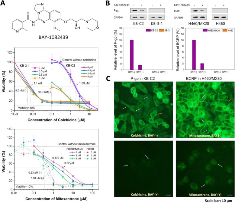
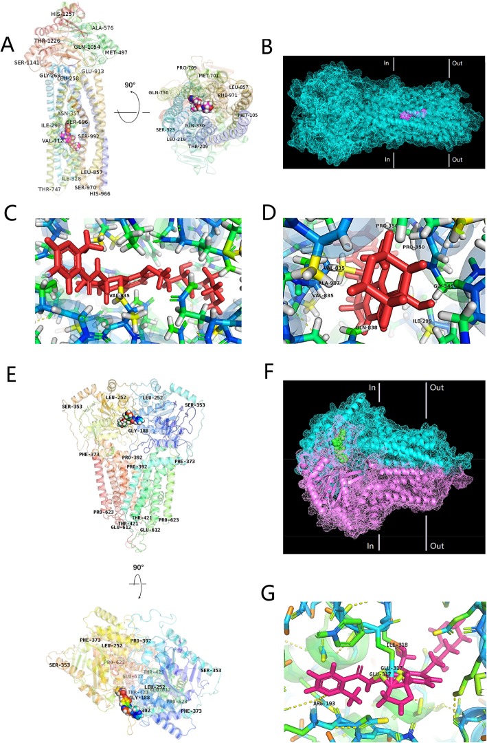
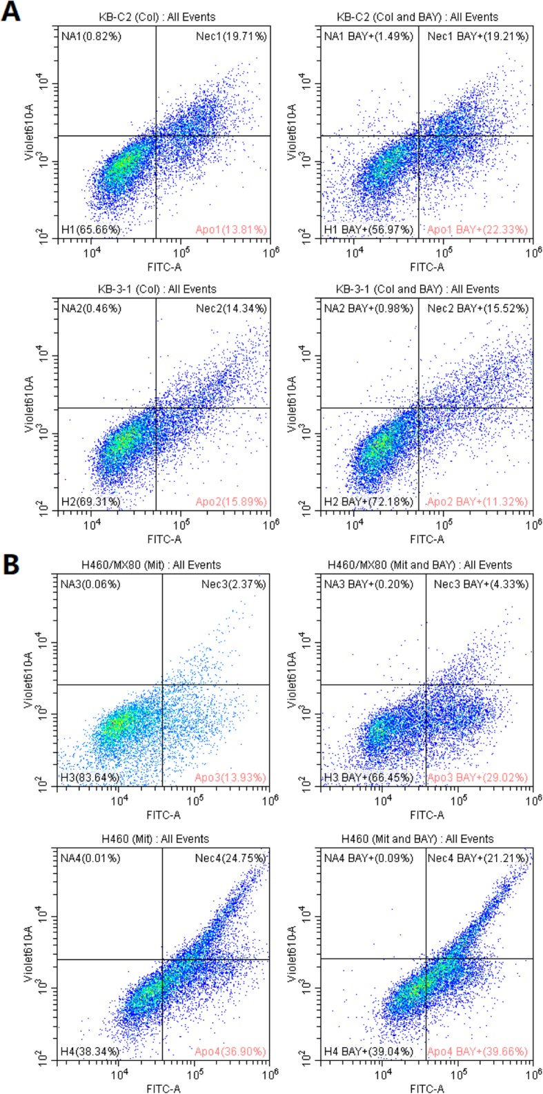
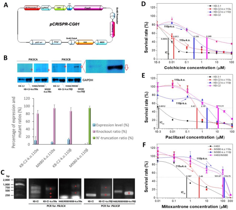
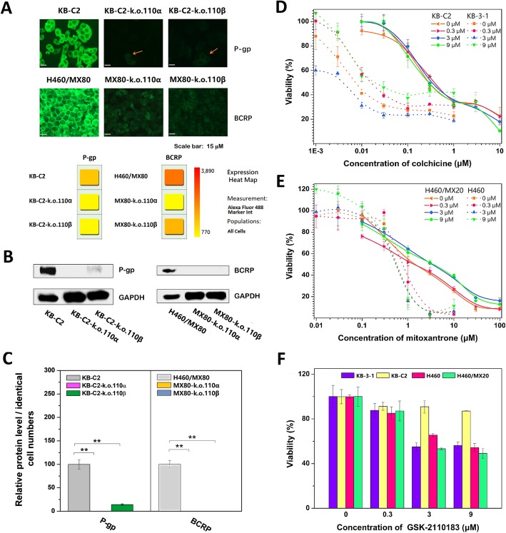
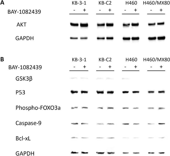
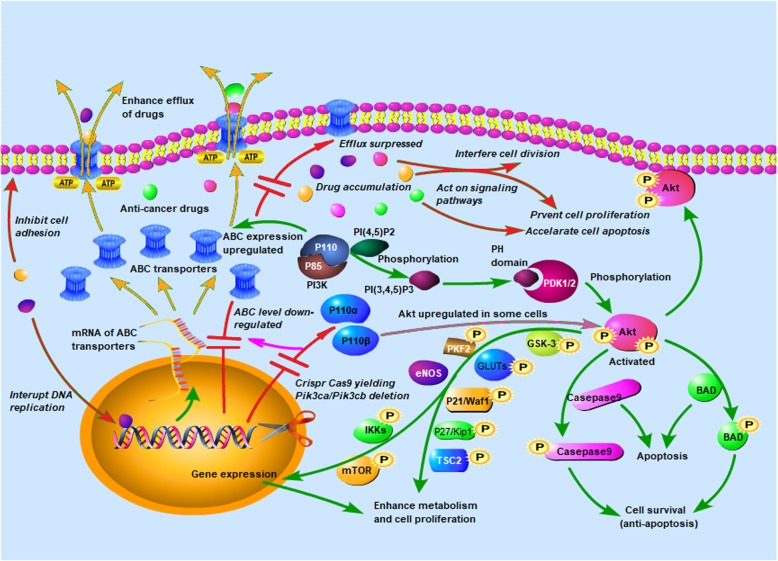
Verification
Claims the paper makes
The claims and statistics of this paper haven’t been checked yet.
Run the check — free, no account needed. It reads the open-access text and checks the sources it cites.
Claims other papers make about this one
Each is checked against this paper’s own text: does it say what the citing paper says it does?
The source backs part of the claim, not all of it — or backs it for a different population, condition or comparison.
In the citing paper
“Multidrug resistance (MDR) has emerged to be a major hindrance in cancer therapy, which contributes to the reduced sensitivity of cancer cells toward chemotherapeutic drugs mainly owing to the over-expression of drug efflux transporters.”
From Nucleolin-Targeting AS1411 Aptamer-Modified Micelle for the Co-Delivery of Doxorubicin and miR-519c to Improve the Therapeutic Efficacy in Hepatocellular Carcinoma Treatment (2021) · cites this paper as [10]
Experimental — accuracy not yet measuredIn the citing paper
“The phenomenon was mainly attributed to the over-expression of ATP-binding cassette (ABC) transporter family in cancer cells, including ABCG1, ABCC1 and ABCG2.8–10”
Self-check
Percentages, sums and reported differences re-computed from the paper’s own numbers.
The self-check needs the paper’s full text.
Citations
Cites
1 retracted
Shi, G., Zhang, H., Yu, Q., Hu, C., & Ji, Y. (2019) · OncoTargets and Therapy · doi:10.2147/ott.s198750 · Cited by 2
Self-citationRetractedPI3K pathway alterations in cancer: variations on a theme
Yuan, T. L., & Cantley, L. C. (2008) · Oncogene · doi:10.1038/onc.2008.245 · Cited by 1,845
Smiley, S. T., Reers, M., Mottola-Hartshorn, C., Lin, M., Chen, A. B., Smith, T. W., Steele, G., & Chen, L. B. (1991) · Proceedings of the National Academy of Sciences · doi:10.1073/pnas.88.9.3671 · Cited by 1,629
Targeting the PI3K pathway in cancer: are we making headway?
Janku, F., Yap, T. A., & Meric-Bernstam, F. (2018) · Nature Reviews Clinical Oncology · doi:10.1038/nrclinonc.2018.28 · Cited by 1,033
Zeng, L., Gupta, P., Chen, Y., Wang, E., Ji, L., Chao, H., & Chen, Z. (2017) · Chemical Society Reviews · doi:10.1039/c7cs00195a · Cited by 1,027
Self-citationKathawala, R. J., Gupta, P., Ashby, C. R., & Chen, Z. (2014) · Drug Resistance Updates · doi:10.1016/j.drup.2014.11.002 · Cited by 798
Self-citationSignalling through phosphoinositide 3-kinases: the lipids take centre stage
Leevers, S. J., Vanhaesebroeck, B., & Waterfield, M. D. (1999) · Current Opinion in Cell Biology · doi:10.1016/s0955-0674(99)80029-5 · Cited by 690
Zhang, Y., Xia, M., Jin, K., Wang, S., Wei, H., Fan, C., Wu, Y., Li, X., Li, X., Li, G., et al. (2018) · Molecular Cancer · doi:10.1186/s12943-018-0796-y · Cited by 593
Self-citationCancer metabolism and the Warburg effect: the role of HIF-1 and PI3K
Courtnay, R., Ngo, D. C., Malik, N., Ververis, K., Tortorella, S. M., & Karagiannis, T. C. (2015) · Molecular Biology Reports · doi:10.1007/s11033-015-3858-x · Cited by 590
PI3K/Akt signaling transduction pathway, erythropoiesis and glycolysis in hypoxia (Review)
Xie, Y., Shi, X., Sheng, K., Han, G., Li, W., Zhao, Q., Jiang, B., Feng, J., Li, J., & Gu, Y. (2018) · Molecular Medicine Reports · doi:10.3892/mmr.2018.9713 · Cited by 505
Cited by
Sorted by citations
Fu, K., Xie, F., Wang, F., & Fu, L. (2022) · Journal of Hematology & Oncology · doi:10.1186/s13045-022-01391-4 · Cited by 432
Activation of PI3K/AKT/mTOR Pathway Causes Drug Resistance in Breast Cancer
Dong, C., Wu, J., Chen, Y., Nie, J., & Chen, C. (2021) · Frontiers in Pharmacology · doi:10.3389/fphar.2021.628690 · Cited by 404
Zou, Y., Zheng, S., Xie, X., Ye, F., Hu, X., Tian, Z., Yang, L., Yang, L., Tang, Y., Tang, Y., et al. (2022) · Nature Communications · doi:10.1038/s41467-022-30217-7 · Cited by 230
Self-citationMechanism of multidrug resistance to chemotherapy mediated by P‑glycoprotein (Review)
Tian, Y., Lei, Y., Wang, Y., Lai, J., Wang, J., & Xia, F. (2023) · International Journal of Oncology · doi:10.3892/ijo.2023.5567 · Cited by 211
Khezri, M. R., Jafari, R., Yousefi, K., & Zolbanin, N. M. (2022) · Experimental and Molecular Pathology · doi:10.1016/j.yexmp.2022.104787 · Cited by 137
Ashrafizadeh, M., Zarrabi, A., Hashemi, F., Zabolian, A., Saleki, H., Bagherian, M., Azami, N., Bejandi, A. K., Hushmandi, K., Ang, H. L., et al. (2020) · Pharmaceutics · doi:10.3390/pharmaceutics12111084 · Cited by 129
CRISPR/Cas9 gene editing: a new approach for overcoming drug resistance in cancer
Vaghari‐Tabari, M., Hassanpour, P., Sadeghsoltani, F., Malakoti, F., Alemi, F., Qujeq, D., Asemi, Z., & Yousefi, B. (2022) · Cellular & Molecular Biology Letters · doi:10.1186/s11658-022-00348-2 · Cited by 137
Gu, J., Huang, W., Wang, X., Zhang, J., Tao, T., Zheng, Y., Liu, S., Yang, J., Chen, Z., Cai, C., et al. (2022) · Molecular Cancer · doi:10.1186/s12943-022-01587-9 · Cited by 113
Self-citationProgress in the studies on the molecular mechanisms associated with multidrug resistance in cancers
Zhang, L., Ye, B., Chen, Z., & Chen, Z. (2022) · Acta Pharmaceutica Sinica B · doi:10.1016/j.apsb.2022.10.002 · Cited by 110
Self-citationWang, D., Yi, H., Geng, S., Jiang, C., Liu, J., Duan, J., Zhang, Z., Shi, J., Song, H., Guo, Z., et al. (2023) · ACS Nano · doi:10.1021/acsnano.3c04002 · Cited by 76
Development of PI3K inhibitors: Advances in clinical trials and new strategies (Review)
Meng, D., He, W. A., Zhang, Y., Liang, Z., Zheng, J., Zhang, X., Zheng, X., Zhan, P., Chen, H., Li, W.-J., et al. (2021) · Pharmacological Research · doi:10.1016/j.phrs.2021.105900 · Cited by 78
Zhang, L., Li, Y., Hu, C., Chen, Y., Chen, Z., Chen, Z., Zhang, J., & Fang, S. (2022) · Molecular Cancer · doi:10.1186/s12943-022-01524-w · Cited by 70
Self-citationRecent advances in targeted drug delivery systems for resistant colorectal cancer
Sharifi-Azad, M., Fathi, M., Cho, W. C., Barzegari, A., Dadashi, H., Dadashpour, M., & Jahanban-Esfahlan, R. (2022) · Cancer Cell International · doi:10.1186/s12935-022-02605-y · Cited by 65
Zhang, S., Peng, X., Li, X., Liu, H., Zhao, B., Elkabets, M., Liu, Y., Wang, W., Wang, R., Zhong, Y., et al. (2021) · Cell Death and Disease · doi:10.1038/s41419-021-03805-6 · Cited by 62
Self-citationMirzaei, S., Gholami, M. H., Mahabady, M. K., Nabavi, N., Zabolian, A., Banihashemi, S. M., Haddadi, A., Entezari, M., Hushmandi, K., Makvandi, P., et al. (2020) · Biomedicine & Pharmacotherapy · doi:10.1016/j.biopha.2020.111077 · Cited by 61
RetractedThe Oncogenic Signaling Disruptor, NDRG1: Molecular and Cellular Mechanisms of Activity
Chekmarev, J., Azad, M. G., & Richardson, D. R. (2021) · Cells · doi:10.3390/cells10092382 · Cited by 54
Gao, L., Meng, F., Yang, Z., Lafuente‐Merchan, M., Fernández, L. M., Cao, Y., Kusamori, K., Nishikawa, M., Itakura, S., Chen, J., et al. (2024) · Biomedicine & Pharmacotherapy · doi:10.1016/j.biopha.2024.117327 · Cited by 59
Di, Y., Jing, X., Hu, K., Wen, X., Ye, L., Zhang, X., Qin, J., Ye, J., Lin, R., Wang, Z., et al. (2022) · Drug Resistance Updates · doi:10.1016/j.drup.2022.100909 · Cited by 53
Erdafitinib Antagonizes ABCB1-Mediated Multidrug Resistance in Cancer Cells
Feng, W., Zhang, M., Wu, Z., Wang, J., Dong, X.-D., Yang, Y., Teng, Q., Chen, X.-Y., Cui, Q., & Yang, D. (2020) · Frontiers in Oncology · doi:10.3389/fonc.2020.00955 · Cited by 51
Self-citationChong, X., Chen, J., Zheng, N., Zhou, Z., Hai, Y., Chen, S., Zhang, Y., Yu, Q., Yu, S., Chen, Z., et al. (2022) · Molecular Cancer · doi:10.1186/s12943-022-01531-x · Cited by 50
Self-citationLiu, R.-M., Xu, P. H., Chen, Q., Feng, S., & Xie, Y. (2020) · Phytomedicine · doi:10.1016/j.phymed.2020.153342 · Cited by 50
Modifications of Plasma Membrane Organization in Cancer Cells for Targeted Therapy
Choromańska, A., Chwiłkowska, A., Kulbacka, J., Baczyńska, D., Rembiałkowska, N., Szewczyk, A., Michel, O., Gajewska-Naryniecka, A., Przystupski, D., & Saczko, J. (2021) · Molecules · doi:10.3390/molecules26071850 · Cited by 53
Peng, X., Zhang, S., Jiao, W., Zhong, Z., Yang, Y., Claret, F. X., Elkabets, M., Wang, F., Wang, R., Zhong, Y., et al. (2021) · Journal of Experimental & Clinical Cancer Research · doi:10.1186/s13046-021-02176-2 · Cited by 48
Self-citationThe PI3K/AKT/mTOR signaling pathway in breast cancer: Review of clinical trials and latest advances
Khorasani, A. B. S., Hafezi, N., Sanaei, M., Jafari‐Raddani, F., Pourbagheri‐Sigaroodi, A., & Bashash, D. (2024) · Cell Biochemistry and Function · doi:10.1002/cbf.3998 · Cited by 47
Ashrafizadeh, M., Zarrabi, A., Orouei, S., Saberifar, S., Salami, S., Hushmandi, K., & Najafi, M. (2020) · Phytotherapy Research · doi:10.1002/ptr.6815 · Cited by 42
Therapeutic resistance in breast cancer cells can result from deregulated EGFR signaling
Steelman, L. S., Chappell, W. H., Akula, S. M., Abrams, S. L., Cocco, L., Manzoli, L., Ratti, S., Martelli, A. M., Montalto, G., Cervello, M., et al. (2020) · Advances in Biological Regulation · doi:10.1016/j.jbior.2020.100758 · Cited by 44
Liang, X., Wang, Y., Shi, H., Dong, M., Han, H., & Li, Q. (2021) · International Journal of Nanomedicine · doi:10.2147/ijn.s304526 · Cited by 41
Mourragui, S. M. C., Loog, M., Vis, D. J., Moore, K., Manjón, A. G., van de Wiel, M. A., Reinders, M. J. T., & Wessels, L. F. A. (2021) · Proceedings of the National Academy of Sciences · doi:10.1073/pnas.2106682118 · Cited by 41
Al-Ouqaili, M. T. S., Ahmad, A., Jwair, N. A., & Al-Marzooq, F. I. (2025) · Frontiers in Cellular and Infection Microbiology · doi:10.3389/fcimb.2025.1588446 · Cited by 47
Yang, Z., Cai, Y., Yang, X., Li, Y., Wu, Q., Yu, Y., Chen, Z., Wei, B., Tian, J., Bao, X., et al. (2023) · Journal of Medicinal Chemistry · doi:10.1021/acs.jmedchem.2c01999 · Cited by 37
Self-citationWu, Z., Yang, Y., Wang, G., Wang, J., Teng, Q., Sun, L., Lei, Z., Lin, L., Chen, Z., & Zou, C. (2020) · Cancer Science · doi:10.1111/cas.14505 · Cited by 37
Self-citationLong noncoding RNAs have pivotal roles in chemoresistance of acute myeloid leukemia
Izadirad, M., Jafari, L., James, A. R., Unfried, J. P., Wu, Z., & Chen, Z. (2021) · Drug Discovery Today · doi:10.1016/j.drudis.2021.03.017 · Cited by 37
Self-citationZhang, S.-L., Ma, L., Zhao, J., You, S., Ma, X., Ye, X., & Liu, T. (2019) · Molecules · doi:10.3390/molecules24183282 · Cited by 35
Yang, X., Wang, Y., Chen, S., Zhang, S., & Cui, C. (2021) · International Journal of Nanomedicine · doi:10.2147/ijn.s332830 · Cited by 35
New emerging targets in osteosarcoma therapy: PTEN and PI3K/Akt crosstalk in carcinogenesis
Sadrkhanloo, M., Paskeh, M. D. A., Hashemi, M., Raesi, R., Bahonar, A., Nakhaee, Z., Entezari, M., Goharrizi, M. A. S. B., Salimimoghadam, S., Ren, J. T., et al. (2023) · Pathology - Research and Practice · doi:10.1016/j.prp.2023.154902 · Cited by 34
Zhou, Y., Chu, P., Wang, Y., Li, N., Gao, Q., Wang, S., Wei, J., Xue, G., Zhao, Y., Jia, H., et al. (2024) · Science Advances · doi:10.1126/sciadv.ado1533 · Cited by 39
Wang, J., Teng, Q., Lei, Z., Ji, N., Cui, Q., Fu, H., Lin, L., Yang, D., Fan, Y., & Chen, Z. (2020) · Frontiers in Cell and Developmental Biology · doi:10.3389/fcell.2020.601400 · Cited by 34
Self-citationYin, L., Yang, Y., Zhu, W., Xian, Y., Han, Z., Huang, H., Peng, L., Zhang, K., & Zhao, Y. (2021) · Frontiers in Oncology · doi:10.3389/fonc.2021.620907 · Cited by 34
Liu, M., Xu, C., Qin, X., Liu, W., Li, D., Jia, H., Gao, X., Wu, Y., Wu, Q., Xu, X., et al. (2022) · Frontiers in Oncology · doi:10.3389/fonc.2022.873649 · Cited by 31
Hou, Y., Pi, C., Feng, X., Wang, Y., Fu, S., Zhang, X., Zhao, L., & Wei, Y. (2020) · Molecular Therapy — Oncolytics · doi:10.1016/j.omto.2020.08.018 · Cited by 29
Comparison of PIK3CA Mutation Prevalence in Breast Cancer Across Predicted Ancestry Populations
Chen, J. W., Murugesan, K., Newberg, J. Y., Sokol, E. S., Savage, H. M., Stout, T. J., Maund, S. L., & Hutchinson, K. E. (2022) · JCO Precision Oncology · doi:10.1200/po.22.00341 · Cited by 26
Xiao, S., Guo, L., Chen, A., Shang, M., Shi, D., Meng, D., Sun, X. Y., Wang, X., Liu, R., Zhao, Y., et al. (2023) · ACS Applied Materials & Interfaces · doi:10.1021/acsami.2c20478 · Cited by 25
Wu, Z., Teng, Q., Yang, Y., Acharekar, N., Wang, J., He, M. Y., Yoganathan, S., Lin, J., Wang, J., & Chen, Z. (2021) · Acta Pharmaceutica Sinica B · doi:10.1016/j.apsb.2021.12.018 · Cited by 25
Self-citationYin, L., Yang, F., Wang, W., Zhang, L., Cao, Z., Shi, H., Pan, K.-H., Wu, L., Xiao, H., & Xing, N. (2025) · Advanced Materials · doi:10.1002/adma.202415322 · Cited by 25
Self-citationCRISPR/Cas9 gene editing: a novel strategy for fighting drug resistance in respiratory disorders
Hussen, B. M., Najmadden, Z. B., Abdullah, S. R., Rasul, M. F., Mustafa, S. A., Ghafouri‐Fard, S., & Taheri, M. N. (2024) · Cell Communication and Signaling · doi:10.1186/s12964-024-01713-8 · Cited by 28
Oh, S., Lee, K. W., Madhi, H., Jeong, J.-W., Park, S., Kim, M.-J., Lee, Y., Han, H.-T., Hwangbo, C., Yoo, J., et al. (2020) · International Journal of Molecular Sciences · doi:10.3390/ijms21051710 · Cited by 23
Yang, Y., Yuan, H., Yang, T., Li, Y., Gao, C., Jiao, T., Cai, Y., & Zhao, S. (2021) · Frontiers in Genetics · doi:10.3389/fgene.2021.691592 · Cited by 23
Self-citationDong, X., Zhang, M., Teng, Q., Lei, Z., Cai, C., Wang, J., Wu, Z., Yang, Y., Chen, X., Guo, H., et al. (2024) · Cancer Letters · doi:10.1016/j.canlet.2024.217309 · Cited by 22
Self-citationFu, H., Wu, Z., Lei, Z., Teng, Q., Yang, Y., Ashby, C. R., Lei, Y., Lian, Y., & Chen, Z. (2022) · Frontiers in Pharmacology · doi:10.3389/fphar.2022.861642 · Cited by 23
Self-citationAbdullah, S., Mukherjee, S., ., S., & Debnath, B. (2024) · Pharmacological Research - Modern Chinese Medicine · doi:10.1016/j.prmcm.2024.100386 · Cited by 22
Zhang, L., Lin, Y., Li, H., Wang, Y., Hu, C., Shangguan, X.-Y., Tang, S., Chen, J., Hu, P., Chen, Z., et al. (2025) · Molecular Cancer · doi:10.1186/s12943-025-02253-6 · Cited by 20
Self-citationSynergistic actions of Alpelisib and Melatonin in breast cancer cell lines with PIK3CA gene mutation
de Godoy, B. L. V., Moschetta‐Pinheiro, M. G., Chuffa, L. G. D. A., Ponde, N. F., Reiter, R. J., Colombo, J., & Zuccari, D. A. P. D. C. (2023) · Life Sciences · doi:10.1016/j.lfs.2023.121708 · Cited by 20
Jiang, S., Zou, J., Dong, J., Shi, H., Chen, J., Li, Y., Duan, X., & Li, W. (2023) · Cell Communication and Signaling · doi:10.1186/s12964-022-01023-x · Cited by 19
Self-citationLin, R., Zhang, L., Ye, B., Wang, Y., Li, Y.-D., Jason, H., Liu, W., Hu, P., Chen, J., Chen, Z., et al. (2023) · Cancer Letters · doi:10.1016/j.canlet.2023.216181 · Cited by 18
Self-citationTargeting kinases that regulate programmed cell death: a new therapeutic strategy for breast cancer
Hong, Y. C., He, J., Deng, D., Liu, Q., Zu, X., & Shen, Y. (2025) · Journal of Translational Medicine · doi:10.1186/s12967-025-06367-9 · Cited by 20
Mahapatra, E., Sengupta, D., Kumar, R., Dehury, B., Das, S., Roy, M., & Mukherjee, S. (2022) · Frontiers in Pharmacology · doi:10.3389/fphar.2022.803114 · Cited by 16
Ma, Y., Zhang, H., Shen, X., Yang, X., Deng, Y. P., Tian, Y., Chen, Z., Pan, Y., Luo, H., Zhong, C., et al. (2024) · Cancer Letters · doi:10.1016/j.canlet.2024.217102 · Cited by 16
Self-citationHuang, L., Liao, Z., Liu, Z., Chen, Y., Huang, T., & Xiao, H. (2022) · Frontiers in Pharmacology · doi:10.3389/fphar.2022.900825 · Cited by 16
Strategies for overcoming ABC transporter-mediated multidrug resistance in colorectal cancer
de Groot, R. A., Reedijk, D., Faucher, Q., Mihaila, S. M., & Masereeuw, R. (2025) · American Journal of Physiology-Cell Physiology · doi:10.1152/ajpcell.00412.2025 · Cited by 19
Narayanan, S., Gujarati, N. A., Wang, J., Wu, Z., Koya, J., Cui, Q., Korlipara, V. L., Ashby, C. R., & Chen, Z. (2021) · International Journal of Molecular Sciences · doi:10.3390/ijms22052463 · Cited by 15
Self-citationZhang, C., Chen, Y., Shi, X., Liu, Y., Zhou, J., Leng, L., Zhang, T., Liu, S., Liu, J., Wang, T., et al. (2026) · Journal of Controlled Release · doi:10.1016/j.jconrel.2026.114654 · Cited by 16
Ma, L., Ai, F., Xiao, H., Wang, F., Shi, L., Bai, X., Zhu, Y., & Ma, W. (2024) · Frontiers in Pharmacology · doi:10.3389/fphar.2024.1367747 · Cited by 16
Guo, L., Shi, H., & Zhu, L. (2021) · Frontiers in Oncology · doi:10.3389/fonc.2021.671382 · Cited by 14
Song, D., Guo, M., Wu, K., Hao, J., Nie, Y., & Fan, D. (2021) · Acta Biochimica et Biophysica Sinica · doi:10.1093/abbs/gmab101 · Cited by 13
Aptamer-functionalized nanomaterials (AFNs) for therapeutic management of hepatocellular carcinoma
Yin, X., Rong, J., Shao, M., Zhang, S., Yin, L., He, Z., & Wang, X. (2024) · Journal of Nanobiotechnology · doi:10.1186/s12951-024-02486-5 · Cited by 17
Zhou, Z., Bai, J., Zhong, S., Zhang, R., Kang, K., Zhang, X., Xu, Y., Zhao, C., & Zhao, M. (2022) · Oxidative Medicine and Cellular Longevity · doi:10.1155/2022/1260161 · Cited by 12
Li, Y., Zhou, H., Huang, Q., Tan, W., Cai, Y., Wang, Z., Zou, J., Li, B., Yoshida, S., & Zhou, Y. (2022) · Frontiers in Immunology · doi:10.3389/fimmu.2022.953812 · Cited by 12
Self-citationTargeting Cardiac Metabolic Pathways: A Role in Ischemic Management
Yehualashet, A. S., Belachew, T. F., Kifle, Z. D., & Abebe, A. M. (2020) · Vascular Health and Risk Management · doi:10.2147/vhrm.s264130 · Cited by 13
Epigenetic regulation of stem cells in lung cancer oncogenesis and therapy resistance
Wu, J., Feng, J., Zhang, Q., He, Y., Xu, C. S., Wang, C., & Li, W. (2023) · Frontiers in Genetics · doi:10.3389/fgene.2023.1120815 · Cited by 13
Zheng, L. T., Chang, R., Liang, B., Wang, Y., Zhu, Y., Jia, Z., Fan, J., Zhang, Z., Du, B., & Kong, D. (2024) · Cancer Drug Resistance · doi:10.20517/cdr.2024.107 · Cited by 11
Ribociclib Inhibits P-gp-Mediated Multidrug Resistance in Human Epidermoid Carcinoma Cells
Zhang, L., Ye, B., Lin, Y., Li, Y.-D., Wang, J., Chen, Z., Ping, F., & Chen, Z.-S. (2022) · Frontiers in Pharmacology · doi:10.3389/fphar.2022.867128 · Cited by 11
Self-citationChang, Y., Fu, Q., Lu, Z., Jin, Q., Jin, T., & Zhang, M. (2024) · Food Science & Nutrition · doi:10.1002/fsn3.4205 · Cited by 10
The Emerging Role of RNA N6-Methyladenosine Modification in Pancreatic Cancer
Hu, X., Lei, X., Guo, J., Fu, W., Sun, W. Y., Lu, Q., Su, W., Xu, Q., & Tu, K. (2022) · Frontiers in Oncology · doi:10.3389/fonc.2022.927640 · Cited by 10
Narayanan, S., Fan, Y., Gujarati, N. A., Teng, Q., Wang, J., Cai, C., Yang, Y., Chintalapati, A. J., Lei, Y., Korlipara, V. L., et al. (2021) · Cancers · doi:10.3390/cancers13184675 · Cited by 10
Self-citationMeng, R., Gao, Y., Peng, Z. Q., Chen, H., Li, Y., Liu, Y., Shen, L., Pan, H., Zheng, L., Cheng, D., et al. (2025) · Molecular Cancer · doi:10.1186/s12943-025-02524-2 · Cited by 9
Self-citationAlves, E., McLeish, E., Blancafort, P., Coudert, J. D., & Gaudieri, S. (2021) · Frontiers in Immunology · doi:10.3389/fimmu.2021.712722 · Cited by 10
[Lung Cancer Stem-like Cells and Drug Resistance].
Pan, Z., Liu, H., & Chen, J. (2022) · PubMed Central · doi:10.3779/j.issn.1009-3419.2022.102.02 · Cited by 9
FDA approved fused-pyrimidines as potential PI3K inhibitors: a computational repurposing approach
Vishwakarma, P., Siddiqui, N. F., Thakur, S., & Jadhav, H. R. (2023) · Journal of Biomolecular Structure and Dynamics · doi:10.1080/07391102.2023.2276315 · Cited by 9
Applications and advancements of CRISPR-Cas in the treatment of lung cancer
Pan, L., Ju, Y., Peng, F., & Luo, J. D. (2023) · Frontiers in Cell and Developmental Biology · doi:10.3389/fcell.2023.1295084 · Cited by 11
Wang, T., Dong, W.-H., Wang, F., Liu, Q., Yang, Y., Guo, P., Li, X., & Wei, B. (2021) · Recent Patents on Anti-Cancer Drug Discovery · doi:10.2174/1574892816666210806154225 · Cited by 9
Self-citationYan, B., Shi, R., Lu, Y.-Y., Fang, D.-D., Ye, M., & Zhou, Q. (2023) · PLoS ONE · doi:10.1371/journal.pone.0279752 · Cited by 9
Cao, J., Zeng, K., Chen, Q., Yang, T., Lu, F., Lin, C., Zhan, J., Ma, W., Zhou, T., Huang, Y., et al. (2024) · Cell Death and Disease · doi:10.1038/s41419-024-06615-8 · Cited by 9
Liu, X., Liu, J., Yan, B., Quan, Z., Wang, X., Ma, Y., Alarfaj, A. A., & Yan, L. (2023) · Environmental Toxicology · doi:10.1002/tox.23981 · Cited by 8
Cen, J., Lu, P., Wang, C., Wu, J., Hu, X., Zhao, H., Li, M., Luo, M., Zhao, S., Li, X., et al. (2025) · Journal of Medicinal Chemistry · doi:10.1021/acs.jmedchem.4c03169 · Cited by 7
Self-citationZha, D., Li, Y., Luo, Y., Liu, Y., Lin, Z., Lin, C., Chen, S., Wu, J., Yu, L., Chen, S., et al. (2022) · RSC Medicinal Chemistry · doi:10.1039/d2md00148a · Cited by 7
Self-citationApplication of CRISPR/Cas9 System to Reverse ABC-Mediated Multidrug Resistance
Wang, W., Liang, Z., Ma, P., Zhao, Q., Dai, M., Zhu, J., Xu, H., Xu, H., Chang, Q., & Zhen, Y. (2021) · Bioconjugate Chemistry · doi:10.1021/acs.bioconjchem.0c00627 · Cited by 7
Predicting clinical drug response from model systems by non-linear subspace-based transfer learningPreprint
Mourragui, S. M. C., Loog, M., Vis, D. J., Moore, K., Manjón, A. G., van de Wiel, M. A., Reinders, M. J. T., & Wessels, L. F. A. (2020) · bioRxiv (Cold Spring Harbor Laboratory) · doi:10.1101/2020.06.29.177139 · Cited by 6
Tumor microenvironment‐responsive drug self‐delivery systems to treat cancer and overcome MDR
Li, L., Xie, Y.-P., Qin, Y., Chu, H.-P., Xie, H., Zang, D., & Liu, T. (2024) · Rare Metals · doi:10.1007/s12598-024-02892-6 · Cited by 6
Garcia, A. R., Mendes, A., Custódia, C., Faria, C. C., Barata, J. T., Malhó, R., Figueira, I., & Brito, M. A. (2023) · Cancers · doi:10.3390/cancers15153973 · Cited by 6
Raza, M., Kumar, N., Nair, U., Luthra, G., Bhattacharyya, U., Jayasundar, S., Jayasundar, R., & Sehrawat, S. (2021) · Molecular and Cellular Biochemistry · doi:10.1007/s11010-021-04149-7 · Cited by 6
Biological significance of extracellular vesicles in innate immune system
Jadamba, B., Park, J., & Lee, H. (2025) · Innate Immunity · doi:10.1177/17534259251355029 · Cited by 5
Sheikhshabani, S. H., Amini‐Farsani, Z., Kazemifard, N., Modarres, P., Farsani, Z. A., Omrani, M. D., & Ghafouri‐Fard, S. (2023) · Cancer Reports · doi:10.1002/cnr2.1884 · Cited by 5
Molecular Mechanisms of Breast Cancer Drug Resistance and CRISPR/Cas9 Strategies to Overcome
Hussen, B. M., Ismael, B. S., Abdulla, S. S., Jamal, N. H., Mustafa, S. A., Najmalddin, Z. B., & Rasul, M. F. (2024) · BioMed Target Journal · doi:10.59786/bmtj.221 · Cited by 4
Huang, Y., Zhu, W., Zhang, J., Zhang, Y., Liu, Y., Wang, G., & Yang, W. (2025) · Biomacromolecules · doi:10.1021/acs.biomac.5c01461 · Cited by 4
Huang, X., Wang, M., Zhu, B., Yu, H., Gao, R., Liu, W., Cheng, J., Hua, G., & Xue, C.-M. (2024) · OncoTargets and Therapy · doi:10.2147/ott.s448636 · Cited by 3
Zhang, M., She, M., Chen, J., Zeng, X.-Q., Xiong, Q.-Q., Cen, Y.-H., Ye, J.-A., Qiu, G., Yang, S.-Y., & Ren, G.-H. (2024) · Biomedicine & Pharmacotherapy · doi:10.1016/j.biopha.2024.117603 · Cited by 3
Liu, B., Lu, Y., Lin, R., Xu, J., Shang, Z., Hou, X., Shao, X., Pan, Z., Yu, T., & Feng, W. (2025) · BMC Cancer · doi:10.1186/s12885-024-13371-7 · Cited by 3
Wang, T., Sang, N., Ménard‐Moyon, C., Miyako, E., & Bianco, A. (2026) · Journal of Controlled Release · doi:10.1016/j.jconrel.2026.114954 · Cited by 3
Liu, J., Luo, D., Chen, X., Liu, J., Chen, J., Shi, M., Dong, H., Xu, Y., Wang, X., Yu, Z., et al. (2024) · Translational Oncology · doi:10.1016/j.tranon.2024.102199 · Cited by 3
Optimization of IgG1 immune complexes to stimulate cytokine production in innate cells
Deb, A., Lott, K., Miceli, A., & Kaplan, B. L. F. (2025) · Journal of Immunological Methods · doi:10.1016/j.jim.2025.113851 · Cited by 3
Metadata
- Authors
- Zhang, L., Li, Y., Wang, Q., Chen, Z., Li, X., Wu, Z., Hu, C., Liao, D., Zhang, W., & Chen, Z.-S.
- Affiliations
- St. John's University (US)
- Chinese Academy of Sciences (CN)
- Fujian Agriculture and Forestry University (CN)
- Weifang Medical University (CN)
- Corresponding author
- Chen, Zhuo
- Chen, Zhe-Sheng
- Venue
- Molecular Cancer
- Published
- 2020-01-17
- Volume
- 19
- Issue
- 1
- Pages
- 10-10
- ISSN
- 1476-4598 (ISSN-L)
- PMC
- PMC6966863
- OpenAlex
- W3004667471
- Cited by
- 152
- References
- 59
- Citations by year
- 2026: 12 · 2025: 20 · 2024: 23 · 2023: 16 · 2022: 45 · 2021: 21 · 2020: 13
- FWCI
- 8.58
- Type
- journal article
- Language
- en
- Open access
- gold
- doi.org
- Locations
- Molecular Cancer (publishedVersion, cc-by)
- PubMed Central (publishedVersion, cc-by)
- PubMed (publishedVersion)
- DOAJ (DOAJ: Directory of Open Access Journals) (submittedVersion, cc-by-sa)
- Funders
- National Institute of Health - USA (1R15GM116043−01)
- China Scholarship Council
- St. John’s University Research Seed Grant (579−1110−7002)
- National Nature Science Foundation of China (21877113, 31300650, 31771723)
- Fujian Provincial Department of Science and Technology (2013N0039, 2017J01456)
- Topics
- Health Sciences › Medicine › Oncology › Drug Transport and Resistance Mechanisms
- Life Sciences › Biochemistry, Genetics and Molecular Biology › Molecular Biology › PI3K/AKT/mTOR signaling in cancer
- Health Sciences › Medicine › Microbiology (medical) › Antibiotic Resistance in Bacteria
- Keywords
- PI3K p110α, PI3K p110β, multidrug resistance, P-glycoprotein, ABCG2, ABC transporters, AKT inhibition, drug resistance reversal
- MeSH terms
- ATP Binding Cassette Transporter, Subfamily G, Member 2, Carcinoma, Squamous Cell, Humans, Neoplasm Proteins, Tumor Cells, Cultured, Biomarkers, Tumor, Gene Expression Regulation, Neoplastic, Apoptosis, Drug Resistance, Multiple, ATP Binding Cassette Transporter, Subfamily B, Drug Resistance, Neoplasm, Protein Kinase Inhibitors, Cell Proliferation, Class I Phosphatidylinositol 3-Kinases
About these checks
Registry, retraction, Cites and statistics results are computed, not judged, with one exception: a printed reference that no index lists is read once by a model before it counts as a citation error. Claims are extracted by a model and kept only when their quote appears verbatim in the text; each cited source is then checked, with a model’s help, for whether it says what the claim relies on. Those verdicts can be wrong — read the claim’s quote and the cited source before relying on one.
Statistics and arithmetic are recomputed, not judged.
Confidence bands are shown only for fields calibrated on our annotated test set. None is calibrated yet, so no band is shown.
What counts as a claim ›How we measure confidence ›How we check this ›
Are you an author, or do you see a problem with this page? Request a review
