

The DOI matches its Crossref record; 46 of the 46 references it cites were found in a registry or an index.
Blood–brain barrier dysfunction developed during normal aging is associated with inflammation and loss of tight junctions but not with leukocyte recruitment
Open accessAbstract
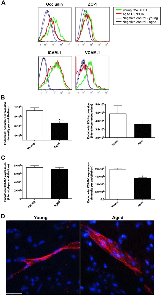
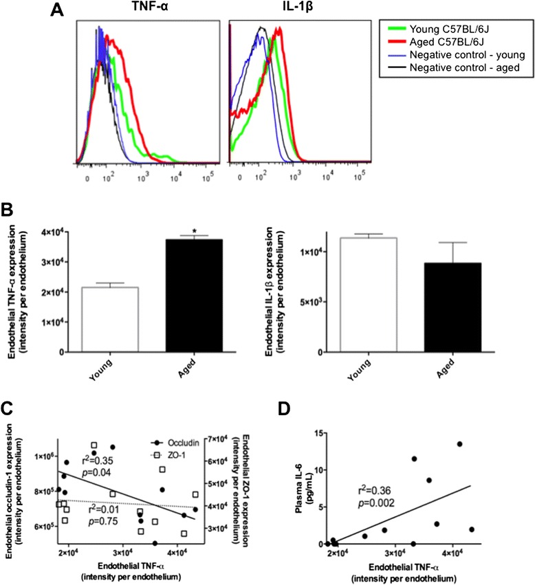
BACKGROUND: Functional loss of blood-brain barrier (BBB) is suggested to be pivotal to pathogenesis and pathology of vascular-based neurodegenerative disorders such as Alzheimer's disease. We recently reported in wild-type mice maintained on standard diets, progressive deterioration of capillary function with aging concomitant with heightened neuroinflammation. However, the mice used in this study were relatively young (12 months of age) and potential mechanisms for loss of capillary integrity were not investigated per se. The current study therefore extended the previous finding to investigate the effect of aging on BBB integrity in aged mice at 24 months and its potential underlying molecular mechanisms. RESULTS: Immunomicroscopy analyses confirmed significantly increased capillary permeability with heightened neuroinflammation in naturally aged 24-month old mice compared to young control at 3 months of age. Aged mice showed significant attenuation in the expression of BBB tight junction proteins, occludin-1 and to lesser extent ZO-1 compared to young mice. In addition, TNF-α in cerebral endothelial cells of aged mice was significantly elevated compared to controls and this was associated with heightened peripheral inflammation. The expression of ICAM-1 and VCAM-1 remained unelevated, and no sign of leukocyte recruitment was observed in aged mice. CONCLUSION: The BBB breakdown that occurs during ordinary aging is associated with inflammation and disruption of tight junction complex assembly but not through leukocyte trafficking.
Overview
The claims and statistics of this paper haven’t been checked yet.
Free: it reads the open-access text and checks the first 5 claims that cite a source we can check. Create a free account to check every claim.
Integrity signals
Registry
Cites
Statistics
Figures

Verification
Claims the paper makes
The claims and statistics of this paper haven’t been checked yet.
Free: it reads the open-access text and checks the first 5 claims that cite a source we can check. Create a free account to check every claim.
Claims other papers make about this one
Each is checked against this paper’s own text: does it say what the citing paper says it does?
The source backs part of the claim, not all of it — or backs it for a different population, condition or comparison.
In the citing paper
“BBB permeability becomes less selective with aging because of a loss of tight junctions and a shift from tightly regulated receptor-mediated transcytosis toward nonspecific transcytosis of plasma proteins (136, 137).”
From Connecting aging biology and inflammation in the omics era (2022) · cites this paper as [136]
Experimental — accuracy not yet measuredThe source backs part of the claim, not all of it — or backs it for a different population, condition or comparison.
In the citing paper
“Circulating TNF-α, IL-1β, and IL-6 bind to receptors in endothelial cells and induce the expression of cellular adhesion proteins (e.g., VCAM1) that further promote inflammation by triggering NF-κB signaling (Figure 3) (136, 140, 141).”
From Connecting aging biology and inflammation in the omics era (2022) · cites this paper as [136]
Experimental — accuracy not yet measured
Self-check
Percentages, sums and reported differences re-computed from the paper’s own numbers.
The self-check needs the paper’s full text.
Citations
Cites
The Blood-Brain Barrier in Health and Chronic Neurodegenerative Disorders
Zlokovic, B. V. (2008) · Neuron · doi:10.1016/j.neuron.2008.01.003 · Cited by 3,280
Neurovascular pathways to neurodegeneration in Alzheimer’s disease and other disorders
Zlokovic, B. V. (2011) · Nature Reviews Neuroscience · doi:10.1038/nrn3114 · Cited by 2,969
Inflammatory mediators in the elderly
Krabbe, K. S., Pedersen, M., & Bruunsgaard, H. (2004) · Experimental Gerontology · doi:10.1016/j.exger.2004.01.009 · Cited by 928
Blood–brain barrier: Ageing and microvascular disease – systematic review and meta-analysis
Farrall, A. J., & Wardlaw, J. M. (2009) · Neurobiology of Aging · doi:10.1016/j.neurobiolaging.2007.07.015 · Cited by 882 · cited 1×
“Several studies have now shown that the function and structure of BBB deteriorate during aging and in the absence of comorbidities [4,5].”
BackgroundThe influence of cytokines on the integrity of the blood-brain barrier in vitro
de Vries, H. E., Blom-Roosemalen, M. C. M., Oosten, M. V., de Boer, A. G., van Berkel, T. J. C., Breimer, D. D., & Kuiper, J. (1996) · Journal of Neuroimmunology · doi:10.1016/0165-5728(95)00148-4 · Cited by 665
Blood brain barrier: ageing and microvascular disease-systematic review and meta-analysis
Farrall, A. J., & Wardlaw, J. M. (2009) · Journal of the Neurological Sciences · doi:10.1016/j.jns.2009.02.089 · Cited by 639
Blood–brain-barriers in aging and in Alzheimer’s disease
Marques, F., Sousa, J. C., Sousa, N., & Palha, J. A. (2013) · Molecular Neurodegeneration · doi:10.1186/1750-1326-8-38 · Cited by 274
The adhesion molecule ICAM-1 and its regulation in relation with the blood–brain barrier
Dietrich, J.-B. (2002) · Journal of Neuroimmunology · doi:10.1016/s0165-5728(02)00114-5 · Cited by 266
Salvioli, S., Capri, M., Valensin, S., Tieri, P., Monti, D., Ottaviani, E., & Franceschi, C. (2006) · Current Pharmaceutical Design · doi:10.2174/138161206777947470 · Cited by 242
Blood-brain barrier alterations in ageing and dementia
Popescu, B. O., Toescu, E. C., Popescu, L. M., Bajenaru, O., Muresanu, D. F., Schultzberg, M., & Bogdanovic, N. (2009) · Journal of the Neurological Sciences · doi:10.1016/j.jns.2009.02.321 · Cited by 236
Cited by
Neuroinflammation and depression: A review
Troubat, R., Barone, P., Leman, S., Desmidt, T., Cressant, A., Atanasova, B., Brizard, B., El Hage, W., Surget, A., Belzung, C., et al. (2021) · European Journal of Neuroscience · doi:10.1111/ejn.14720 · Cited by 1,107
Blood-brain barrier dysfunction and recovery after ischemic stroke
Jiang, X., Andjelkovic, A. V., Zhu, L., Yang, T., Bennett, M. V. L., Chen, J., Keep, R. F., & Shi, Y. (2018) · Progress in Neurobiology · doi:10.1016/j.pneurobio.2017.10.001 · Cited by 936
The Gut Microbiota and Alzheimer’s Disease
Jiang, C., Li, G., Huang, P., Liu, Z., & Zhao, B. (2017) · Journal of Alzheimer's Disease · doi:10.3233/jad-161141 · Cited by 852
The blood-brain barrier in aging and neurodegeneration
Knox, E. G., Aburto, M. R., Clarke, G., Cryan, J. F., & O’Driscoll, C. M. (2022) · Molecular Psychiatry · doi:10.1038/s41380-022-01511-z · Cited by 738
“Aged mice also have hallmarks of BBB breakdown compared to young mice with increased leakage of IgG into the brain parenchyma, a reduction of the tight junction protein occludin, and a reduction in pericyte coverage and pericyte induced expression of genes in the endothelium [54, 90, 91].”
BackgroundZhao, Z., Ning, J., Bao, X.-Q., Shang, M., Ma, J., Li, G., & Zhang, D. (2021) · Microbiome · doi:10.1186/s40168-021-01107-9 · Cited by 616
“As a result, neuroinflammation and cell death of dopaminergic neurons can occur in the SN [20, 24, 25].”
Background
Metadata
- Authors
- Elahy, M., Jackaman, C., Mamo, J. C., Lam, V., Dhaliwal, S. S., Giles, C., Nelson, D., & Takechi, R.
- Affiliations
- Curtin University (AU)
- Corresponding author
- Takechi, Ryusuke
- ORCID
- Venue
- Immunity & Ageing
- Published
- 2015-03-07
- Volume
- 12
- Issue
- 1
- Pages
- 2-2
- ISSN
- 1742-4933 (ISSN-L)
- PMC
- PMC4362825
- OpenAlex
- W2099448948
- Cited by
- 300
- References
- 46
- Citations by year
- 2026: 19 · 2025: 23 · 2024: 27 · 2023: 35 · 2022: 27 · 2021: 38 · 2020: 23 · 2019: 25 · 2018: 29 · 2017: 28 · 2016: 20 · 2015: 6
- FWCI
- 14.79
- Type
- journal article
- Language
- en
- Open access
- gold
- doi.org
- Locations
- Immunity & Ageing (publishedVersion, cc-by)
- PubMed Central (publishedVersion, cc-by)
- PubMed (publishedVersion)
- eSpace (Curtin University) (submittedVersion, cc-by)
- Minerva Access (University of Melbourne) (submittedVersion, cc-by)
- Funders
- Curtin University of Technology
- Medical Research Council
- National Health and Medical Research Council
- Topics
- Life Sciences › Neuroscience › Neurology › Barrier Structure and Function Studies
- Life Sciences › Neuroscience › Neurology › Neuroinflammation and Neurodegeneration Mechanisms
- Health Sciences › Medicine › Oncology › Drug Transport and Resistance Mechanisms
- Keywords
- blood-brain barrier dysfunction, aging, neuroinflammation, tight junctions, occludin-1, ZO-1, capillary permeability, TNF-α, brain endothelial cells, peripheral inflammation, leukocyte recruitment, ICAM-1, VCAM-1, Alzheimer's disease
About these checks
Registry, retraction, Cites and statistics results are computed, not judged, with one exception: a printed reference that no index lists is read once by a model before it counts as a citation error. Claims are extracted by a model and kept only when their quote appears verbatim in the text; each cited source is then checked, with a model’s help, for whether it says what the claim relies on. Those verdicts can be wrong — read the claim’s quote and the cited source before relying on one.
Statistics and arithmetic are recomputed, not judged.
Confidence bands are shown only for fields calibrated on our annotated test set. None is calibrated yet, so no band is shown.
What counts as a claim ›How we measure confidence ›How we check this ›
Are you an author, or do you see a problem with this page? Request a review
How to cite
Cite CoChat for the checks on this page, and the paper for its own findings.
- This page
- CoChat. (2026). Citation check: Blood–brain barrier dysfunction developed during normal aging is associated with inflammation and loss of tight junctions but not with leukocyte recruitment. https://app.cochat.ai/papers/10.1186/s12979-015-0029-9
- The paper
- Elahy, M., Jackaman, C., Mamo, J. C., Lam, V., Dhaliwal, S. S., Giles, C., Nelson, D., & Takechi, R. (2015). Blood–brain barrier dysfunction developed during normal aging is associated with inflammation and loss of tight junctions but not with leukocyte recruitment. Immunity & Ageing. https://doi.org/10.1186/s12979-015-0029-9
Data sources
Bibliographic data: Semantic Scholar, OpenAlex, Crossref.
Contains information from the Semantic Scholar Academic Graph Datasets, made available under the ODC Attribution License (ODC-BY 1.0).
Semantic Scholar’s data is described in: Kinney, R., et al. (2023). The Semantic Scholar Open Data Platform. arXiv:2301.10140.
