

The DOI matches its Crossref record; 170 of the 172 references it cites were found in a registry or an index, and 2 are not checked yet.
Endo-lysosomal dysregulations and late-onset Alzheimer’s disease: impact of genetic risk factors
Open accessAbstract
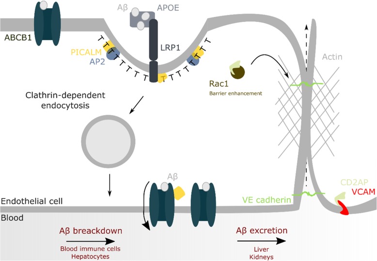
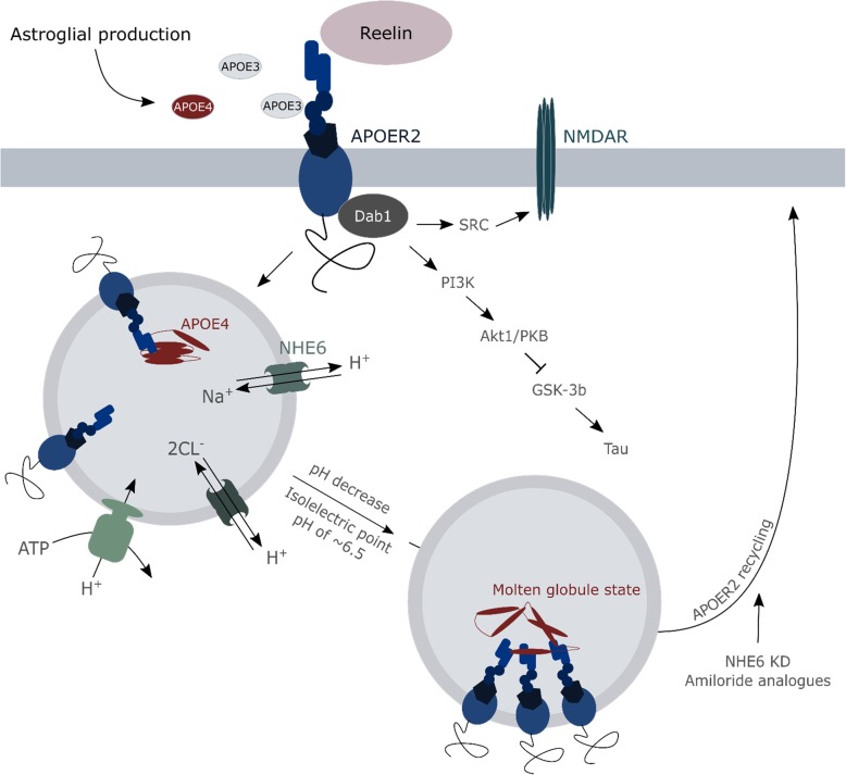
Increasing evidence supports that cellular dysregulations in the degradative routes contribute to the initiation and progression of neurodegenerative diseases, including Alzheimer's disease. Autophagy and endolysosomal homeostasis need to be maintained throughout life as they are major cellular mechanisms involved in both the production of toxic amyloid peptides and the clearance of misfolded or aggregated proteins. As such, alterations in endolysosomal and autophagic flux, as a measure of degradation activity in these routes or compartments, may directly impact as well on disease-related mechanisms such as amyloid-β clearance through the blood-brain-barrier and the interneuronal spreading of amyloid-β and/or Tau seeds, affecting synaptic function, plasticity and metabolism. The emerging of several genetic risk factors for late-onset Alzheimer's disease that are functionally related to endocytic transport regulation, including cholesterol metabolism and clearance, supports the notion that in particular the autophagy/lysosomal flux might become more vulnerable during ageing thereby contributing to disease onset. In this review we discuss our current knowledge of the risk genes APOE4, BIN1, CD2AP, PICALM, PLD3 and TREM2 and their impact on endolysosomal (dys)regulations in the light of late-onset Alzheimer's disease pathology.
Overview
The claims and statistics of this paper haven’t been checked yet.
Run the check — free, no account needed. It reads the open-access text and checks the sources it cites.
Integrity signals
Registry
Cites
Statistics
Figures
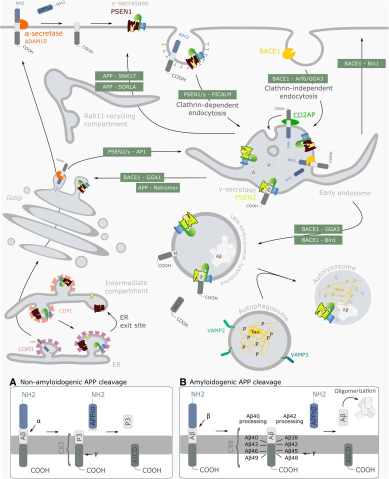
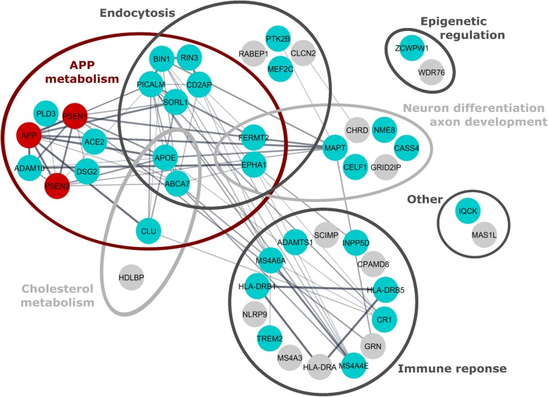
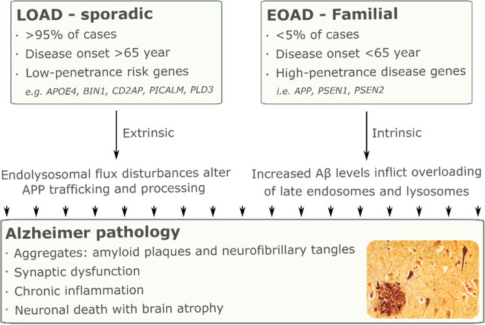
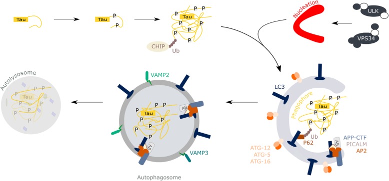
Verification
Claims the paper makes
The claims and statistics of this paper haven’t been checked yet.
Run the check — free, no account needed. It reads the open-access text and checks the sources it cites.
Claims other papers make about this one
Each is checked against this paper’s own text: does it say what the citing paper says it does?
The cited source says what the claim attributes to it.
In the citing paper
“multiple genetic risk alleles associated with AD are thought to be active in this pathway [34].”
From Microglia specific deletion of miR-155 in Alzheimer’s disease mouse models reduces amyloid-β pathology but causes hyperexcitability and seizures (2023) · cites this paper as [34]
The cited source says what the claim attributes to it.
In the citing paper
“Aβ results from the proteolysis of APP, a type I transmembrane protein targeted first at the plasma membrane, then rapidly endocytosed to endosomes to be metabolized to Aβ or subsequently sent to the lysosomal compartment for degradation (Wang X. et al., 2017; Van Acker et al., 2019).”
From Emerging Alternative Proteinases in APP Metabolism and Alzheimer’s Disease Pathogenesis: A Focus on MT1-MMP and MT5-MMP (2019) · cites this paper as Van Acker et al., 2019
Self-check
Percentages, sums and reported differences re-computed from the paper’s own numbers.
The self-check needs the paper’s full text.
Citations
Cites
Meta-Analysis of 74,046 Individuals Identifies 11 New Susceptibility Loci for Alzheimer’s Disease
(EADI), E. A. D. I., Lambert, J.-C., (GERAD), G. A. E. R. I. A. D., Alzheimer's Disease Genetic Consortium (ADGC), (CHARGE), C. F. H. A. A. R. I. G. E., Ibrahim-Verbaas, C. A., Harold, D., Naj, A. C., Sims, R., Bellenguez, C., et al. (2013) · Nature Genetics · doi:10.1038/ng.2802 · Cited by 4,336
Blood–brain barrier breakdown in Alzheimer disease and other neurodegenerative disorders
Sweeney, M. D., Sagare, A. P., & Zlokovic, B. V. (2018) · Nature Reviews Neurology · doi:10.1038/nrneurol.2017.188 · Cited by 3,076
TREM2 Variants in Alzheimer's Disease
Guerreiro, R., Wojtas, A., Bras, J., Carrasquillo, M., Rogaeva, E., Majounie, E., Cruchaga, C., Sassi, C., Kauwe, J. S. K., Younkin, S., et al. (2013) · New England Journal of Medicine · doi:10.1056/nejmoa1211851 · Cited by 3,020
Krasemann, S., Madore, C., Cialic, R., Baufeld, C., Calcagno, N., El Fatimy, R., Beckers, L., O’Loughlin, E., Xu, Y., Fanek, Z., et al. (2017) · Immunity · doi:10.1016/j.immuni.2017.08.008 · Cited by 2,979
Alzheimer Disease Genetics Consortium (ADGC), Kunkle, B. W., The European Alzheimer’s Disease Initiative (EADI), Cohorts for Heart and Aging Research in Genomic Epidemiology Consortium (CHARGE), Genetic and Environmental Risk in AD/Defining Genetic, P. A. E. R. F. A. D. C. (., Grenier-Boley, B., Sims, R., Bis, J. C., Damotte, V., Naj, A. C., et al. (2019) · Nature Genetics · doi:10.1038/s41588-019-0358-2 · Cited by 2,946
Variant of TREM2 Associated with the Risk of Alzheimer's Disease
Jonsson, T., Stefansson, H., Steinberg, S., Jonsdottir, I., Jonsson, P. V., Snaedal, J., Bjornsson, S., Huttenlocher, J., Levey, A. I., Lah, J. J., et al. (2013) · New England Journal of Medicine · doi:10.1056/nejmoa1211103 · Cited by 2,558
Jansen, I. E., Savage, J. E., Watanabe, K., Bryois, J., Williams, D. M., Steinberg, S., Sealock, J., Karlsson, I. K., Hägg, S., Athanasiu, L., et al. (2019) · Nature Genetics · doi:10.1038/s41588-018-0311-9 · Cited by 2,277
The cholinergic system in the pathophysiology and treatment of Alzheimer’s disease
Hampel, H., Mesulam, M.-M., Cuello, A. C., Farlow, M. R., Giacobini, E., Grossberg, G. T., Khachaturian, A. S., Vergallo, A., Cavedo, E., Snyder, P. J., et al. (2018) · Brain · doi:10.1093/brain/awy132 · Cited by 1,918
Depletion of microglia and inhibition of exosome synthesis halt tau propagation
Asai, H., Ikezu, S., Tsunoda, S., Medalla, M., Luebke, J., Haydar, T., Wolozin, B., Butovsky, O., Kügler, S., & Ikezu, T. (2015) · Nature Neuroscience · doi:10.1038/nn.4132 · Cited by 1,591
Brain Infarction and the Clinical Expression of Alzheimer Disease<subtitle>The Nun Study</subtitle>
Snowdon, D. A. (1997) · JAMA: The Journal of the American Medical Association · doi:10.1001/jama.1997.03540340047031 · Cited by 1,590
Cited by
Sorted by citations
Knopman, D. S., Amieva, H., Petersen, R. C., Chételat, G., Holtzman, D. M., Hyman, B. T., Nixon, R. A., & Jones, D. T. (2021) · Nature Reviews Disease Primers · doi:10.1038/s41572-021-00269-y · Cited by 2,606
ApoE in Alzheimer’s disease: pathophysiology and therapeutic strategies
Raulin, A.-C., Doss, S. V., Trottier, Z. A., Ikezu, T. C., Bu, G., & Liu, C.-C. (2022) · Molecular Neurodegeneration · doi:10.1186/s13024-022-00574-4 · Cited by 756
ApoE Cascade Hypothesis in the pathogenesis of Alzheimer’s disease and related dementias
Martens, Y. A., Zhao, N., Liu, C.-C., Kanekiyo, T., Yang, A. J., Goate, A. M., Holtzman, D. M., & Bu, G. (2022) · Neuron · doi:10.1016/j.neuron.2022.03.004 · Cited by 316
Fernández-Calle, R., Konings, S. C., Frontiñán-Rubio, J., García-Revilla, J., Camprubí-Ferrer, L., Svensson, M., Martinson, I., Boza-Serrano, A., Venero, J. L., Nielsen, H. M., et al. (2022) · Molecular Neurodegeneration · doi:10.1186/s13024-022-00566-4 · Cited by 278
Lysosomal dysfunction in neurodegeneration: emerging concepts and methods
Udayar, V., Chen, Y., Sidransky, E., & Jagasia, R. (2022) · Trends in Neurosciences · doi:10.1016/j.tins.2021.12.004 · Cited by 255
Metadata
- Authors
- Van Acker, Z. P., Bretou, M., & Annaert, W.
- Affiliations
- VIB-KU Leuven Center for Brain & Disease Research (BE)
- Corresponding author
- Annaert, Wim
- Venue
- Molecular Neurodegeneration
- Published
- 2019-06-03
- Volume
- 14
- Issue
- 1
- Pages
- 20-20
- ISSN
- 1750-1326 (ISSN-L)
- PMC
- PMC6547588
- OpenAlex
- W2948656839
- Cited by
- 222
- References
- 171
- Citations by year
- 2026: 10 · 2025: 31 · 2024: 53 · 2023: 26 · 2022: 26 · 2021: 40 · 2020: 31 · 2019: 5
- FWCI
- 3.00
- Type
- journal article
- Language
- en
- Open access
- gold
- doi.org
- Locations
- Molecular Neurodegeneration (publishedVersion, cc-by)
- PubMed Central (publishedVersion, cc-by)
- PubMed (publishedVersion)
- Lirias (KU Leuven) (submittedVersion, other-oa)
- DOAJ (DOAJ: Directory of Open Access Journals) (submittedVersion, cc-by-sa)
- Funders
- Onderzoeksraad, KU Leuven (C16/15/073)
- FWO (S006617N, G078117N, G05601N)
- Hercules (AKUL/11/30, AKUL/13/39)
- SAO (2917/033, S#20180020)
- Alzheimer Association (AARF-18-564825)
- Topics
- Life Sciences › Neuroscience › Cellular and Molecular Neuroscience › Alzheimer's disease research and treatments
- Life Sciences › Biochemistry, Genetics and Molecular Biology › Cell Biology › Autophagy in Disease and Therapy
- Life Sciences › Biochemistry, Genetics and Molecular Biology › Cell Biology › Cellular transport and secretion
- Keywords
- late-onset Alzheimer's disease, endolysosomal dysfunction, autophagy, autophagic flux, lysosomal homeostasis, genetic risk factors, APOE ε4, BIN1, CD2AP, PICALM, PLD3, TREM2, amyloid-beta clearance, blood-brain barrier, cholesterol metabolism, endocytic transport, aging, neurodegeneration
- MeSH terms
- Alzheimer Disease, Animals, Autophagy, Humans, Lysosomes, Membrane Glycoproteins, Risk Factors, Amyloid beta-Protein Precursor
About these checks
Registry, retraction, Cites and statistics results are computed, not judged, with one exception: a printed reference that no index lists is read once by a model before it counts as a citation error. Claims are extracted by a model and kept only when their quote appears verbatim in the text; each cited source is then checked, with a model’s help, for whether it says what the claim relies on. Those verdicts can be wrong — read the claim’s quote and the cited source before relying on one.
Statistics and arithmetic are recomputed, not judged.
Confidence bands are shown only for fields calibrated on our annotated test set. None is calibrated yet, so no band is shown.
What counts as a claim ›How we measure confidence ›How we check this ›
Are you an author, or do you see a problem with this page? Request a review
