

The DOI matches its Crossref record; 354 of the 354 references it cites were found in a registry or an index.
Basal lamina changes in neurodegenerative disorders
Open accessAbstract
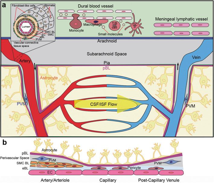
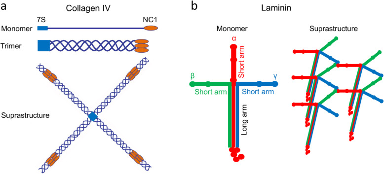
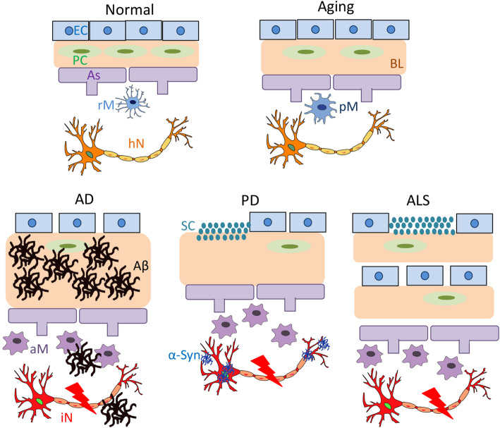
Background: Neurodegenerative disorders are a group of age-associated diseases characterized by progressive degeneration of the structure and function of the CNS. Two key pathological features of these disorders are blood-brain barrier (BBB) breakdown and protein aggregation. Main body: The BBB is composed of various cell types and a non-cellular component---the basal lamina (BL). Although how different cells affect the BBB is well studied, the roles of the BL in BBB maintenance and function remain largely unknown. In addition, located in the perivascular space, the BL is also speculated to regulate protein clearance via the meningeal lymphatic/glymphatic system. Recent studies from our laboratory and others have shown that the BL actively regulates BBB integrity and meningeal lymphatic/glymphatic function in both physiological and pathological conditions, suggesting that it may play an important role in the pathogenesis and/or progression of neurodegenerative disorders. In this review, we focus on changes of the BL and its major components during aging and in neurodegenerative disorders, including Alzheimer’s disease (AD), Parkinson’s disease (PD), and amyotrophic lateral sclerosis (ALS). First, we introduce the vascular and lymphatic systems in the CNS. Next, we discuss the BL and its major components under homeostatic conditions, and summarize their changes during aging and in AD, PD, and ALS in both rodents and humans. The functional significance of these alterations and potential therapeutic targets are also reviewed. Finally, key challenges in the field and future directions are discussed. Conclusions: Understanding BL changes and the functional significance of these changes in neurodegenerative disorders will fill the gap of knowledge in the field. Our goal is to provide a clear and concise review of the complex relationship between the BL and neurodegenerative disorders to stimulate new hypotheses and further research in this field.
Overview
The claims and statistics of this paper haven’t been checked yet.
Run the check — free, no account needed. It reads the open-access text and checks the sources it cites.
Integrity signals
Registry
Cites
Statistics
Figures
Verification
Claims the paper makes
The claims and statistics of this paper haven’t been checked yet.
Run the check — free, no account needed. It reads the open-access text and checks the sources it cites.
Claims other papers make about this one
Each is checked against this paper’s own text: does it say what the citing paper says it does?
The cited source says what the claim attributes to it.
In the citing paper
“Breakdown of the blood-brain barrier (BBB) is an early biomarker of human cognitive decline”
From ApoE4 reduction: An emerging and promising therapeutic strategy for Alzheimer's disease (2022) · cites this paper as Nguyen et al 2021
Self-check
Percentages, sums and reported differences re-computed from the paper’s own numbers.
The self-check needs the paper’s full text.
Citations
Cites
1 retracted
Montagne, A., Nikolakopoulou, A. M., Zhao, Z., Sagare, A. P., Si, G., Lazic, D., Barnes, S. R., Daianu, M., Ramanathan, A., Go, A., et al. (2018) · Nature Medicine · doi:10.1038/nm.4482 · Cited by 344
RetractedMutation in the α-Synuclein Gene Identified in Families with Parkinson's Disease
Polymeropoulos, M. H., Lavedan, C., Leroy, E., Ide, S. E., Dehejia, A., Dutra, A., Pike, B., Root, H., Rubenstein, J., Boyer, R., et al. (1997) · Science · doi:10.1126/science.276.5321.2045 · Cited by 8,240
Rosen, D. R., Siddique, T., Patterson, D., Figlewicz, D. A., Sapp, P., Hentati, A., Donaldson, D., Goto, J., O'Regan, J. P., Deng, H.-X., et al. (1993) · Nature · doi:10.1038/362059a0 · Cited by 7,062
Iliff, J. J., Wang, M., Liao, Y., Plogg, B. A., Peng, W., Gundersen, G. A., Benveniste, H., Vates, G. E., Deane, R., Goldman, S. A., et al. (2012) · Science Translational Medicine · doi:10.1126/scitranslmed.3003748 · Cited by 5,824
Sleep Drives Metabolite Clearance from the Adult Brain
Xie, L., Kang, H., Xu, Q., Chen, M. J., Liao, Y., Thiyagarajan, M., O’Donnell, J., Christensen, D. J., Nicholson, C., Iliff, J. J., et al. (2013) · Science · doi:10.1126/science.1241224 · Cited by 5,411
2016 Alzheimer's disease facts and figures
Alzheimer's Association (2016) · Alzheimer's & Dementia · doi:10.1016/j.jalz.2016.03.001 · Cited by 5,069
Structural and functional features of central nervous system lymphatic vessels
Louveau, A., Smirnov, I., Keyes, T. J., Eccles, J. D., Rouhani, S. J., Peske, J. D., Derecki, N. C., Castle, D., Mandell, J. W., Lee, K. S., et al. (2015) · Nature · doi:10.1038/nature14432 · Cited by 4,413
AGEING AND PARKINSON'S DISEASE: SUBSTANTIA NIGRA REGIONAL SELECTIVITY
FEARNLEY, J. M., & LEES, A. J. (1991) · Brain · doi:10.1093/brain/114.5.2283 · Cited by 3,529
The Extracellular Matrix: Not Just Pretty Fibrils
Hynes, R. O. (2009) · Science · doi:10.1126/science.1176009 · Cited by 3,527
The Blood-Brain Barrier in Health and Chronic Neurodegenerative Disorders
Zlokovic, B. V. (2008) · Neuron · doi:10.1016/j.neuron.2008.01.003 · Cited by 3,280
Cited by
Sorted by citations
ApoE4 reduction: An emerging and promising therapeutic strategy for Alzheimer's disease
Li, Y., Macyczko, J. R., Liu, C.-C., & Bu, G. (2022) · Neurobiology of Aging · doi:10.1016/j.neurobiolaging.2022.03.011 · Cited by 61
Metadata
- Authors
- Nguyen, B., Bix, G., & Yao, Y.
- Affiliations
- University of Georgia (US)
- Tulane University (US)
- Corresponding author
- Yao, Yao
- Venue
- Molecular Neurodegeneration
- Published
- 2021-12-07
- Volume
- 16
- Issue
- 1
- Pages
- 81-81
- ISSN
- 1750-1326 (ISSN-L)
- PMC
- PMC8650282
- OpenAlex
- W4200551731
- Cited by
- 75
- References
- 354
- Citations by year
- 2026: 9 · 2025: 23 · 2024: 16 · 2023: 13 · 2022: 14
- FWCI
- 1.27
- Type
- journal article
- Language
- en
- Open access
- gold
- doi.org
- Locations
- Molecular Neurodegeneration (publishedVersion, cc-by)
- PubMed Central (publishedVersion, cc-by)
- PubMed (publishedVersion)
- DOAJ (DOAJ: Directory of Open Access Journals) (submittedVersion, cc-by-sa)
- Funders
- National Heart, Lung, and Blood Institute (R01HL146574)
- National Institute on Aging (R21AG064422, R21AG073862, RF1AG065345)
- American Heart Association (16SDG29320001)
- Topics
- Life Sciences › Neuroscience › Neurology › Barrier Structure and Function Studies
- Health Sciences › Medicine › Clinical Neurology › Cerebrospinal fluid and hydrocephalus
- Life Sciences › Neuroscience › Neurology › Neuroinflammation and Neurodegeneration Mechanisms
- Keywords
- basement membrane, neurodegenerative diseases, blood-brain barrier, Alzheimer's disease, Parkinson's disease, amyotrophic lateral sclerosis, aging, meningeal lymphatic system, glymphatic system, protein clearance, protein aggregation, perivascular spaces, therapeutic target
- MeSH terms
- Glymphatic System, Alzheimer Disease, Basement Membrane, Blood-Brain Barrier, Humans, Neurodegenerative Diseases
About these checks
Registry, retraction, Cites and statistics results are computed, not judged, with one exception: a printed reference that no index lists is read once by a model before it counts as a citation error. Claims are extracted by a model and kept only when their quote appears verbatim in the text; each cited source is then checked, with a model’s help, for whether it says what the claim relies on. Those verdicts can be wrong — read the claim’s quote and the cited source before relying on one.
Statistics and arithmetic are recomputed, not judged.
Confidence bands are shown only for fields calibrated on our annotated test set. None is calibrated yet, so no band is shown.
What counts as a claim ›How we measure confidence ›How we check this ›
Are you an author, or do you see a problem with this page? Request a review
