

The DOI matches its Crossref record; 52 of the 52 references it cites were found in a registry or an index; 27 of the 41 sources its claims cite were checked for support.
Alzheimer’s disease phospholipase C-gamma-2 (PLCG2) protective variant is a functional hypermorph
Open accessPublished version of a preprint
Abstract
BACKGROUND: Recent Genome Wide Association Studies (GWAS) have identified novel rare coding variants in immune genes associated with late onset Alzheimer's disease (LOAD). Amongst these, a polymorphism in phospholipase C-gamma 2 (PLCG2) P522R has been reported to be protective against LOAD. PLC enzymes are key elements in signal transmission networks and are potentially druggable targets. PLCG2 is highly expressed in the hematopoietic system. Hypermorphic mutations in PLCG2 in humans have been reported to cause autoinflammation and immune disorders, suggesting a key role for this enzyme in the regulation of immune cell function. METHODS: We assessed PLCG2 distribution in human and mouse brain tissue via immunohistochemistry and in situ hybridization. We transfected heterologous cell systems (COS7 and HEK293T cells) to determine the effect of the P522R AD-associated variant on enzymatic function using various orthogonal assays, including a radioactive assay, IP-One ELISA, and calcium assays. RESULTS: PLCG2 expression is restricted primarily to microglia and granule cells of the dentate gyrus. Plcg2 mRNA is maintained in plaque-associated microglia in the cerebral tissue of an AD mouse model. Functional analysis of the p.P522R variant demonstrated a small hypermorphic effect of the mutation on enzyme function. CONCLUSIONS: The PLCG2 P522R variant is protective against AD. We show that PLCG2 is expressed in brain microglia, and the p.P522R polymorphism weakly increases enzyme function. These data suggest that activation of PLCγ2 and not inhibition could be therapeutically beneficial in AD. PLCγ2 is therefore a potential target for modulating microglia function in AD, and a small molecule drug that weakly activates PLCγ2 may be one potential therapeutic approach.
Overview
AI snapshot
Written from the paper’s full text.
TL;DR
PLCG2 is expressed primarily in brain microglia and dentate gyrus granule cells, and its mRNA is maintained in plaque-associated microglia in an AD mouse model. The AD-protective PLCG2 P522R variant acts as a weak functional hypermorph in cell-based assays, suggesting that PLCγ2 activation rather than inhibition may be therapeutically beneficial.
Key findings
- PLCG2 expression was primarily restricted to microglia and granule cells of the dentate gyrus; in cortical gray and white matter, all IBA1-immunoreactive microglia cell bodies contained Plcg2 transcripts (100%), with only occasional co-expression with astrocyte (5.8 ± 3.8%), endothelial (9.6 ± 1.44%), and oligodendrocyte-lineage (1.6 ± 2.2%) markers.
- Plcg2 mRNA was maintained in microglia surrounding plaques in 6-month-old TgAPPswe/ind mice, similar to non-transgenic littermates, and Plcg2 co-localized with Trem2 transcripts in cortical and hippocampal microglia.
- The AD-associated P522R variant showed a small hypermorphic effect: in a radioactive assay, PLC activity after EGF stimulation increased 1.24 ± 0.06 fold over WT, compared with 2.27 ± 0.43 fold for the Ali5 D993G mutation.
- IP-One ELISA showed higher IP1 production for P522R than WT PLCγ2, and calcium flux assays showed a small but clear increase in intracellular Ca2+ release for P522R versus WT, with no significant difference in extracellular Ca2+ entry.
Methods
- Human post-mortem frontal cortex and mouse brain tissue (WT C57BL6 and TgCRND8/TgAPPswe/ind mice) were assessed by immunohistochemistry and RNAScope in situ hybridization; RNAScope reactions were quantified using CellProfiler.
- COS7 and HEK293T cells were transfected with PLCG2 constructs (common/WT, P522R, and D993G Ali5) and analyzed with orthogonal functional assays: radioactive inositol phosphate formation, IP-One ELISA, and Fura-2 calcium flux, with Western blotting for expression.
- Statistical analysis used Prism 6, with one-way ANOVA plus Tukey’s multiple comparison test and two-way ANOVA with Bonferroni multiple comparisons.
Limitations
- The paper states a technical limitation: mRNA and protein co-expression could not be confirmed in the same cells, and antibody technical difficulties occurred (one antibody no longer commercially available; others gave unspecific or no signal on fixed human post-mortem brain or mouse tissue).
- The paper states that nuclear staining used to identify cell boundaries does not allow assessment of transcript quantification in processes.
- Evident from the text: functional characterization was performed in heterologous cell systems (COS7 and HEK293T cells), not in microglia; the molecular mechanism of the P522R hypermorphic effect is unknown, and further experiments are needed to address functional consequences on immune cell phenotype.
Model-written summary. Check it against the paper before relying on it.
Integrity signals
Registry
Cites
Statistics
Figures
Verification
Claims the paper makes
30 Own result · 11 Own interpretation · 9 Supported · 6 Partly supported · 6 No full text of the sources · 3 Supported so far · 3 Own method · 2 Couldn’t confirm · accuracy not yet measured
The sources with a verdict support this claim; the rest weren’t checked yet, have no full text we could read, or their verdicts aren’t rated yet.
In the paper
“Hypermorphic mutations in PLCG2 in humans have been reported to cause autoinflammation and immune disorders, suggesting a key role for this enzyme in the regulation of immune cell function.”
- Also stated in: § Background · plain
“PLCG2 variants cause inherited immune disorders designated as PLAID (PLCG2-associated antibody deficiency and immune dysregulation [12] and APLAID (autoinflammatory PLAID [13]).”
§ Alzheimer’s disease phospholipase C-gamma-2 (PLCG2) protective variant is a functional hypermorph > Abstract > Background
What the sources say
- Supportedcites [12] Ombrello MJ, Remmers EF, Sun G, Freeman AF, Datta S, Torabi-Parizi P, et al. Cold Urticaria, immunodeficiency, and autoimmunity related to PLCG2 deletions. N Engl J Med. 2012;366:330–8. https://doi.org/10.1056/NEJMoa1102140 . source
The Discussion (page not provided) reports that PLCG2 deletions caused a dominantly inherited syndrome of cold urticaria and immune dysregulation, and that the deletions produced constitutive phospholipase activity. It also links mutant PLCγ2 to leukocyte function, supporting the citing sentence’s inference about immune-cell regulation. The paper establishes this for PLAID; it does not identify APLAID. (LLM, conf=0.90)
- Not checked — no full text of the sourcecites [13] Zhou Q, Lee GS, Brady J, Datta S, Katan M, Sheikh A, et al. A hypermorphic missense mutation in PLCG2, encoding phospholipase Cγ2, causes a dominantly inherited autoinflammatory disease with immunodeficiency. Am J Hum Genet. 2012;91:713–20. https://doi.org/10.1016/j.ajhg.2012.08.006 . source
plaindirect- Asserted: passes — The authors put it forward as true.
- Checkable: passes — It can be checked against evidence.
- Load-bearing: unclear — It is unclear whether the paper’s conclusions depend on it.
- Based: passes — The paper gives a reason why it holds.
- Also stated in: § Background · plain
Every source the paper cites for this claim was checked and supports it.
In the paper
“Amongst these, a polymorphism in phospholipase C-gamma 2 (PLCG2) P522R has been reported to be protective against LOAD.”
- Also stated in: § Background · plain
“Notably, the PLCG2 missense variant Pro522Arg (P = 5.38 × 10⁻¹⁰, OR = 0.68) was associated with decreased risk of LOAD [6].”
- Also stated in: § Conclusions · plain
“The PLCG2 P522R variant is protective against AD.”
§ Alzheimer’s disease phospholipase C-gamma-2 (PLCG2) protective variant is a functional hypermorph > Abstract > Background
What the sources say
- Supportedcites [6] Sims R, Van Der Lee SJ, Naj AC, Bellenguez C, Badarinarayan N, Jakobsdottir J, et al. Rare coding variants in PLCG2, ABI3, and TREM2 implicate microglial-mediated innate immunity in Alzheimer’s disease. Nat Genet. 2017;49:1373–84. source
The passage on the PLCG2 result (labeled Abstract in our copy; no page given) explicitly reports that p.P522R is associated with decreased risk of late-onset Alzheimer’s disease, with an odds ratio of 0.68. This supports the citing sentence’s description of the variant as protective against LOAD. (LLM, conf=0.95)
plaincorrelational- Asserted: passes — The authors put it forward as true.
- Checkable: passes — It can be checked against evidence.
- Load-bearing: passes — The paper’s conclusions depend on it.
- Based: passes — The paper gives a reason why it holds.
- Also stated in: § Background · plain
The cited sources support only part of this claim — read each source’s verdict below.
In the paper
“However, normalization to microglia-specific genes (e.g., Abi3) and data from single-cell profiling of brain immune cells [6, 36, 37], suggest that apparent PLCG2 transcript upregulation in bulk tissue is mostly related to microgliosis occurring as a consequence of neurodegeneration.”
§ Alzheimer’s disease phospholipase C-gamma-2 (PLCG2) protective variant is a functional hypermorph > Results > PLCG2 in microglia in Alzheimer’s disease
What the sources say
- Partly supportedcites [6] Sims R, Van Der Lee SJ, Naj AC, Bellenguez C, Badarinarayan N, Jakobsdottir J, et al. Rare coding variants in PLCG2, ABI3, and TREM2 implicate microglial-mediated innate immunity in Alzheimer’s disease. Nat Genet. 2017;49:1373–84. sourceExperimental — accuracy not yet measured
The Gene Expression section reports that PLCG2 expression is higher in AD temporal cortex before adjustment for cell-type markers, but not after, and attributes the apparent elevation most likely to microgliosis. It does not establish that the microgliosis is a consequence of neurodegeneration; the paper separately argues that the microglial response in Alzheimer’s disease is not simply a downstream consequence of neurodegeneration. (LLM, conf=0.86)
- Supportedcites [36] Keren-Shaul H, Spinrad A, Weiner A, Matcovitch-Natan O, Dvir-Szternfeld R, Ulland TK, et al. A unique microglia type associated with restricting development of Alzheimer’s disease. Cell. 2017;169:1276–1290.e17. https://doi.org/10.1016/j.cell.2017.05.018 . source
The citation identifies a source of single-cell profiling data on brain immune cells, rather than attributing the citing authors’ conclusion about bulk PLCG2 upregulation to this paper alone. The Discussion describes single-cell RNA sequencing of immune cells from the brain, and the Results report microglial states in the AD model. No page numbers were provided in our copy. (LLM, conf=0.90)
- Partly supportedcites [37] Mathys H, Adaikkan C, Gao F, Young JZ, Manet E, Hemberg M, et al. Temporal tracking of microglia activation in neurodegeneration at single-cell resolution. Cell Rep. 2017;21:366–80. https://doi.org/10.1016/j.celrep.2017.09.039 . sourceExperimental — accuracy not yet measured
The Results section on microglial proliferation reports increased microglial density in the hippocampus of neurodegenerating CK-p25 mice. The Discussion also says that increased immune-related transcripts in tissue could reflect either changes in microglial expression or expansion of the microglial population, and finds evidence for both. Our copy does not establish that bulk-tissue PLCG2 upregulation is mostly due to microgliosis; searches for PLCG2 and Abi3 found no passage addressing that specific inference. (LLM, conf=0.82)
hedgeddirect- Asserted: passes — The authors put it forward as true.
- Checkable: passes — It can be checked against evidence.
- Load-bearing: passes — The paper’s conclusions depend on it.
- Based: passes — The paper gives a reason why it holds.
- Partly supported
Every source the paper cites for this claim was checked and supports it.
In the paper
“Other mutations in PLCG2 that lead to immune dysfunction are known to be activating [7].”
§ Alzheimer’s disease phospholipase C-gamma-2 (PLCG2) protective variant is a functional hypermorph > Discussion
What the sources say
- Supportedcites [7] Koss H, Bunney TD, Behjati S, Katan M. Dysfunction of phospholipase Cγ in immune disorders and cancer. Trends Biochem Sci. 2014;39:603–11. https://doi.org/10.1016/j.tibs.2014.09.004 . source
The “PLC γ mutations in immune diseases” section (pp. 3–4) describes PLCG2 mutations associated with immune disorders as activating: PLAID deletions cause constitutive PLC activation, and the APLAID S707Y substitution increases enzymatic function. The paper notes that PLAID can nevertheless diminish downstream signalling, which does not negate the claim about activation. (LLM, conf=0.94)
boosteddirect- Asserted: passes — The authors put it forward as true.
- Checkable: passes — It can be checked against evidence.
- Load-bearing: passes — The paper’s conclusions depend on it.
- Based: passes — The paper gives a reason why it holds.
- Supported
The cited sources support only part of this claim — read each source’s verdict below.
In the paper
“The protective variant P522R is certainly located within a region of the enzyme that has a regulatory function (Fig. 4d, [7])”
§ Alzheimer’s disease phospholipase C-gamma-2 (PLCG2) protective variant is a functional hypermorph > Discussion
What the sources say
- Partly supportedcites [7] Koss H, Bunney TD, Behjati S, Katan M. Dysfunction of phospholipase Cγ in immune disorders and cancer. Trends Biochem Sci. 2014;39:603–11. https://doi.org/10.1016/j.tibs.2014.09.004 . sourceExperimental — accuracy not yet measured
Box 1 (p. 17) describes the PLCγ-specific array as involved in interactions with regulatory proteins, and the Figures (p. 16) describe regulatory interactions involving its domains. Our copy does not mention P522R or locate that variant within the array, so it supports the region’s regulatory role but not the claimed location of P522R. (LLM, conf=0.78)
boosted- Asserted: passes — The authors put it forward as true.
- Checkable: passes — It can be checked against evidence.
- Load-bearing: passes — The paper’s conclusions depend on it.
- Based: passes — The paper gives a reason why it holds.
- Partly supported
The cited sources support only part of this claim — read each source’s verdict below.
In the paper
“In comparison to previous studies which are aligned to the concept that suppressing microglial function may be beneficial in the setting of AD [49], the data described here, together with studies on TREM2 and other AD-associated variants, suggest that a different directionality may be therapeutically useful [6, 46, 50].”
§ Alzheimer’s disease phospholipase C-gamma-2 (PLCG2) protective variant is a functional hypermorph > Conclusion
What the sources say
- Partly supportedcites [6] Sims R, Van Der Lee SJ, Naj AC, Bellenguez C, Badarinarayan N, Jakobsdottir J, et al. Rare coding variants in PLCG2, ABI3, and TREM2 implicate microglial-mediated innate immunity in Alzheimer’s disease. Nat Genet. 2017;49:1373–84. sourceExperimental — accuracy not yet measured
The paper’s discussion of TREM2 says that risk variants may reduce TREM2 signaling, which participates in microglial activation, and its concluding passage identifies PLCγ2 as a druggable target. These passages support investigating microglial signaling in AD, but the paper does not establish that activating microglia, rather than suppressing them, would be therapeutically useful. (LLM, conf=0.82)
- Not checked — no full text of the sourcecites [46] Ulland TK, Colonna M. TREM2—a key player in microglial biology and Alzheimer disease. Nat Rev Neurol. 2018;14:667–75. source
- Supportedcites [50] Wang Y, Cella M, Mallinson K, Ulrich JD, Young KL, Robinette ML, et al. TREM2 lipid sensing sustains the microglial response in an Alzheimer’s disease model. Cell. 2015;160:1061–71. https://doi.org/10.1016/j.cell.2015.01.049 . source
The Discussion reports that TREM2-supported microglial expansion and activation promotes Aβ removal and reduces neuronal damage in the 5XFAD mouse model. This supports the citing authors’ hedged suggestion that sustaining, rather than suppressing, a microglial response may be useful; the paper does not test a therapy in people with AD. (LLM, conf=0.86)
hedgedconditional- Asserted: passes — The authors put it forward as true.
- Checkable: passes — It can be checked against evidence.
- Load-bearing: passes — The paper’s conclusions depend on it.
- Based: passes — The paper gives a reason why it holds.
- Partly supported
A result the paper reports from its own data — it cites no other source for it, so there is none to check. The quote shows it is in the paper.
In the paper
“We showed that microglia are the main cell brain population expressing Plcg2.”
§ Alzheimer’s disease phospholipase C-gamma-2 (PLCG2) protective variant is a functional hypermorph > Discussion
boosted- Asserted: passes — The authors put it forward as true.
- Checkable: passes — It can be checked against evidence.
- Load-bearing: passes — The paper’s conclusions depend on it.
- Based: passes — The paper gives a reason why it holds.
A result the paper reports from its own data — it cites no other source for it, so there is none to check. The quote shows it is in the paper.
In the paper
“Functional analysis of the p.P522R variant demonstrated a small hypermorphic effect of the mutation on enzyme function.”
- Also stated in: § Conclusions · plain
“We show that PLCG2 is expressed in brain microglia, and the p.P522R polymorphism weakly increases enzyme function.”
§ Alzheimer’s disease phospholipase C-gamma-2 (PLCG2) protective variant is a functional hypermorph > Abstract > Results
boosteddirect- Asserted: passes — The authors put it forward as true.
- Checkable: passes — It can be checked against evidence.
- Load-bearing: passes — The paper’s conclusions depend on it.
- Based: passes — The paper gives a reason why it holds.
- Also stated in: § Conclusions · plain
A result the paper reports from its own data — it cites no other source for it, so there is none to check. The quote shows it is in the paper.
In the paper
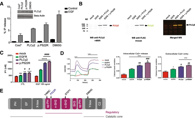
“For comparison, the inflammation-related Ali5 p.D993G mutation exhibited a larger (2.27 ± 0.43 fold, mean value ± SD) increase in enzyme function upon EGF stimulation (Fig. 4a).”
§ Alzheimer’s disease phospholipase C-gamma-2 (PLCG2) protective variant is a functional hypermorph > Results > AD associated PLCG2 p.P522R variant shows a weak hypermorphic activity
plaindirect- Asserted: passes — The authors put it forward as true.
- Checkable: passes — It can be checked against evidence.
- Load-bearing: unclear — It is unclear whether the paper’s conclusions depend on it.
- Based: passes — The paper gives a reason why it holds.
A result the paper reports from its own data — it cites no other source for it, so there is none to check. The quote shows it is in the paper.
In the paper
“endothelial cells (Pecam1, platelet and endothelial cell adhesion molecule 1, 9.6 ± 1.44%, Additional file 2b)”
§ Alzheimer’s disease phospholipase C-gamma-2 (PLCG2) protective variant is a functional hypermorph > Results > PLCG2 is expressed in microglia cells throughout the brain
plaincorrelational- Asserted: passes — The authors put it forward as true.
- Checkable: passes — It can be checked against evidence.
- Load-bearing: unclear — It is unclear whether the paper’s conclusions depend on it.
- Based: passes — The paper gives a reason why it holds.
A result the paper reports from its own data — it cites no other source for it, so there is none to check. The quote shows it is in the paper.
In the paper
“p.P522R enzyme activity was higher than PLCγ2, as measured by IP1 production (Fig. 4c).”
§ Alzheimer’s disease phospholipase C-gamma-2 (PLCG2) protective variant is a functional hypermorph > Results > AD associated PLCG2 p.P522R variant shows a weak hypermorphic activity
plaindirect- Asserted: passes — The authors put it forward as true.
- Checkable: passes — It can be checked against evidence.
- Load-bearing: passes — The paper’s conclusions depend on it.
- Based: passes — The paper gives a reason why it holds.
A result the paper reports from its own data — it cites no other source for it, so there is none to check. The quote shows it is in the paper.
In the paper
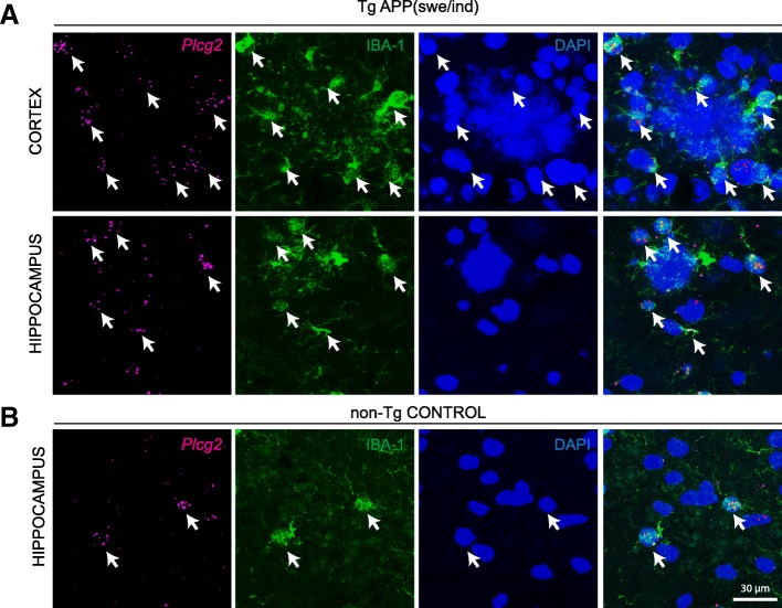
“b RNAScope for Plcg2 in adult mouse hippocampus of a non-transgenic control mouse. Arrows point to co-labeling of IBA-1 and Plcg2”
§ Alzheimer’s disease phospholipase C-gamma-2 (PLCG2) protective variant is a functional hypermorph > Results > PLCG2 in microglia in Alzheimer’s disease
plaincorrelationalSingle case- Asserted: passes — The authors put it forward as true.
- Checkable: passes — It can be checked against evidence.
- Load-bearing: unclear — It is unclear whether the paper’s conclusions depend on it.
- Based: passes — The paper gives a reason why it holds.
A result the paper reports from its own data — it cites no other source for it, so there is none to check. The quote shows it is in the paper.
In the paper
“we found intense Plcg2 RNAScope reaction product in the granule cell layer of the hippocampal dentate gyrus, where the Plcg2 signal co-localizes with the neuronal marker NeuN (Additional file 2d).”
§ Alzheimer’s disease phospholipase C-gamma-2 (PLCG2) protective variant is a functional hypermorph > Results > PLCG2 is expressed in microglia cells throughout the brain
plaincorrelational- Asserted: passes — The authors put it forward as true.
- Checkable: passes — It can be checked against evidence.
- Load-bearing: unclear — It is unclear whether the paper’s conclusions depend on it.
- Based: passes — The paper gives a reason why it holds.
A result the paper reports from its own data — it cites no other source for it, so there is none to check. The quote shows it is in the paper.
In the paper
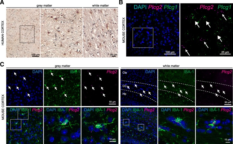
“In these regions, all IBA1-immunoreactive microglia cell bodies contained transcripts for Plcg2 (100%, Fig. 1c).”
- Also stated in: § PLCG2 is expressed in microglia cells throughout the brain · boosted
“we confirmed a high degree of co-localization between Plcg2 mRNA and the microglia-specific marker IBA-1 in cortical gray and white matter of the adult mouse brain.”
§ Alzheimer’s disease phospholipase C-gamma-2 (PLCG2) protective variant is a functional hypermorph > Results > PLCG2 is expressed in microglia cells throughout the brain
boostedcorrelational- Asserted: passes — The authors put it forward as true.
- Checkable: passes — It can be checked against evidence.
- Load-bearing: passes — The paper’s conclusions depend on it.
- Based: passes — The paper gives a reason why it holds.
- Also stated in: § PLCG2 is expressed in microglia cells throughout the brain · boosted
A result the paper reports from its own data — it cites no other source for it, so there is none to check. The quote shows it is in the paper.
In the paper
“We detected PLCγ2 immunoreactivity in putative microglia of both white and gray matter, in the human frontal cortex (Fig. 1a).”
§ Alzheimer’s disease phospholipase C-gamma-2 (PLCG2) protective variant is a functional hypermorph > Results > PLCG2 is expressed in microglia cells throughout the brain
hedged- Asserted: passes — The authors put it forward as true.
- Checkable: passes — It can be checked against evidence.
- Load-bearing: passes — The paper’s conclusions depend on it.
- Based: passes — The paper gives a reason why it holds.
A result the paper reports from its own data — it cites no other source for it, so there is none to check. The quote shows it is in the paper.
In the paper
“oligodendrocyte-lineage cells (Olig2, oligodendrocyte transcription factor 2, 1.6 ± 2.2% Additional file 2c).”
§ Alzheimer’s disease phospholipase C-gamma-2 (PLCG2) protective variant is a functional hypermorph > Results > PLCG2 is expressed in microglia cells throughout the brain
plaincorrelational- Asserted: passes — The authors put it forward as true.
- Checkable: passes — It can be checked against evidence.
- Load-bearing: unclear — It is unclear whether the paper’s conclusions depend on it.
- Based: passes — The paper gives a reason why it holds.
A result the paper reports from its own data — it cites no other source for it, so there is none to check. The quote shows it is in the paper.
In the paper
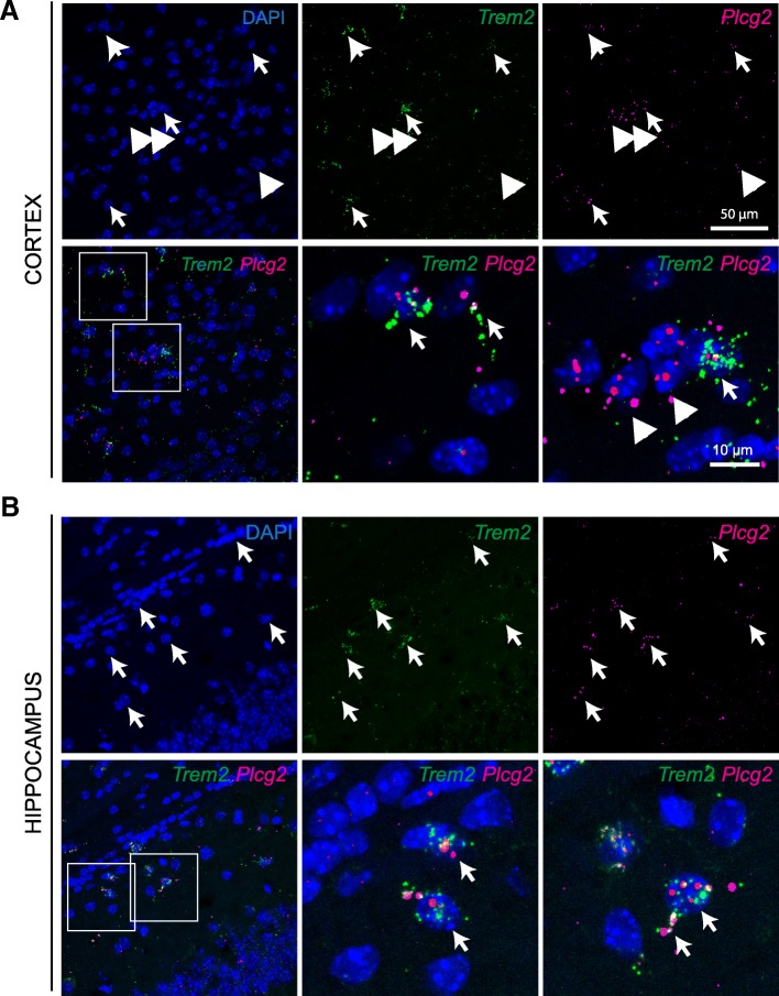
“Indeed, we detected co-expression of Plcg2 and Trem2 mRNAs in the cell soma and processes of putative cortical and hippocampal microglia (Fig. 3).”
§ Alzheimer’s disease phospholipase C-gamma-2 (PLCG2) protective variant is a functional hypermorph > Results > PLCG2 in microglia in Alzheimer’s disease
hedgedcorrelational- Asserted: passes — The authors put it forward as true.
- Checkable: passes — It can be checked against evidence.
- Load-bearing: passes — The paper’s conclusions depend on it.
- Based: passes — The paper gives a reason why it holds.
A result the paper reports from its own data — it cites no other source for it, so there is none to check. The quote shows it is in the paper.
In the paper
“and that its expression is maintained in microglia in close proximity to plaques in the cerebral tissue of an APP mouse model.”
- Also stated in: § Discussion · plain
“We found Plcg2 expression in microglia surrounding amyloid plaques in a mouse model of AD”
§ Alzheimer’s disease phospholipase C-gamma-2 (PLCG2) protective variant is a functional hypermorph > Conclusion
plaincorrelational- Asserted: passes — The authors put it forward as true.
- Checkable: passes — It can be checked against evidence.
- Load-bearing: passes — The paper’s conclusions depend on it.
- Based: passes — The paper gives a reason why it holds.
- Also stated in: § Discussion · plain
A result the paper reports from its own data — it cites no other source for it, so there is none to check. The quote shows it is in the paper.
In the paper
“Plcg2 mRNA is maintained in plaque-associated microglia in the cerebral tissue of an AD mouse model.”
- Also stated in: § PLCG2 in microglia in Alzheimer’s disease · hedged
“Plcg2 appeared to be equally expressed in microglia surrounding the plaques in the cortical regions of 6 months old transgenic mice (Fig. 2a) as compared to localization in microglia in control non-transgenic littermates (Fig. 2b).”
§ Alzheimer’s disease phospholipase C-gamma-2 (PLCG2) protective variant is a functional hypermorph > Abstract > Results
plain- Asserted: passes — The authors put it forward as true.
- Checkable: passes — It can be checked against evidence.
- Load-bearing: unclear — It is unclear whether the paper’s conclusions depend on it.
- Based: passes — The paper gives a reason why it holds.
- Also stated in: § PLCG2 in microglia in Alzheimer’s disease · hedged
A result the paper reports from its own data — it cites no other source for it, so there is none to check. The quote shows it is in the paper.
In the paper
“Note expression of Plcg2 in IBA-1-labeled microglia surrounding the plaques (DAPI).”
- Also stated in: § PLCG2 in microglia in Alzheimer’s disease · plain
“Arrows point to co-labeling of IBA-1 and Plcg2.”
§ Alzheimer’s disease phospholipase C-gamma-2 (PLCG2) protective variant is a functional hypermorph > Results > PLCG2 in microglia in Alzheimer’s disease
plaincorrelational- Asserted: passes — The authors put it forward as true.
- Checkable: passes — It can be checked against evidence.
- Load-bearing: passes — The paper’s conclusions depend on it.
- Based: passes — The paper gives a reason why it holds.
- Also stated in: § PLCG2 in microglia in Alzheimer’s disease · plain
A result the paper reports from its own data — it cites no other source for it, so there is none to check. The quote shows it is in the paper.
In the paper
“We found only occasional co-expression with markers for astrocytes (Glast GLutamate ASpartate Transporter/Slc1a3 Solute Carrier family 1 member 3, 5.8 ± 3.8%, Additional file 2a)”
§ Alzheimer’s disease phospholipase C-gamma-2 (PLCG2) protective variant is a functional hypermorph > Results > PLCG2 is expressed in microglia cells throughout the brain
plaincorrelational- Asserted: passes — The authors put it forward as true.
- Checkable: passes — It can be checked against evidence.
- Load-bearing: unclear — It is unclear whether the paper’s conclusions depend on it.
- Based: passes — The paper gives a reason why it holds.
A result the paper reports from its own data — it cites no other source for it, so there is none to check. The quote shows it is in the paper.
In the paper
“In summary, we show that PLCG2 is expressed in human and mouse brain microglia”
§ Alzheimer’s disease phospholipase C-gamma-2 (PLCG2) protective variant is a functional hypermorph > Conclusion
plain- Asserted: passes — The authors put it forward as true.
- Checkable: passes — It can be checked against evidence.
- Load-bearing: passes — The paper’s conclusions depend on it.
- Based: passes — The paper gives a reason why it holds.
A result the paper reports from its own data — it cites no other source for it, so there is none to check. The quote shows it is in the paper.
In the paper
“We found that Plcg1 and Plcg2 mRNAs show complementary expression patterns, with very little degree of overlap in the adult mouse brain (Fig. 1b).”
§ Alzheimer’s disease phospholipase C-gamma-2 (PLCG2) protective variant is a functional hypermorph > Results > PLCG2 is expressed in microglia cells throughout the brain
plaincorrelational- Asserted: passes — The authors put it forward as true.
- Checkable: passes — It can be checked against evidence.
- Load-bearing: unclear — It is unclear whether the paper’s conclusions depend on it.
- Based: passes — The paper gives a reason why it holds.
A result the paper reports from its own data — it cites no other source for it, so there is none to check. The quote shows it is in the paper.
In the paper
“PLC activity after stimulation with EGF showed a 1.24 ± 0.06 fold increase (mean value ± SD over 3 independent experiments, normalized to PLCγ2 WT activity) upon transfection with the P522R construct over the WT.”
§ Alzheimer’s disease phospholipase C-gamma-2 (PLCG2) protective variant is a functional hypermorph > Results > AD associated PLCG2 p.P522R variant shows a weak hypermorphic activity
plaindirect- Asserted: passes — The authors put it forward as true.
- Checkable: passes — It can be checked against evidence.
- Load-bearing: passes — The paper’s conclusions depend on it.
- Based: passes — The paper gives a reason why it holds.
A result the paper reports from its own data — it cites no other source for it, so there is none to check. The quote shows it is in the paper.
In the paper
“In a modest, but significant number of cells in the mouse cortex Plcg2 co-localized with a marker of endothelial cells.”
§ Alzheimer’s disease phospholipase C-gamma-2 (PLCG2) protective variant is a functional hypermorph > Discussion
plaincorrelational- Asserted: passes — The authors put it forward as true.
- Checkable: passes — It can be checked against evidence.
- Load-bearing: unclear — It is unclear whether the paper’s conclusions depend on it.
- Based: passes — The paper gives a reason why it holds.
A result the paper reports from its own data — it cites no other source for it, so there is none to check. The quote shows it is in the paper.
In the paper
“as well as in “homeostatic” microglia.”
§ Alzheimer’s disease phospholipase C-gamma-2 (PLCG2) protective variant is a functional hypermorph > Discussion
plain- Asserted: passes — The authors put it forward as true.
- Checkable: passes — It can be checked against evidence.
- Load-bearing: unclear — It is unclear whether the paper’s conclusions depend on it.
- Based: passes — The paper gives a reason why it holds.
A result the paper reports from its own data — it cites no other source for it, so there is none to check. The quote shows it is in the paper.
In the paper
“Stimulation of mock transfected cells with EGF failed to increase IP1 level (Fig. 4c).”
§ Alzheimer’s disease phospholipase C-gamma-2 (PLCG2) protective variant is a functional hypermorph > Results > AD associated PLCG2 p.P522R variant shows a weak hypermorphic activity
plain- Asserted: passes — The authors put it forward as true.
- Checkable: passes — It can be checked against evidence.
- Load-bearing: unclear — It is unclear whether the paper’s conclusions depend on it.
- Based: passes — The paper gives a reason why it holds.
A result the paper reports from its own data — it cites no other source for it, so there is none to check. The quote shows it is in the paper.
In the paper
“We found Plcg2 expression in microglia surrounding amyloid plaques in a mouse model of AD as well as in “homeostatic” microglia.”
§ Alzheimer’s disease phospholipase C-gamma-2 (PLCG2) protective variant is a functional hypermorph > Discussion
plaincorrelational- Asserted: passes — The authors put it forward as true.
- Checkable: passes — It can be checked against evidence.
- Load-bearing: unclear — It is unclear whether the paper’s conclusions depend on it.
- Based: passes — The paper gives a reason why it holds.
A result the paper reports from its own data — it cites no other source for it, so there is none to check. The quote shows it is in the paper.
In the paper
“Quantification of the intracellular Ca2+ release showed a small but clear increase for p.P522R variant as compared to WT PLCγ2 (Fig. 4d, middle panel).”
§ Alzheimer’s disease phospholipase C-gamma-2 (PLCG2) protective variant is a functional hypermorph > Results > AD associated PLCG2 p.P522R variant shows a weak hypermorphic activity
boosteddirect- Asserted: passes — The authors put it forward as true.
- Checkable: passes — It can be checked against evidence.
- Load-bearing: passes — The paper’s conclusions depend on it.
- Based: passes — The paper gives a reason why it holds.
A result the paper reports from its own data — it cites no other source for it, so there is none to check. The quote shows it is in the paper.
In the paper
“and PLCG2 transcripts were detected in similarly sparsely distributed cells, reminiscent of microglia.”
§ Alzheimer’s disease phospholipase C-gamma-2 (PLCG2) protective variant is a functional hypermorph > Discussion
hedgedcorrelational- Asserted: passes — The authors put it forward as true.
- Checkable: passes — It can be checked against evidence.
- Load-bearing: passes — The paper’s conclusions depend on it.
- Based: passes — The paper gives a reason why it holds.
A result the paper reports from its own data — it cites no other source for it, so there is none to check. The quote shows it is in the paper.
In the paper
“However, in human cortical tissue, we were able to show PLCγ2 immunoreactivity in sparsely located cells with microglia-like morphology”
§ Alzheimer’s disease phospholipase C-gamma-2 (PLCG2) protective variant is a functional hypermorph > Discussion
plain- Asserted: passes — The authors put it forward as true.
- Checkable: passes — It can be checked against evidence.
- Load-bearing: passes — The paper’s conclusions depend on it.
- Based: passes — The paper gives a reason why it holds.
A result the paper reports from its own data — it cites no other source for it, so there is none to check. The quote shows it is in the paper.
In the paper
“Expression of EGFR induced significant intracellular Ca2+ changes after addition of EGF, as measured by increase of the 340/380 nm excitation ratio (Fig. 4d, left panel).”
§ Alzheimer’s disease phospholipase C-gamma-2 (PLCG2) protective variant is a functional hypermorph > Results > AD associated PLCG2 p.P522R variant shows a weak hypermorphic activity
plaindirect- Asserted: passes — The authors put it forward as true.
- Checkable: passes — It can be checked against evidence.
- Load-bearing: unclear — It is unclear whether the paper’s conclusions depend on it.
- Based: passes — The paper gives a reason why it holds.
In the paper
“No Ca2+ changes were seen in mock HEK293T cells stimulated with EGF (Fig. 4d), confirming our previous ELISA IP-One results, and as previously observed [13].”
§ Alzheimer’s disease phospholipase C-gamma-2 (PLCG2) protective variant is a functional hypermorph > Results > AD associated PLCG2 p.P522R variant shows a weak hypermorphic activity
What the sources say
- Not checked — no full text of the sourcecites [13] Zhou Q, Lee GS, Brady J, Datta S, Katan M, Sheikh A, et al. A hypermorphic missense mutation in PLCG2, encoding phospholipase Cγ2, causes a dominantly inherited autoinflammatory disease with immunodeficiency. Am J Hum Genet. 2012;91:713–20. https://doi.org/10.1016/j.ajhg.2012.08.006 . source
plain- Asserted: passes — The authors put it forward as true.
- Checkable: passes — It can be checked against evidence.
- Load-bearing: unclear — It is unclear whether the paper’s conclusions depend on it.
- Based: passes — The paper gives a reason why it holds.
A result the paper reports from its own data — it cites no other source for it, so there is none to check. The quote shows it is in the paper.
In the paper
“The expression of EGFR or EGFR + PLCγ2 significantly increased the amount of IP1 confirming an activation of the receptor and PLCγ2.”
§ Alzheimer’s disease phospholipase C-gamma-2 (PLCG2) protective variant is a functional hypermorph > Results > AD associated PLCG2 p.P522R variant shows a weak hypermorphic activity
plaindirect- Asserted: passes — The authors put it forward as true.
- Checkable: passes — It can be checked against evidence.
- Load-bearing: passes — The paper’s conclusions depend on it.
- Based: passes — The paper gives a reason why it holds.
A result the paper reports from its own data — it cites no other source for it, so there is none to check. The quote shows it is in the paper.
In the paper
“Ca2+ level monitoring show no significant differences in the extracellular Ca2+ entry between cells expressing EGFR + PLCγ2 and EGFR + p.P522R (Fig. 4d, right panel).”
§ Alzheimer’s disease phospholipase C-gamma-2 (PLCG2) protective variant is a functional hypermorph > Results > AD associated PLCG2 p.P522R variant shows a weak hypermorphic activity
plain- Asserted: passes — The authors put it forward as true.
- Checkable: passes — It can be checked against evidence.
- Load-bearing: unclear — It is unclear whether the paper’s conclusions depend on it.
- Based: passes — The paper gives a reason why it holds.
A conclusion the paper draws from its own results — it cites no other source for it, so there is none to check. The quote shows it is in the paper.
In the paper
“suggesting a key role for this enzyme in the regulation of immune cell function.”
§ Alzheimer’s disease phospholipase C-gamma-2 (PLCG2) protective variant is a functional hypermorph > Abstract > Background
hedgeddirect- Asserted: passes — The authors put it forward as true.
- Checkable: passes — It can be checked against evidence.
- Load-bearing: passes — The paper’s conclusions depend on it.
- Based: passes — The paper gives a reason why it holds.
A conclusion the paper draws from its own results — it cites no other source for it, so there is none to check. The quote shows it is in the paper.
In the paper
“These data suggest that activation of PLCγ2 and not inhibition could be therapeutically beneficial in AD.”
§ Alzheimer’s disease phospholipase C-gamma-2 (PLCG2) protective variant is a functional hypermorph > Abstract > Conclusions
hedgedconditional- Asserted: passes — The authors put it forward as true.
- Checkable: passes — It can be checked against evidence.
- Load-bearing: passes — The paper’s conclusions depend on it.
- Based: passes — The paper gives a reason why it holds.
A conclusion the paper draws from its own results — it cites no other source for it, so there is none to check. The quote shows it is in the paper.
In the paper
“PLCγ2 is therefore a potential target for modulating microglia function in AD”
- Also stated in: § Background · hedged
“PLCγ2 is therefore a potential target for modulating microglia function in AD, and a small molecule drug that activates PLCγ2 may be one potential therapeutic approach.”
§ Alzheimer’s disease phospholipase C-gamma-2 (PLCG2) protective variant is a functional hypermorph > Abstract > Conclusions
hedgedconditional- Asserted: passes — The authors put it forward as true.
- Checkable: passes — It can be checked against evidence.
- Load-bearing: passes — The paper’s conclusions depend on it.
- Based: passes — The paper gives a reason why it holds.
- Also stated in: § Background · hedged
A conclusion the paper draws from its own results — it cites no other source for it, so there is none to check. The quote shows it is in the paper.
In the paper
“Together with the data of Plcg2 expression in mouse tissue, our results strongly point to co-expression of protein and mRNA, at least in microglia in the mammalian cortex.”
§ Alzheimer’s disease phospholipase C-gamma-2 (PLCG2) protective variant is a functional hypermorph > Discussion
boostedcorrelational- Asserted: passes — The authors put it forward as true.
- Checkable: passes — It can be checked against evidence.
- Load-bearing: passes — The paper’s conclusions depend on it.
- Based: passes — The paper gives a reason why it holds.
A conclusion the paper draws from its own results — it cites no other source for it, so there is none to check. The quote shows it is in the paper.
In the paper
“This pathway, and in particular PLCγ2 activity, could be critical for microglia function in neurodegenerative conditions.”
§ Alzheimer’s disease phospholipase C-gamma-2 (PLCG2) protective variant is a functional hypermorph > Discussion
hedgedconditional- Asserted: passes — The authors put it forward as true.
- Checkable: passes — It can be checked against evidence.
- Load-bearing: passes — The paper’s conclusions depend on it.
- Based: passes — The paper gives a reason why it holds.
A conclusion the paper draws from its own results — it cites no other source for it, so there is none to check. The quote shows it is in the paper.
In the paper
“AD associated PLCG2 p.P522R variant shows a weak hypermorphic activity”
- Also stated in: § Discussion · plain
“Our functional analyses of the p.P522R variant reveal a small hypermorphic effect on enzyme activity.”
- Also stated in: § Conclusion · plain
“The rare-coding AD protective variant shows a small hypermorphic activity upon stimulation in various orthogonal cell-based assays.”
§ Alzheimer’s disease phospholipase C-gamma-2 (PLCG2) protective variant is a functional hypermorph > Results > AD associated PLCG2 p.P522R variant shows a weak hypermorphic activity
plaindirect- Asserted: passes — The authors put it forward as true.
- Checkable: passes — It can be checked against evidence.
- Load-bearing: passes — The paper’s conclusions depend on it.
- Based: passes — The paper gives a reason why it holds.
- Also stated in: § Discussion · plain
A conclusion the paper draws from its own results — it cites no other source for it, so there is none to check. The quote shows it is in the paper.
In the paper
“and provide evidence that a limited activation of this enzyme may have a beneficial therapeutic effect.”
§ Alzheimer’s disease phospholipase C-gamma-2 (PLCG2) protective variant is a functional hypermorph > Conclusion
hedgedconditional- Asserted: passes — The authors put it forward as true.
- Checkable: passes — It can be checked against evidence.
- Load-bearing: passes — The paper’s conclusions depend on it.
- Based: passes — The paper gives a reason why it holds.
A conclusion the paper draws from its own results — it cites no other source for it, so there is none to check. The quote shows it is in the paper.
In the paper
“which is consistent with this polymorphism modulating enzyme activation.”
§ Alzheimer’s disease phospholipase C-gamma-2 (PLCG2) protective variant is a functional hypermorph > Discussion
hedgedconditional- Asserted: passes — The authors put it forward as true.
- Checkable: passes — It can be checked against evidence.
- Load-bearing: unclear — It is unclear whether the paper’s conclusions depend on it.
- Based: passes — The paper gives a reason why it holds.
- Probably not a substantial claim. 25
- It may state two separate claims as one. 9
The cited sources that could be checked couldn’t confirm or dispute this claim.
In the paper
“PLCG2 mRNA upregulation has been reported in cortical tissue of LOAD patients (FC, 1.35; p = 0.0028, [33])”
§ Alzheimer’s disease phospholipase C-gamma-2 (PLCG2) protective variant is a functional hypermorph > Results > PLCG2 in microglia in Alzheimer’s disease
What the sources say
- Could not verifycites [33] Allen M, Carrasquillo MM, Funk C, Heavner BD, Zou F, Younkin CS, et al. Human whole genome genotype and transcriptome data for Alzheimer’s and other neurodegenerative diseases. Sci Data. 2016;3:160089. https://doi.org/10.1038/sdata.2016.89 . sourceExperimental — accuracy not yet measured
The Methods (Study Populations and Molecular Data; page unavailable) describe transcriptome measurements from temporal cortex samples of AD and non-AD subjects. Searches for PLCG2, the reported fold change of 1.35, and p = 0.0028 found no passage reporting the claimed upregulation in our copy. (LLM, conf=0.74)
plaincorrelational- Asserted: passes — The authors put it forward as true.
- Checkable: passes — It can be checked against evidence.
- Load-bearing: fails — The paper’s conclusions may not depend on it.
- Based: passes — The paper gives a reason why it holds.
Probably not a substantial claim. The paper’s conclusions may not depend on it.
- Could not verify
Every source the paper cites for this claim was checked and supports it.
In the paper
“In osteoclasts, activation downstream Trem2 initiates a cascade of events, including phosphorylation and activation of PLCγ2, amongst other proteins [17].”
§ Alzheimer’s disease phospholipase C-gamma-2 (PLCG2) protective variant is a functional hypermorph > Background
What the sources say
- Supportedcites [17] Peng Q, Malhotra S, Torchia JA, Kerr WG, Coggeshall KM, Humphrey MB. TREM2- and DAP12-dependent activation of PI3K requires DAP10 and is inhibited by SHIP1. Sci Signal. 2010;3:1–16. source
The Discussion states that TREM2 cross-linking induced PLC-γ2 phosphorylation alongside phosphorylation of other signaling proteins, and identifies PLC-γ2 with cell activation. The Results section on TREM2 and DAP12 signaling in preosteoclasts also reports PLC-γ2 recruitment and phosphorylation after activation. (LLM, conf=0.92)
plaindirect- Asserted: passes — The authors put it forward as true.
- Checkable: passes — It can be checked against evidence.
- Load-bearing: passes — The paper’s conclusions depend on it.
- Based: passes — The paper gives a reason why it holds.
Probably not a substantial claim.
- Supported
The cited sources support only part of this claim — read each source’s verdict below.
In the paper
“PLCγ2 belongs to the same interaction network as TREM2 [6] and may be directly involved in its signaling pathway [15, 16].”
§ Alzheimer’s disease phospholipase C-gamma-2 (PLCG2) protective variant is a functional hypermorph > Background
What the sources say
- Supportedcites [6] Sims R, Van Der Lee SJ, Naj AC, Bellenguez C, Badarinarayan N, Jakobsdottir J, et al. Rare coding variants in PLCG2, ABI3, and TREM2 implicate microglial-mediated innate immunity in Alzheimer’s disease. Nat Genet. 2017;49:1373–84. source
The paper’s Abstract describes a 56-gene interaction network that includes both PLCG2 and TREM2, supporting the clause cited to this source. The separate suggestion that PLCγ2 may be directly involved in TREM2 signaling is attached to citations [15, 16], not this one. (LLM, conf=0.94)
- Could not verifycites [15] Ford JW, McVicar DW. TREM and TREM-like receptors in inflammation and disease. Curr Opin Immunol. 2009;21:38–46. sourceExperimental — accuracy not yet measured
The citation to this paper is attached to the suggestion that PLCγ2 may be involved in TREM2 signaling; the interaction-network statement has a separate citation. The TREM Signaling section describes TREM2 signaling through DAP12 and NTAL/LAB, but does not mention PLCγ2. Searches for “PLCγ2” and “phospholipase C gamma 2” found no passage connecting it to TREM2. (LLM, conf=0.73)
- Not matchedcites [16] Xing J, Titus AR, Humphrey MB. The TREM2-DAP12 signaling pathway in Nasu–Hakola disease: a molecular genetics perspective. Res Rep Biochem. 2015;5:89–100.
Couldn’t be matched to a paper in Crossref, so it can’t be checked.
hedgedconditional- Asserted: passes — The authors put it forward as true.
- Checkable: passes — It can be checked against evidence.
- Load-bearing: passes — The paper’s conclusions depend on it.
- Based: passes — The paper gives a reason why it holds.
It may state two separate claims as one.
- Supported
Every source the paper cites for this claim was checked and supports it.
In the paper
“Recent findings point to microglia heterogeneity in neurodegeneration [36, 38].”
§ Alzheimer’s disease phospholipase C-gamma-2 (PLCG2) protective variant is a functional hypermorph > Results > PLCG2 in microglia in Alzheimer’s disease
What the sources say
- Supportedcites [36] Keren-Shaul H, Spinrad A, Weiner A, Matcovitch-Natan O, Dvir-Szternfeld R, Ulland TK, et al. A unique microglia type associated with restricting development of Alzheimer’s disease. Cell. 2017;169:1276–1290.e17. https://doi.org/10.1016/j.cell.2017.05.018 . source
The Results identify distinct disease-associated microglial states in an Alzheimer’s disease model that were not observed in wild-type mice. The Introduction describes Alzheimer’s disease as a neurodegenerative disease, so these findings support the broad statement that microglia are heterogeneous in neurodegeneration. (LLM, conf=0.91)
- Supportedcites [38] Krasemann S, Madore C, Cialic R, Baufeld C, Calcagno N, El Fatimy R, et al. The TREM2-APOE pathway drives the transcriptional phenotype of dysfunctional microglia in neurodegenerative diseases. Immunity. 2017;47:566–581.e9. https://doi.org/10.1016/j.immuni.2017.08.008 . source
The Results section on the MGnD microglia subset (Figure 2A; page not provided) identifies three microglial subsets in an Alzheimer’s disease mouse model, distinguished by their markers and proximity to plaques. This directly supports the broad claim that findings point to microglial heterogeneity in neurodegeneration. (LLM, conf=0.93)
hedged- Asserted: passes — The authors put it forward as true.
- Checkable: passes — It can be checked against evidence.
- Load-bearing: fails — The paper’s conclusions may not depend on it.
- Based: passes — The paper gives a reason why it holds.
Probably not a substantial claim. The paper’s conclusions may not depend on it.
- Supported
Every source the paper cites for this claim was checked and supports it.
In the paper
“PLCG2 is highly expressed in the hematopoietic system.”
- Also stated in: § Background · plain
“PLCγ2 is predominantly expressed in the bone marrow and lymphoid organs (Human Protein Atlas available from www.proteinatlas.org [11])”
§ Alzheimer’s disease phospholipase C-gamma-2 (PLCG2) protective variant is a functional hypermorph > Abstract > Background
What the sources say
- Supportedcites [11] Mao D, Epple H, Uthgenannt B, Novack DV, Faccio R. PLCγ2 regulates osteoclastogenesis via its interaction with ITAM proteins and GAB2. J Clin Invest. 2006;116:2869–79. source
The Introduction states that, unlike the widely distributed PLCγ1, PLCγ2 is primarily limited to cells of hematopoietic lineage. This supports the citing sentence’s statement that PLCG2 is highly expressed in the hematopoietic system. (LLM, conf=0.93)
plain- Asserted: passes — The authors put it forward as true.
- Checkable: passes — It can be checked against evidence.
- Load-bearing: fails — The paper’s conclusions may not depend on it.
- Based: passes — The paper gives a reason why it holds.
Probably not a substantial claim. The paper’s conclusions may not depend on it.
- Also stated in: § Background · plain
Every source the paper cites for this claim was checked and supports it.
In the paper
“PLCγ2 shares high structural and mechanistic overlap with the other member of the PLCγ family, PLCγ1 [7].”
§ Alzheimer’s disease phospholipase C-gamma-2 (PLCG2) protective variant is a functional hypermorph > Background
What the sources say
- Supportedcites [7] Koss H, Bunney TD, Behjati S, Katan M. Dysfunction of phospholipase Cγ in immune disorders and cancer. Trends Biochem Sci. 2014;39:603–11. https://doi.org/10.1016/j.tibs.2014.09.004 . source
Box 1 (p. 17) states that PLCγ1 and PLCγ2 have the same domain organisation and high sequence similarity across all domains. It also describes activation of both downstream of tyrosine kinases, while Figure 2 (p. 16) depicts a shared autoinhibitory interface, supporting the claimed mechanistic overlap. (LLM, conf=0.91)
plaincorrelational- Asserted: passes — The authors put it forward as true.
- Checkable: passes — It can be checked against evidence.
- Load-bearing: fails — The paper’s conclusions may not depend on it.
- Based: passes — The paper gives a reason why it holds.
Probably not a substantial claim. The paper’s conclusions may not depend on it.
- Supported
Every source the paper cites for this claim was checked and supports it.
In the paper
“TREM2 is a transmembrane receptor expressed on the membrane of myeloid cells and an important susceptibility gene for AD [5].”
§ Alzheimer’s disease phospholipase C-gamma-2 (PLCG2) protective variant is a functional hypermorph > Background
What the sources say
- Supportedcites [5] Guerreiro R, Wojtas A, Bras J, Carrasquillo M, Rogaeva E, Majounie E, et al. TREM2 variants in Alzheimer’s disease. N Engl J Med. 2013;368:117–27. https://doi.org/10.1056/NEJMoa1211851 . source
The Discussion describes TREM2 as a single-pass membrane protein involved in receptor signaling in macrophages and dendritic cells, and reports its expression on microglia. The Discussion also states that rare TREM2 variants predispose to Alzheimer’s disease, supported by the association results reported in Results. (LLM, conf=0.92)
plaincorrelational- Asserted: passes — The authors put it forward as true.
- Checkable: passes — It can be checked against evidence.
- Load-bearing: fails — The paper’s conclusions may not depend on it.
- Based: passes — The paper gives a reason why it holds.
Probably not a substantial claim. The paper’s conclusions may not depend on it. It may state two separate claims as one.
- Supported
The cited sources support only part of this claim — read each source’s verdict below.
In the paper
“and in transgenic mice carrying mutations associated with early AD (APP KM670/671 NL, PNSEN1 M146V) or overexpressing the human tau-4R/2N isoform (P301L) [34, 35].”
§ Alzheimer’s disease phospholipase C-gamma-2 (PLCG2) protective variant is a functional hypermorph > Results > PLCG2 in microglia in Alzheimer’s disease
What the sources say
- Partly supportedcites [34] Castillo E, Leon J, Mazzei G, Abolhassani N, Haruyama N, Saito T, et al. Comparative profiling of cortical gene expression in Alzheimer’s disease patients and mouse models demonstrates a link between amyloidosis and neuroinflammation. Sci Rep. 2017;7:17762. https://doi.org/10.1038/s41598-017-17999-3 . sourceExperimental — accuracy not yet measured
The Results report significantly increased Plcg2 expression in the cortex of App NL-G-F/NL-G-F mice carrying pathogenic App mutations. But this is an App knock-in model, not the APP/PSEN1/MAPT transgenic model described in the citing sentence. For that 3xTg-AD-H model, the Results report only Trem2—not Plcg2—as significantly increased. (LLM, conf=0.88)
- Could not verifycites [35] Matarin M, Salih DA, Yasvoina M, Cummings DM, Guelfi S, Liu W, et al. A genome-wide gene-expression analysis and database in transgenic mice during development of amyloid or tau pathology. Cell Rep. 2015;10:633–45. https://doi.org/10.1016/j.celrep.2014.12.041 . sourceExperimental — accuracy not yet measured
The Introduction (p. 2) identifies the APP, PSEN1, and tau P301L mouse lines, and the Genome-wide Expression Analysis (p. 3) reports gene-expression changes in these models. However, searches for PLCG2 upregulation and phospholipase C gamma 2 found no passage in our copy reporting that gene's expression, so the specific claim cannot be verified. (LLM, conf=0.72)
plaincorrelational- Asserted: passes — The authors put it forward as true.
- Checkable: passes — It can be checked against evidence.
- Load-bearing: fails — The paper’s conclusions may not depend on it.
- Based: passes — The paper gives a reason why it holds.
Probably not a substantial claim. The paper’s conclusions may not depend on it.
- Partly supported
Every source the paper cites for this claim was checked and supports it.
In the paper
“In the brain, microglia activation of Plcg2 might regulate intracellular calcium release, as shown for B cells [12, 45], and contribute to the modulation of specific immune responses such as phagocytosis, migration, proliferation, and survival.”
§ Alzheimer’s disease phospholipase C-gamma-2 (PLCG2) protective variant is a functional hypermorph > Discussion
What the sources say
- Supportedcites [12] Ombrello MJ, Remmers EF, Sun G, Freeman AF, Datta S, Torabi-Parizi P, et al. Cold Urticaria, immunodeficiency, and autoimmunity related to PLCG2 deletions. N Engl J Med. 2012;366:330–8. https://doi.org/10.1056/NEJMoa1102140 . source
The citation is attached to the B-cell comparison. The Results (Linkage and Mutational Analysis) explain that PLCγ2 signaling produces IP3, which releases calcium from the endoplasmic reticulum, and identify a prominent role for PLCγ2 in B cells. The Results (Functional Lymphocyte Studies) also report defective calcium flux in affected B cells. (LLM, conf=0.91)
- Supportedcites [45] Hashimoto A, Takeda K, Inaba M, Sekimata M, Kaisho T, Ikehara S, et al. Cutting edge: essential role of phospholipase C-2 in B cell development and function. J Immunol. 2000;165:1738–42. https://doi.org/10.4049/jimmunol.165.4.1738 . source
The citation is attached to the B-cell comparison. The paper explains that PLC-γ2 signaling produces IP3, which mediates calcium release from intracellular stores (section E), and reports reduced calcium mobilization in PLC-γ2-deficient B cells (Results). It does not test the proposed effects in microglia. (LLM, conf=0.92)
hedgedconditional- Asserted: passes — The authors put it forward as true.
- Checkable: passes — It can be checked against evidence.
- Load-bearing: passes — The paper’s conclusions depend on it.
- Based: passes — The paper gives a reason why it holds.
Probably not a substantial claim. It may state two separate claims as one.
- Supported
Every source the paper cites for this claim was checked and supports it.
In the paper
“Indeed, human induced pluripotent stem cell microglia-like cells harboring mutations in TREM2 show distinct defects in phagocytosis [47].”
§ Alzheimer’s disease phospholipase C-gamma-2 (PLCG2) protective variant is a functional hypermorph > Discussion
What the sources say
- Supportedcites [47] Garcia-Reitboeck P, Phillips A, Piers TM, Villegas-Llerena C, Butler M, Mallach A, et al. Human induced pluripotent stem cell-derived microglia-like cells harboring TREM2 missense mutations show specific deficits in phagocytosis. Cell Rep. 2018;24:2300–11. source
The Abstract reports that TREM2 missense mutations in human iPSC-derived microglia-like cells markedly impaired phagocytosis of apoptotic bodies. It also specifies that phagocytosis of E. coli and zymosan substrates was not impaired, consistent with the claim's description of distinct defects. (LLM, conf=0.94)
plaindirect- Asserted: passes — The authors put it forward as true.
- Checkable: passes — It can be checked against evidence.
- Load-bearing: fails — The paper’s conclusions may not depend on it.
- Based: passes — The paper gives a reason why it holds.
Probably not a substantial claim. The paper’s conclusions may not depend on it.
- Supported
The sources with a verdict support this claim; the rest weren’t checked yet, have no full text we could read, or their verdicts aren’t rated yet.
In the paper
“One receptor acting upstream of Plcg2 and evoking such responses in innate immune cells might be Trem2 [17, 46].”
§ Alzheimer’s disease phospholipase C-gamma-2 (PLCG2) protective variant is a functional hypermorph > Discussion
What the sources say
- Supportedcites [17] Peng Q, Malhotra S, Torchia JA, Kerr WG, Coggeshall KM, Humphrey MB. TREM2- and DAP12-dependent activation of PI3K requires DAP10 and is inhibited by SHIP1. Sci Signal. 2010;3:1–16. source
The Discussion reports that cross-linking TREM2 induced PLC-γ2 phosphorylation and that TREM2 ligation produced cellular responses in osteoclasts, including cytoskeletal reorganization and increased multinucleation. This supports the hedged claim that TREM2 might act upstream of PLC-γ2 and evoke such responses; the demonstrated responses were in osteoclasts. (LLM, conf=0.90)
- Not checked — no full text of the sourcecites [46] Ulland TK, Colonna M. TREM2—a key player in microglial biology and Alzheimer disease. Nat Rev Neurol. 2018;14:667–75. source
hedgedconditional- Asserted: passes — The authors put it forward as true.
- Checkable: passes — It can be checked against evidence.
- Load-bearing: fails — The paper’s conclusions may not depend on it.
- Based: passes — The paper gives a reason why it holds.
Probably not a substantial claim. The paper’s conclusions may not depend on it.
- Supported
The cited sources support only part of this claim — read each source’s verdict below.
In the paper
“By analogy with Plcg1 function in neurons, Plcg2 in granule cells may be relevant for synaptic transmission and plasticity via induction of hippocampal long-term potentiation LTP [10, 44].”
§ Alzheimer’s disease phospholipase C-gamma-2 (PLCG2) protective variant is a functional hypermorph > Discussion
What the sources say
- Not checked — no full text of the sourcecites [10] Yang YR, Jung JH, Kim S-J, Hamada K, Suzuki A, Kim HJ, et al. Forebrain-specific ablation of phospholipase Cγ1 causes manic-like behavior. Mol Psychiatry. 2017;22:1473–82. https://doi.org/10.1038/mp.2016.261 . source
- Partly supportedcites [44] Horn KE, Glasgow SD, Gobert D, Bull SJ, Luk T, Girgis J, et al. DCC expression by neurons regulates synaptic plasticity in the adult brain. Cell Rep. 2013;3:173–85. sourceExperimental — accuracy not yet measured
The section “DCC Regulates Src, Phospholipase C γ, and Phosphorylated Src Family Kinase Expression” (p. 7) reports reduced PLCγ1 activation in DCC-deficient hippocampi, and “Src Is Essential for LTP Induction” (p. 10) links PLC signaling to LTP. These passages support the Plcg1 analogy, but the paper does not establish a role for Plcg2 in granule cells or show that it induces LTP. (LLM, conf=0.83)
hedgedconditional- Asserted: passes — The authors put it forward as true.
- Checkable: passes — It can be checked against evidence.
- Load-bearing: fails — The paper’s conclusions may not depend on it.
- Based: passes — The paper gives a reason why it holds.
Probably not a substantial claim. The paper’s conclusions may not depend on it.
The cited sources that could be checked couldn’t confirm or dispute this claim.
In the paper
“Moreover, increased levels of Trem2 have been detected in disease-associated microglia compared to microglia in a homeostatic state (37).”
§ Alzheimer’s disease phospholipase C-gamma-2 (PLCG2) protective variant is a functional hypermorph > Discussion
What the sources say
- Could not verifycites [37] Mathys H, Adaikkan C, Gao F, Young JZ, Manet E, Hemberg M, et al. Temporal tracking of microglia activation in neurodegeneration at single-cell resolution. Cell Rep. 2017;21:366–80. https://doi.org/10.1016/j.celrep.2017.09.039 . sourceExperimental — accuracy not yet measured
The Discussion section, “Two Distinct Reactive Microglia Phenotypes in Neurodegeneration” (page unavailable), describes a Trem2-dependent transition to a disease-associated microglia stage and lists genes upregulated in those cells, but does not list Trem2 among them. A search for Trem2 found no passage comparing its levels in disease-associated and homeostatic microglia. (LLM, conf=0.76)
plaincorrelational- Asserted: passes — The authors put it forward as true.
- Checkable: passes — It can be checked against evidence.
- Load-bearing: fails — The paper’s conclusions may not depend on it.
- Based: passes — The paper gives a reason why it holds.
Probably not a substantial claim. The paper’s conclusions may not depend on it.
- Could not verify
The sources with a verdict support this claim; the rest weren’t checked yet, have no full text we could read, or their verdicts aren’t rated yet.
In the paper
“Mutations in TREM2 have been associated with increased risk of AD [5, 18].”
§ Alzheimer’s disease phospholipase C-gamma-2 (PLCG2) protective variant is a functional hypermorph > Discussion
What the sources say
- Supportedcites [5] Guerreiro R, Wojtas A, Bras J, Carrasquillo M, Rogaeva E, Majounie E, et al. TREM2 variants in Alzheimer’s disease. N Engl J Med. 2013;368:117–27. https://doi.org/10.1056/NEJMoa1211851 . source
The Conclusions state that rare heterozygous TREM2 variants are associated with a significant increase in Alzheimer’s disease risk. The Results also report significant associations for specific variants, including R47H. (LLM, conf=0.95)
- Not checked — no full text of the sourcecites [18] Colonna M, Wang Y. TREM2 variants: new keys to decipher Alzheimer disease pathogenesis. Nat Rev Neurosci. 2016;17:201–7. https://doi.org/10.1038/nrn.2016.7 . source
plaincorrelational- Asserted: passes — The authors put it forward as true.
- Checkable: passes — It can be checked against evidence.
- Load-bearing: fails — The paper’s conclusions may not depend on it.
- Based: passes — The paper gives a reason why it holds.
Probably not a substantial claim. The paper’s conclusions may not depend on it.
- Supported
A result the paper reports from its own data — it cites no other source for it, so there is none to check. The quote shows it is in the paper.
In the paper
“PLCG2 expression is restricted primarily to microglia and granule cells of the dentate gyrus.”
- Also stated in: § PLCG2 is expressed in microglia cells throughout the brain · plain
“In summary, Plcg2 transcripts are primarily restricted to microglia cells across the mouse brain, with the exception of the pronounced expression seen in granule cells in the dentate gyrus.”
§ Alzheimer’s disease phospholipase C-gamma-2 (PLCG2) protective variant is a functional hypermorph > Abstract > Results
plain- Asserted: passes — The authors put it forward as true.
- Checkable: passes — It can be checked against evidence.
- Load-bearing: passes — The paper’s conclusions depend on it.
- Based: passes — The paper gives a reason why it holds.
It may state two separate claims as one.
- Also stated in: § PLCG2 is expressed in microglia cells throughout the brain · plain
A result the paper reports from its own data — it cites no other source for it, so there is none to check. The quote shows it is in the paper.
In the paper
“Another interesting finding is the detection of Plcg2 in the granule cell layer of the dentate gyrus.”
§ Alzheimer’s disease phospholipase C-gamma-2 (PLCG2) protective variant is a functional hypermorph > Discussion
plain- Asserted: passes — The authors put it forward as true.
- Checkable: passes — It can be checked against evidence.
- Load-bearing: fails — The paper’s conclusions may not depend on it.
- Based: passes — The paper gives a reason why it holds.
Probably not a substantial claim. The paper’s conclusions may not depend on it.
In the paper
“In microglia, these pathways promote several cellular responses including survival, proliferation, phagocytosis, enhanced secretion of cytokines, and chemokines [18] and are potentially involved in neurodegenerative processes.”
§ Alzheimer’s disease phospholipase C-gamma-2 (PLCG2) protective variant is a functional hypermorph > Background
What the sources say
- Not checked — no full text of the sourcecites [18] Colonna M, Wang Y. TREM2 variants: new keys to decipher Alzheimer disease pathogenesis. Nat Rev Neurosci. 2016;17:201–7. https://doi.org/10.1038/nrn.2016.7 . source
hedgedconditional- Asserted: passes — The authors put it forward as true.
- Checkable: passes — It can be checked against evidence.
- Load-bearing: fails — The paper’s conclusions may not depend on it.
- Based: passes — The paper gives a reason why it holds.
Probably not a substantial claim. The paper’s conclusions may not depend on it.
A conclusion the paper draws from its own results — it cites no other source for it, so there is none to check. The quote shows it is in the paper.
In the paper
“a small molecule drug that weakly activates PLCγ2 may be one potential therapeutic approach.”
- Also stated in: § Background · hedged
“a small molecule drug that activates PLCγ2 may be one potential therapeutic approach.”
§ Alzheimer’s disease phospholipase C-gamma-2 (PLCG2) protective variant is a functional hypermorph > Abstract > Conclusions
hedgedconditional- Asserted: passes — The authors put it forward as true.
- Checkable: passes — It can be checked against evidence.
- Load-bearing: passes — The paper’s conclusions depend on it.
- Based: passes — The paper gives a reason why it holds.
Probably not a substantial claim.
- Also stated in: § Background · hedged
In the paper
“In microglia, these pathways promote several cellular responses including survival, proliferation, phagocytosis, enhanced secretion of cytokines, and chemokines [18] and are potentially involved in neurodegenerative processes.”
§ Alzheimer’s disease phospholipase C-gamma-2 (PLCG2) protective variant is a functional hypermorph > Background
What the sources say
- Not checked — no full text of the sourcecites [18] Colonna M, Wang Y. TREM2 variants: new keys to decipher Alzheimer disease pathogenesis. Nat Rev Neurosci. 2016;17:201–7. https://doi.org/10.1038/nrn.2016.7 . source
plaindirect- Asserted: passes — The authors put it forward as true.
- Checkable: passes — It can be checked against evidence.
- Load-bearing: fails — The paper’s conclusions may not depend on it.
- Based: passes — The paper gives a reason why it holds.
Probably not a substantial claim. The paper’s conclusions may not depend on it. It may state two separate claims as one.
In the paper
“In peripheral immune cells, PLCγ2 has been implicated in signaling pathways downstream of the B cell receptor, and it is thought to modulate the functions of macrophages, platelets, mast cells, neutrophils, and NK cells through the Fc receptor [14].”
§ Alzheimer’s disease phospholipase C-gamma-2 (PLCG2) protective variant is a functional hypermorph > Background
What the sources say
- Not checked — no full text of the sourcecites [14] Wang D, Feng J, Wen R, Marine J-C, Sangster MY, Parganas E, et al. Phospholipase Cγ2 is essential in the functions of B cell and several Fc receptors. Immunity. 2000;13:25–35. https://doi.org/10.1016/S1074-7613(00)00005-4 . source
hedgedconditional- Asserted: passes — The authors put it forward as true.
- Checkable: passes — It can be checked against evidence.
- Load-bearing: fails — The paper’s conclusions may not depend on it.
- Based: passes — The paper gives a reason why it holds.
Probably not a substantial claim. The paper’s conclusions may not depend on it. It may state two separate claims as one.
A claim is checked only against the cited paper’s full text, never its abstract, and none is available for its sources.
In the paper
“PLCG1 is ubiquitously distributed, and mutations are associated with some forms of cancers, such as cutaneous T cell lymphoma [8, 9], but also neuropsychiatric disorders [10].”
§ Alzheimer’s disease phospholipase C-gamma-2 (PLCG2) protective variant is a functional hypermorph > Background
What the sources say
- Not checked — no full text of the sourcecites [8] Bunney TD, Katan M. Phosphoinositide signalling in cancer: beyond PI3K and PTEN. Nat Rev Cancer. 2010;10:342–52. https://doi.org/10.1038/nrc2842 . source
- Not checked — no full text of the sourcecites [9] Vaqué JP, Gómez-López G, Monsálvez V, Varela I, Martínez N, Pérez C, et al. PLCG1 mutations in cutaneous T-cell lymphomas. Blood. 2014;123:2034–43. https://doi.org/10.1182/blood-2013-05-504308 . source
- Not checked — no full text of the sourcecites [10] Yang YR, Jung JH, Kim S-J, Hamada K, Suzuki A, Kim HJ, et al. Forebrain-specific ablation of phospholipase Cγ1 causes manic-like behavior. Mol Psychiatry. 2017;22:1473–82. https://doi.org/10.1038/mp.2016.261 . source
plaincorrelational- Asserted: passes — The authors put it forward as true.
- Checkable: passes — It can be checked against evidence.
- Load-bearing: fails — The paper’s conclusions may not depend on it.
- Based: passes — The paper gives a reason why it holds.
Probably not a substantial claim. The paper’s conclusions may not depend on it. It may state two separate claims as one.
A conclusion the paper draws from its own results — it cites no other source for it, so there is none to check. The quote shows it is in the paper.
In the paper
“Given the involvement of the dentate gyrus in pattern separation—distinction of closely related memories—Plcg2 expression might be important for mnemonic functions.”
§ Alzheimer’s disease phospholipase C-gamma-2 (PLCG2) protective variant is a functional hypermorph > Discussion
hedgedconditional- Asserted: passes — The authors put it forward as true.
- Checkable: passes — It can be checked against evidence.
- Load-bearing: fails — The paper’s conclusions may not depend on it.
- Based: passes — The paper gives a reason why it holds.
Probably not a substantial claim. The paper’s conclusions may not depend on it.
A conclusion the paper draws from its own results — it cites no other source for it, so there is none to check. The quote shows it is in the paper.
In the paper
“We found apparent co-localization of Plcg2 and Trem2 transcripts in microglia processes, which suggests a possible requirement of local translation for quick responses to environmental changes, and a mechanism to detect neural tissue damage.”
§ Alzheimer’s disease phospholipase C-gamma-2 (PLCG2) protective variant is a functional hypermorph > Discussion
hedgedconditional- Asserted: passes — The authors put it forward as true.
- Checkable: passes — It can be checked against evidence.
- Load-bearing: fails — The paper’s conclusions may not depend on it.
- Based: passes — The paper gives a reason why it holds.
Probably not a substantial claim. The paper’s conclusions may not depend on it. It may state two separate claims as one.
What the paper did — it cites no source for this, so there is none to check. The quote shows it is in the paper.
In the paper
“The data are representative for 3 independent experiments.”
§ Alzheimer’s disease phospholipase C-gamma-2 (PLCG2) protective variant is a functional hypermorph > Results > AD associated PLCG2 p.P522R variant shows a weak hypermorphic activity
plainn = 3- Asserted: passes — The authors put it forward as true.
- Checkable: passes — It can be checked against evidence.
- Load-bearing: passes — The paper’s conclusions depend on it.
- Based: passes — The paper gives a reason why it holds.
Probably not a substantial claim.
What the paper did — it cites no source for this, so there is none to check. The quote shows it is in the paper.
In the paper
“We confirmed expression of the WT, the p.P522R variant, and EGFR (Fig. 4b, Additional file 3).”
§ Alzheimer’s disease phospholipase C-gamma-2 (PLCG2) protective variant is a functional hypermorph > Results > AD associated PLCG2 p.P522R variant shows a weak hypermorphic activity
plain- Asserted: passes — The authors put it forward as true.
- Checkable: passes — It can be checked against evidence.
- Load-bearing: passes — The paper’s conclusions depend on it.
- Based: passes — The paper gives a reason why it holds.
Probably not a substantial claim. It may state two separate claims as one.
In the paper
“According to a transcriptomic analysis of cells in the vasculature, Plcg2 seems to be expressed in certain subtypes of endothelial cells, although at lower level than compared to microglia (http://betsholtzlab.org/VascularSingleCells/database.html [43]).”
§ Alzheimer’s disease phospholipase C-gamma-2 (PLCG2) protective variant is a functional hypermorph > Discussion
What the sources say
- Not checked — no full text of the sourcecites [43] Vanlandewijck M, He L, Mäe MA, Andrae J, Ando K, Del Gaudio F, et al. A molecular atlas of cell types and zonation in the brain vasculature. Nature. 2018;554:475–80. https://doi.org/10.1038/nature25739 . source
hedgedcorrelational- Asserted: passes — The authors put it forward as true.
- Checkable: passes — It can be checked against evidence.
- Load-bearing: fails — The paper’s conclusions may not depend on it.
- Based: passes — The paper gives a reason why it holds.
Probably not a substantial claim. The paper’s conclusions may not depend on it.
What the paper did — it cites no source for this, so there is none to check. The quote shows it is in the paper.
In the paper
“One technical limitation of this study is that we could not confirm mRNA and protein co-expression in the same cells.”
§ Alzheimer’s disease phospholipase C-gamma-2 (PLCG2) protective variant is a functional hypermorph > Discussion
plain- Asserted: passes — The authors put it forward as true.
- Checkable: passes — It can be checked against evidence.
- Load-bearing: fails — The paper’s conclusions may not depend on it.
- Based: passes — The paper gives a reason why it holds.
Probably not a substantial claim. The paper’s conclusions may not depend on it.
The paper relies on this without support.
The effect of the P522R polymorphism on PLCγ2 enzyme function is unknown.
“The P522R polymorphism lies within the regulatory domain of PLCγ2, but its effect on enzyme function is unknown.”
The paper relies on this without support.
PLCγ2 spatial expression in resident brain immune cells and in AD is unexplored.
“PLCγ2 spatial expression in resident brain immune cells and in AD is unexplored.”
The paper relies on this without support.
Single-cell transcriptomics of human and mouse cortex suggest enrichment of PLCG2 transcripts in the microglia population.
“Although single-cell transcriptomics of the human and mouse cortex suggest enrichment of PLCG2 transcripts in the microglia population”
The paper relies on this without support.
No spatial analysis of PLCG2 in human and mouse tissue has been described so far.
“no spatial analysis has been described for human and mouse tissue so far.”
Claims other papers make about this one
Each is checked against this paper’s own text: does it say what the citing paper says it does?
The cited source doesn’t support the claim as written.
In the citing paper
“In addition, depletion of intracellular calcium stores activates a calcium flux across the plasma membrane, referred to as store‐operated calcium entry (SOCE), which is also moderately increased by the P522R variant in HEK293T cells. 10”
From The P522R protective variant of PLCG2 promotes the expression of antigen presentation genes by human microglia in an Alzheimer's disease mouse model (2022) · cites this paper as [10]
Checked by a reasoning model — may be wrong.
Experimental — accuracy not yet measuredThe cited source doesn’t support the claim as written.
In the citing paper
“PLCG2 is exclusively expressed in microglia within the central nervous system”
From Heterozygous expression of the Alzheimer’s disease-protective PLCγ2 P522R variant enhances Aβ clearance while preserving synapses (2022) · cites this paper as [11]
Checked by a reasoning model — may be wrong.
Experimental — accuracy not yet measuredThe source backs part of the claim, not all of it — or backs it for a different population, condition or comparison.
In the citing paper
“Several studies have examined the role of PLCG2P522R in myeloid cells and reported that this variant modulates phagocytosis, inflammatory responses, lipid profiles, and mitochondrial function. 11 , 19 , 24 , 25 , 26 , 27 , 28 , 29 , 30”
From Alzheimer's disease–associated PLCG2 variants alter microglial state and function in human induced pluripotent stem cell–derived microglia‐like cells (2025) · cites this paper as [11]
Experimental — accuracy not yet measuredThe source backs part of the claim, not all of it — or backs it for a different population, condition or comparison.
In the citing paper
“In vitro studies have demonstrated that the P522R variant induces a slight increase in cytosolic calcium, both basally and upon stimulation, indicating mild hypermorphic activity. 10 , 11 , 12”
From The P522R protective variant of PLCG2 promotes the expression of antigen presentation genes by human microglia in an Alzheimer's disease mouse model (2022) · cites this paper as [10]
Experimental — accuracy not yet measuredThe source backs part of the claim, not all of it — or backs it for a different population, condition or comparison.
In the citing paper
“The P522R mutation has been shown to be hypermorphic, indicating an enhancement of immune cell functions [12].”
From Alzheimer’s disease-associated protective variant Plcg2-P522R modulates peripheral macrophage function in a sex-dimorphic manner (2024) · cites this paper as [12]
Experimental — accuracy not yet measured
The source backs part of the claim, not all of it — or backs it for a different population, condition or comparison.
In the citing paper
“Alterations in TREM2 and PLCG2 expression are associated with significant transcriptional shifts, including in pathways associated with immunity, metabolism, proliferation, and autophagy. 12 , 16 , 25 , 39 , 43”
From PLCG2 modulates TREM2 expression and signaling in response to Alzheimer's disease pathology (2025) · cites this paper as [25]
Experimental — accuracy not yet measuredThe source backs part of the claim, not all of it — or backs it for a different population, condition or comparison.
In the citing paper
“Plcg2/PLCG2 is expressed in the mouse and human DG24”
From PLCG2 downregulation impairs synaptic function and increases Alzheimer’s disease hallmarks in neuronal cultures (2026) · cites this paper as [24]
Experimental — accuracy not yet measuredThe source backs part of the claim, not all of it — or backs it for a different population, condition or comparison.
In the citing paper
“In AD, the effect of PLCG2 might depend on the stage of neurodegeneration, the gene expression, the enzymatic activity, and the formation of intracellular signaling complexes in microglia [10, 13–16].”
From PLCG2 is associated with the inflammatory response and is induced by amyloid plaques in Alzheimer’s disease (2022) · cites this paper as [10]
Experimental — accuracy not yet measuredThe source backs part of the claim, not all of it — or backs it for a different population, condition or comparison.
In the citing paper
“a hypermorphic variant in PLCG2, rs72824905, is protective against AD risk [9–11].”
From PLCG2 is associated with the inflammatory response and is induced by amyloid plaques in Alzheimer’s disease (2022) · cites this paper as [10]
Experimental — accuracy not yet measuredThe source backs part of the claim, not all of it — or backs it for a different population, condition or comparison.
In the citing paper
“Although not covered in this review, the importance of aberrant functions of different PLCs and PI3Ks in disease development is well established and continues to expand [11,62,67,72,134–138].”
From Phosphatidylinositol(4,5)bisphosphate: diverse functions at the plasma membrane (2020) · cites this paper as [134]
Experimental — accuracy not yet measuredThe source backs part of the claim, not all of it — or backs it for a different population, condition or comparison.
In the citing paper
“In AD mice, Plcg2 is highly expressed in plaque‐associated microglia, increasing in abundance along the pathological timeline (Tsai et al. 2022; Magno et al. 2019).”
From Alzheimer's Protection by PLCγ2 Compacts Plaques, Redistributes Microglia, and Protects Synapses in App NL ‐G‐F Mice (2026) · cites this paper as Magno et al. 2019
Experimental — accuracy not yet measuredThe source backs part of the claim, not all of it — or backs it for a different population, condition or comparison.
In the citing paper
“We and others have shown that PLCγ2R522 is hyperfunctional, enhancing intracellular Ca2+ signaling and modulating microglial endocytosis and phagocytosis (Maguire et al. 2021; Magno et al. 2019).”
From Alzheimer's Protection by PLCγ2 Compacts Plaques, Redistributes Microglia, and Protects Synapses in App NL ‐G‐F Mice (2026) · cites this paper as Magno et al. 2019
Experimental — accuracy not yet measuredThe source backs part of the claim, not all of it — or backs it for a different population, condition or comparison.
In the citing paper
“While missense hypermorphic variants of PLCG2 confer a reduced risk for AD, the mechanistic role that PLCγ2 plays in AD pathogenesis is unclear [44–48].”
From Microglial function, INPP5D/SHIP1 signaling, and NLRP3 inflammasome activation: implications for Alzheimer’s disease (2023) · cites this paper as [44]
Experimental — accuracy not yet measuredThe source backs part of the claim, not all of it — or backs it for a different population, condition or comparison.
In the citing paper
“the activity of the AD-protective variant (measured by P3/1 accumulation and intracellular calcium flux) was elevated in comparison to the WT PLCγ2 upon EGF receptor stimulation, but not at basal levels”
From TREM2/PLCγ2 signalling in immune cells: function, structural insight, and potential therapeutic modulation (2021) · cites this paper as [26]
Experimental — accuracy not yet measuredThe cited source says what the claim attributes to it.
In the citing paper
“PLCG2 encodes an enzyme predominately expressed in microglia within the central nervous system. 11 , 12”
From Alzheimer's disease–associated PLCG2 variants alter microglial state and function in human induced pluripotent stem cell–derived microglia‐like cells (2025) · cites this paper as [11]
The cited source says what the claim attributes to it.
In the citing paper
“The phospholipase C-γ−2 (PLCγ2) P522R protective variant is a functional hypermorph (12–16), while it is predicted that M28L is a loss-of-function variant (17).”
From Multi-omics analyses reveal novel effects of PLCγ2 deficiency in the mouse brain (2023) · cites this paper as [12]
The cited source says what the claim attributes to it.
In the citing paper
“These results were surprising given that microglia are the main cell type that express Plcg2 in the brain, with relatively low expression in oligodendrocytes, the cells that produce myelin (12).”
From Multi-omics analyses reveal novel effects of PLCγ2 deficiency in the mouse brain (2023) · cites this paper as [12]
The cited source says what the claim attributes to it.
In the citing paper
“we and others found that PLCG2 is also expressed in neurons22–24”
From PLCG2 downregulation impairs synaptic function and increases Alzheimer’s disease hallmarks in neuronal cultures (2026) · cites this paper as [24]
The cited source says what the claim attributes to it.
In the citing paper
“This is consistent with the fact that the functionally hypermorphic PLCG2 p.P522R variant is protective against AD24.”
From PLCG2 downregulation impairs synaptic function and increases Alzheimer’s disease hallmarks in neuronal cultures (2026) · cites this paper as [24]
The cited source says what the claim attributes to it.
In the citing paper
“In the brain, PLCγ2 is expressed primarily in microglia, the resident immune cells of the brain [2, 3].”
From The protective PLCγ2-P522R variant mitigates Alzheimer’s disease-associated pathologies by enhancing beneficial microglial functions (2025) · cites this paper as [3]
The cited source says what the claim attributes to it.
In the citing paper
“PLCγ2-P522R is a functional hypermorph that mildly increases PLCγ2 enzyme activity [3, 6, 7].”
From The protective PLCγ2-P522R variant mitigates Alzheimer’s disease-associated pathologies by enhancing beneficial microglial functions (2025) · cites this paper as [3]
The cited source says what the claim attributes to it.
In the citing paper
“PLCG2 is highly expressed in microglia [3].”
From The protective PLCγ2-P522R variant mitigates Alzheimer’s disease-associated pathologies by enhancing beneficial microglial functions (2025) · cites this paper as [3]
The cited source says what the claim attributes to it.
In the citing paper
“Recent studies have suggested that the P522R variant is a mild hypermorph [15–17], serving to potentiate the enzymatic activity of the protein”
From Heterozygous expression of the Alzheimer’s disease-protective PLCγ2 P522R variant enhances Aβ clearance while preserving synapses (2022) · cites this paper as [16]
The cited source says what the claim attributes to it.
In the citing paper
“A rare coding variant, P522R, in the phospholipase C gamma 2 (PLCG2) gene has been identified as protective against late-onset Alzheimer’s disease (AD)”
From Heterozygous expression of the Alzheimer’s disease-protective PLCγ2 P522R variant enhances Aβ clearance while preserving synapses (2022) · cites this paper as [11]
The cited source says what the claim attributes to it.
In the citing paper
“Since the P522R variant appears to be a mild hypermorph [16], it is likely that mild changes induced by the presence of this variant are masked by the larger impact of PLCγ2 over-expression.”
From Heterozygous expression of the Alzheimer’s disease-protective PLCγ2 P522R variant enhances Aβ clearance while preserving synapses (2022) · cites this paper as [16]
The cited source says what the claim attributes to it.
In the citing paper
“Mutations in the gene cause the immune disorder PLCG2-associated antibody deficiency and immune dysregulation (PLAID) and its autoimmune version APLAID, which result in immunodeficiency [44].”
From Microglial function, INPP5D/SHIP1 signaling, and NLRP3 inflammasome activation: implications for Alzheimer’s disease (2023) · cites this paper as [44]
The cited source says what the claim attributes to it.
In the citing paper
“On a functional level, the P522R mutation shows an increased enzymatic activity in cell lines compared with WT enzyme, acting as a functional hypermorph (164).”
From The role of PLCγ2 in immunological disorders, cancer, and neurodegeneration (2021) · cites this paper as [164]
The cited source says what the claim attributes to it.
In the citing paper
“Although PLCγ2 is active in various cell types and tissues as highlighted in this article, within the brain, PLCγ2 is, interestingly, primarily expressed in microglia, in both humans and mice (164) (Fig. 4).”
From The role of PLCγ2 in immunological disorders, cancer, and neurodegeneration (2021) · cites this paper as [164]
The cited source says what the claim attributes to it.
In the citing paper
“On the other hand, PLCγ2 is more broadly present in myeloid cells [5, 25, 26], neutrophils [25, 27], mast cells [25], NK cells [5, 25], B-cells and platelets [5, 28, 29]”
From TREM2/PLCγ2 signalling in immune cells: function, structural insight, and potential therapeutic modulation (2021) · cites this paper as [26]
The cited source says what the claim attributes to it.
In the citing paper
“iii) in vivo work has validated mRNA co-expression in subcellular domains [26]”
From TREM2/PLCγ2 signalling in immune cells: function, structural insight, and potential therapeutic modulation (2021) · cites this paper as [26]
The cited source says what the claim attributes to it.
In the citing paper
“the PLCγ2 variant p.P522R has been identified as a protective gain of function (GOF) mutation [1, 26]”
From TREM2/PLCγ2 signalling in immune cells: function, structural insight, and potential therapeutic modulation (2021) · cites this paper as [26]
The cited source says what the claim attributes to it.
In the citing paper
“p.P522R has been shown to be a functional hypermorph in assays using human cell lines overexpressing it [26]”
From TREM2/PLCγ2 signalling in immune cells: function, structural insight, and potential therapeutic modulation (2021) · cites this paper as [26]
The cited source says what the claim attributes to it.
In the citing paper
“PLCG2 P522R shows slightly elevated activity 11,16,34”
From Genetic variants of phospholipase C-γ2 alter the phenotype and function of microglia and confer differential risk for Alzheimer’s disease (2023) · cites this paper as [34]
Self-check
Percentages, sums and reported differences re-computed from the paper’s own numbers.
Each contradiction between two passages listed here was confirmed by an independent check.
Rated minor: it would not touch a result — a value no result rests on, a p-value that stays on the same side of the significance level, or a conflict that is clerical, resolved elsewhere in the paper, or outside its own findings.
In the paper
§ Alzheimer’s disease phospholipase C-gamma-2 (PLCG2) protective variant is a functional hypermorph > Results > AD associated PLCG2 p.P522R variant shows a weak hypermorphic activity
“we firstly measured production of 3[H] inositol phosphates in a radioactive assay as described [26] upon transient transfection of COS7 cells with pTriEx4-PLCG2”
§ Alzheimer’s disease phospholipase C-gamma-2 (PLCG2) protective variant is a functional hypermorph > Results > AD associated PLCG2 p.P522R variant shows a weak hypermorphic activity
“Measurement of % IP release in non-transfected COS7 cells (COS7), COS7 cells transfected with pEGFPC1-PLCG2 constructs”
Rated minor: it would not touch a result — a value no result rests on, a p-value that stays on the same side of the significance level, or a conflict that is clerical, resolved elsewhere in the paper, or outside its own findings.
In the paper
§ Alzheimer’s disease phospholipase C-gamma-2 (PLCG2) protective variant is a functional hypermorph > Discussion
“The protective variant P522R is certainly located within a region of the enzyme that has a regulatory function (Fig. 4d, [7])”
§ Alzheimer’s disease phospholipase C-gamma-2 (PLCG2) protective variant is a functional hypermorph > Results > AD associated PLCG2 p.P522R variant shows a weak hypermorphic activity
“e Linear representation of the domains of PLCG2 with locations of the point mutations (Y495C, Ali14; P522R, AD protective variant; S707Y, APLAID; D993G, Ali5)”
Rated minor: it would not touch a result — a value no result rests on, a p-value that stays on the same side of the significance level, or a conflict that is clerical, resolved elsewhere in the paper, or outside its own findings.
In the paper
§ Alzheimer’s disease phospholipase C-gamma-2 (PLCG2) protective variant is a functional hypermorph > Results > AD associated PLCG2 p.P522R variant shows a weak hypermorphic activity
“Figure: Fig. 4 PLCγ2 protective variant p.P552R shows a slightly hypermorphic activity by increasing intracellular calcium release.”
§ Alzheimer’s disease phospholipase C-gamma-2 (PLCG2) protective variant is a functional hypermorph > Results > AD associated PLCG2 p.P522R variant shows a weak hypermorphic activity
“a PLC activity of the p.P522R variant under basal and stimulated conditions.”
Citations
Cites
A Unique Microglia Type Associated with Restricting Development of Alzheimer’s Disease
Keren-Shaul, H., Spinrad, A., Weiner, A., Matcovitch-Natan, O., Dvir-Szternfeld, R., Ulland, T. K., David, E., Baruch, K., Lara-Astaiso, D., Toth, B., et al. (2017) · Cell · doi:10.1016/j.cell.2017.05.018 · Cited by 5,493
CellProfiler: image analysis software for identifying and quantifying cell phenotypes
Carpenter, A. E., Jones, T. R., Lamprecht, M. R., Clarke, C., Kang, I. H., Friman, O., Guertin, D. A., Chang, J. H., Lindquist, R. A., Moffat, J., et al. (2006) · Genome biology · doi:10.1186/gb-2006-7-10-r100 · Cited by 5,212
Cell types in the mouse cortex and hippocampus revealed by single-cell RNA-seq
Zeisel, A., Muñoz-Manchado, A. B., Codeluppi, S., Lönnerberg, P., La Manno, G., Juréus, A., Marques, S., Munguba, H., He, L., Betsholtz, C., et al. (2015) · Science · doi:10.1126/science.aaa1934 · Cited by 3,124
TREM2 Variants in Alzheimer's Disease
Guerreiro, R., Wojtas, A., Bras, J., Carrasquillo, M., Rogaeva, E., Majounie, E., Cruchaga, C., Sassi, C., Kauwe, J. S. K., Younkin, S., et al. (2013) · New England Journal of Medicine · doi:10.1056/nejmoa1211851 · Cited by 3,020
Self-citationKrasemann, S., Madore, C., Cialic, R., Baufeld, C., Calcagno, N., El Fatimy, R., Beckers, L., O’Loughlin, E., Xu, Y., Fanek, Z., et al. (2017) · Immunity · doi:10.1016/j.immuni.2017.08.008 · Cited by 2,979
A model for receptor-regulated calcium entry
Putney, J. W. (1986) · Cell Calcium · doi:10.1016/0143-4160(86)90026-6 · Cited by 2,575 · cited 1×
“Depletion of the intracellular Ca2+ stores activates a Ca2+ flux across the plasma membrane, referred to as store-operated Ca2+ entry or capacitative Ca2+ entry [41, 42].”
(2012) · doi:10.1016/j.jmoldx.2011.08.002 · Cited by 2,567
Inflammation as a central mechanism in Alzheimer's disease
Kinney, J. W., Bemiller, S. M., Murtishaw, A. S., Leisgang, A. M., Salazar, A. M., & Lamb, B. T. (2018) · Alzheimer's & Dementia: Translational Research & Clinical Interventions · doi:10.1016/j.trci.2018.06.014 · Cited by 2,253
A molecular atlas of cell types and zonation in the brain vasculature
Vanlandewijck, M., He, L., Mäe, M. A., Andrae, J., Ando, K., Del Gaudio, F., Nahar, K., Lebouvier, T., Laviña, B., Gouveia, L., et al. (2018) · Nature · doi:10.1038/nature25739 · Cited by 1,892
New tools for studying microglia in the mouse and human CNS
Bennett, M. L., Bennett, F. C., Liddelow, S. A., Ajami, B., Zamanian, J. L., Fernhoff, N. B., Mulinyawe, S. B., Bohlen, C. J., Adil, A., Tucker, A., et al. (2016) · Proceedings of the National Academy of Sciences · doi:10.1073/pnas.1525528113 · Cited by 1,804
Cited by
Sorted by citations
The probabilistic model of Alzheimer disease: the amyloid hypothesis revised
Frisoni, G. B., Altomare, D., Thal, D. R., Ribaldi, F., van der Kant, R., Ossenkoppele, R., Blennow, K., Cummings, J., van Duijn, C., Nilsson, P. M., et al. (2022) · Nature Reviews Neuroscience · doi:10.1038/s41583-021-00533-w · Cited by 513
The multiplex model of the genetics of Alzheimer's disease
Sims, R., Hill, M., & Williams, J. (2020) · Nature Neuroscience · doi:10.1038/s41593-020-0599-5 · Cited by 466
Fernández-Calle, R., Konings, S. C., Frontiñán-Rubio, J., García-Revilla, J., Camprubí-Ferrer, L., Svensson, M., Martinson, I., Boza-Serrano, A., Venero, J. L., Nielsen, H. M., et al. (2022) · Molecular Neurodegeneration · doi:10.1186/s13024-022-00566-4 · Cited by 278
Andreone, B. J., Przybyla, L., Llapashtica, C., Rana, A., Davis, S. S., van Lengerich, B., Lin, K., Shi, J., Mei, Y., Astarita, G., et al. (2020) · Nature Neuroscience · doi:10.1038/s41593-020-0650-6 · Cited by 233
van Lengerich, B., Zhan, L., Xia, D., Chan, D., Joy, D., Park, J. I., Tatarakis, D., Calvert, M., Hummel, S., Lianoglou, S., et al. (2023) · Nature Neuroscience · doi:10.1038/s41593-022-01240-0 · Cited by 233
Metadata
- Authors
- Magno, L., Lessard, C. B., Martins, M., Lang, V., Cruz, P., Asi, Y., Katan, M., Bilsland, J., Lashley, T., Chakrabarty, P., Golde, T. E., & Whiting, P. J.
- Affiliations
- Alzheimer’s Research UK (GB)
- University of Florida Health (US)
- Instituto de Medicina Molecular João Lobo Antunes
- National Hospital for Neurology and Neurosurgery (GB)
- University College London (GB)
- Corresponding author
- Magno, Lorenza
- Whiting, Paul J.
- ORCID
- Venue
- Alzheimer's Research & Therapy
- Published
- 2019-02-02
- Volume
- 11
- Issue
- 1
- Pages
- 16-16
- ISSN
- 1758-9193 (ISSN-L)
- PMC
- PMC6359863
- OpenAlex
- W2892300592
- Semantic Scholar
- e664a981934b5b9e7429e48c2bc75211c87a6a26
- Cited by
- 143
- References
- 52
- Citations by year
- 2026: 10 · 2025: 14 · 2024: 13 · 2023: 14 · 2022: 33 · 2021: 21 · 2020: 29 · 2019: 7
- FWCI
- 12.96
- Type
- journal article
- Language
- en
- Open access
- gold
- doi.org
- Locations
- Alzheimer s Research & Therapy (publishedVersion, cc-by)
- bioRxiv (Cold Spring Harbor Laboratory) (acceptedVersion, cc-by-nc-nd)
- PubMed (publishedVersion)
- Europe PMC (PubMed Central) (submittedVersion, other-oa)
- PubMed Central (submittedVersion, cc-by)
- DOAJ (DOAJ: Directory of Open Access Journals) (submittedVersion, cc-by-sa)
- UCL Discovery (University College London) (submittedVersion)
- Portuguese National Funding Agency for Science, Research and Technology (RCAAP Project by FCT) (submittedVersion, other-oa)
- Funders
- National Institutes of Health (U01 AG046139, R01 AG018454, P50 AG047266)
- Florida Department of Health (8AZ16)
- Alzheimer's Research UK
- Topics
- Life Sciences › Neuroscience › Cellular and Molecular Neuroscience › Alzheimer's disease research and treatments
- Life Sciences › Neuroscience › Neurology › Neuroinflammation and Neurodegeneration Mechanisms
- Health Sciences › Medicine › Psychiatry and Mental health › Dementia and Cognitive Impairment Research
- Keywords
- Alzheimer's disease, PLCγ2, late-onset Alzheimer's disease, microglia, granule cells, dentate gyrus, enzyme activation, rare coding variants, autoinflammation, therapeutic target, AD mouse model
- MeSH terms
- Alzheimer Disease, Animals, Female, Frontal Lobe, Humans, Male, Mice, Inbred C3H, Mice, Inbred C57BL, Mice, Transgenic, Genetic Variation, Mice, Phospholipase C gamma, HEK293 Cells
About these checks
Registry, retraction, Cites and statistics results are computed, not judged, with one exception: a printed reference that no index lists is read once by a model before it counts as a citation error. Claims are extracted by a model and kept only when their quote appears verbatim in the text; each cited source is then checked, with a model’s help, for whether it says what the claim relies on. Those verdicts can be wrong — read the claim’s quote and the cited source before relying on one.
A language model drew the claims from the paper on 2026-10-18, under version 3 of the claim definition (model deepseek-v4.1-flash, prompt extract-v3).
A second model labelled each claim on 2026-10-18 (model jev-1.13-20260917, definition revision gold-guideline-v0.2).
Cited sources are checked with a language model’s help (gpt-6-sol).
Statistics and arithmetic are recomputed, not judged.
Confidence bands are shown only for fields calibrated on our annotated test set. None is calibrated yet, so no band is shown.
What counts as a claim ›How we measure confidence ›How we check this ›
Are you an author, or do you see a problem with this page? Request a review
