

The DOI matches its Crossref record; 40 of the 40 references it cites were found in a registry or an index.
Antioxidants inhibit cell senescence and preserve stemness of adipose tissue-derived stem cells by reducing ROS generation during long-term in vitro expansion
Open accessAbstract
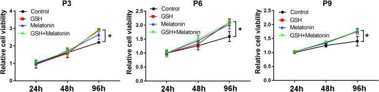
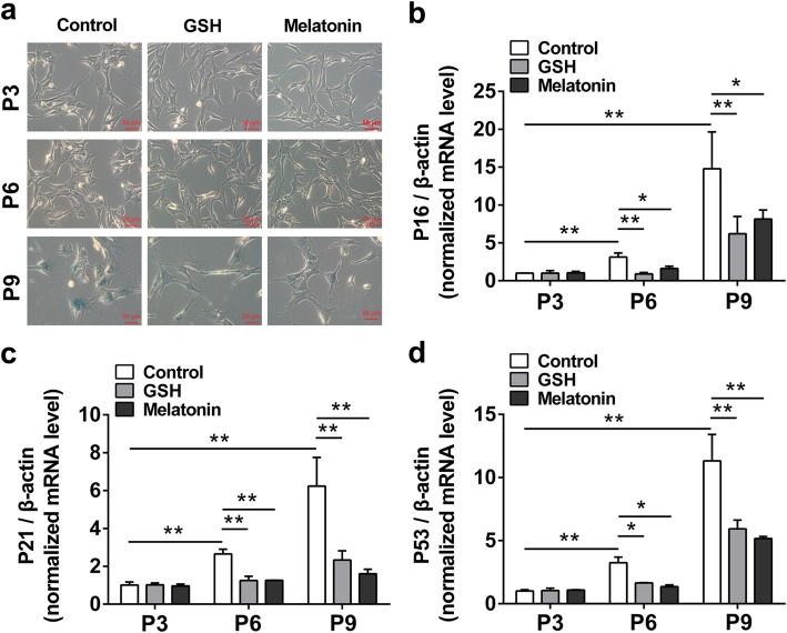
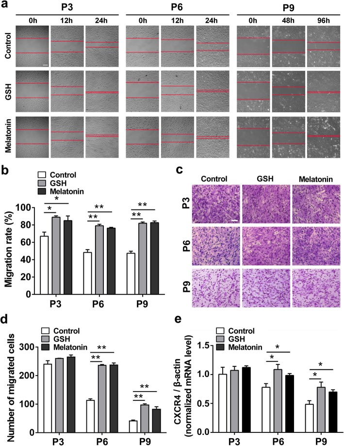
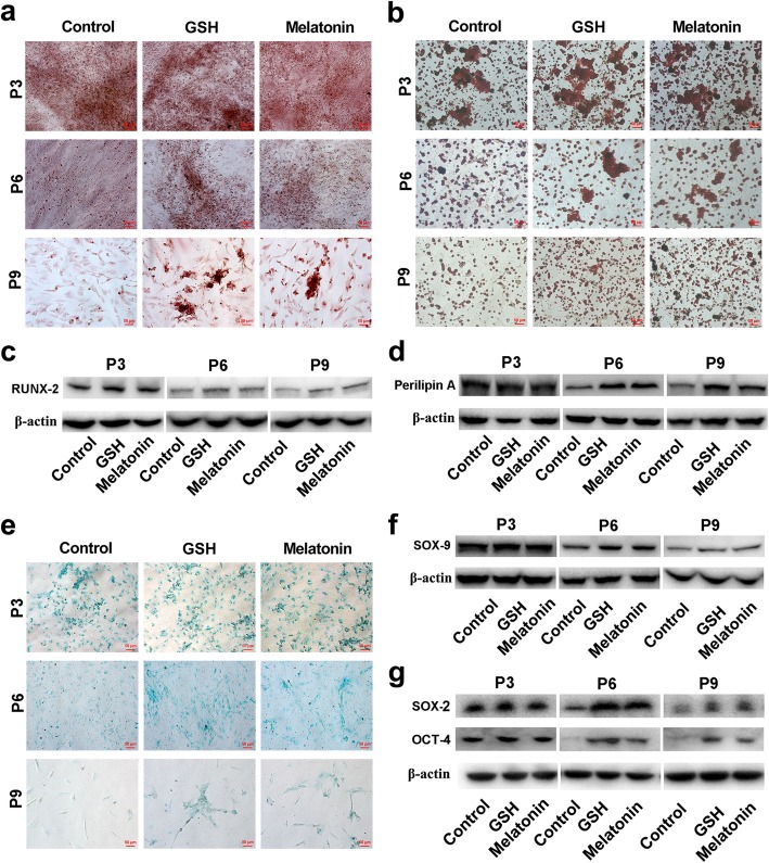
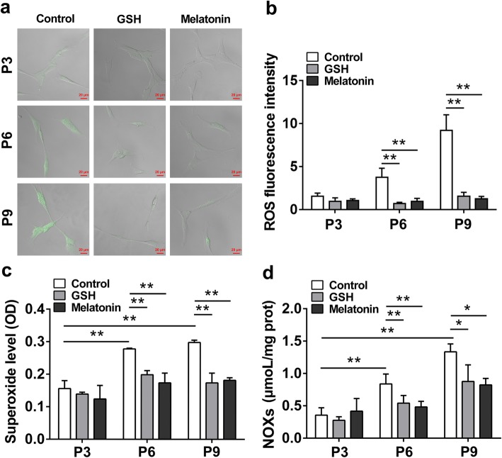
Background: Adipose tissue-derived mesenchymal stem cells (ADSCs) are promising candidates for regenerative medicine. However, long-term in vitro passaging leads to stemness loss and cell senescence of ADSCs, resulting in failure of ADSC-based therapy. Methods: In this study, ADSCs were treated with low dose of antioxidants (reduced glutathione and melatonin) with anti-aging and stem cell protection properties in the in vitro passaging, and the cell functions including stem cell senescence, cell migration, cell multidirectional differentiation potential, and ROS content were carefully analyzed. Results: We found that GSH and melatonin could maintain ADSC cell functions through reducing cell senescence and promoting cell migration, as well as by preserving stemness and multidirectional differentiation potential, through inhibiting ROS generation during long-term expansion of ADSCs. Conclusions: Our results suggested that antioxidant treatment could efficiently prevent the dysfunction and preserve cell functions of ADSCs after long-term passaging, providing a practical strategy to facilitate ADSC-based therapy.
Overview
The claims and statistics of this paper haven’t been checked yet.
Run the check — free, no account needed. It reads the open-access text and checks the sources it cites.
Integrity signals
Registry
Cites
Statistics
Figures
Verification
Claims the paper makes
The claims and statistics of this paper haven’t been checked yet.
Run the check — free, no account needed. It reads the open-access text and checks the sources it cites.
Claims other papers make about this one
Each is checked against this paper’s own text: does it say what the citing paper says it does?
In the citing paper
“In this context, decreased cellular senescence, preserved self-renewal and activation of antioxidant defence pathways have all been observed after melatonin supplementation [460,461].”
From The Role of Antioxidants in the Interplay between Oxidative Stress and Senescence (2022) · cites this paper as [460]
In the citing paper
“Regarding MSCs, it is known that long term culture stimulates ROS generation, thus promoting oxidative stress-induced senescence [460].”
From The Role of Antioxidants in the Interplay between Oxidative Stress and Senescence (2022) · cites this paper as [460]
Self-check
Percentages, sums and reported differences re-computed from the paper’s own numbers.
The self-check needs the paper’s full text.
Citations
Cites
Cellular mechanisms and physiological consequences of redox-dependent signalling
Holmström, K. M., & Finkel, T. (2014) · Nature Reviews Molecular Cell Biology · doi:10.1038/nrm3801 · Cited by 2,093
No place like home: anatomy and function of the stem cell niche
Jones, D. L., & Wagers, A. J. (2007) · Nature Reviews Molecular Cell Biology · doi:10.1038/nrm2319 · Cited by 830
NADPH Oxidases, Reactive Oxygen Species, and the Kidney
Sedeek, M. H., Nasrallah, R., Touyz, R. M., & Hébert, R. (2013) · Journal of the American Society of Nephrology · doi:10.1681/asn.2012111112 · Cited by 476
Manufacturing of primed mesenchymal stromal cells for therapy
Yin, J. Q., Zhu, J., & Ankrum, J. A. (2019) · Nature Biomedical Engineering · doi:10.1038/s41551-018-0325-8 · Cited by 349
Reactive Oxygen Species in Cancer Stem Cells
Shi, X., Zhang, Y., Zheng, J., & Pan, J. (2012) · Antioxidants and Redox Signaling · doi:10.1089/ars.2012.4529 · Cited by 335
Tarocco, A., Caroccia, N., Morciano, G., Wieckowski, M. R., Ancora, G., Garani, G., & Pinton, P. (2019) · Cell Death and Disease · doi:10.1038/s41419-019-1556-7 · Cited by 307
Redox homeostasis: the linchpin in stem cell self-renewal and differentiation
Wang, K., Zhang, T., Dong, Q. G., Nice, E. C., Huang, C., & Wei, Y. (2013) · Cell Death and Disease · doi:10.1038/cddis.2013.50 · Cited by 298
Structure and mechanisms of ROS generation by NADPH oxidases
Magnani, F., & Mattevi, A. (2019) · Current Opinion in Structural Biology · doi:10.1016/j.sbi.2019.03.001 · Cited by 297
Targeted and Persistent 8-Oxoguanine Base Damage at Telomeres Promotes Telomere Loss and Crisis
Fouquerel, E., Barnes, R. P., Uttam, S., Watkins, S. C., Bruchez, M. P., & Opresko, P. L. (2019) · Molecular Cell · doi:10.1016/j.molcel.2019.04.024 · Cited by 275
Radio, N. M., Doctor, J. S., & Witt‐Enderby, P. A. (2006) · Journal of Pineal Research · doi:10.1111/j.1600-079x.2006.00318.x · Cited by 226
Cited by
Sorted by citations
Wang, Y., Cheng, H., Wang, T., Zhang, K., Zhang, Y., & Kang, X. (2023) · Cell Proliferation · doi:10.1111/cpr.13448 · Cited by 243
Self-citationThe Role of Antioxidants in the Interplay between Oxidative Stress and Senescence
Varesi, A., Chirumbolo, S., Campagnoli, L. I. M., Pierella, E., Piccini, G. B., Carrara, A., Ricevuti, G., Scassellati, C., Bonvicini, C., & Pascale, A. (2022) · Antioxidants · doi:10.3390/antiox11071224 · Cited by 145
The Link between Chronic Stress and Accelerated Aging
Yegorov, Y. E., Poznyak, A. V., Nikiforov, N. G., Sobenin, I. A., & Orekhov, A. N. (2020) · Biomedicines · doi:10.3390/biomedicines8070198 · Cited by 147
Role of Nrf2 in cell senescence regulation
Yuan, H., Xu, Y., Luo, Y., Wang, N.-X., & Xiao, J. (2020) · Molecular and Cellular Biochemistry · doi:10.1007/s11010-020-03901-9 · Cited by 104
Liao, N., Su, L., Zheng, Y., Zhao, B., Wu, M., Zhang, D., Yang, H., Liu, X., & Song, J. (2021) · Angewandte Chemie International Edition · doi:10.1002/anie.202106730 · Cited by 94
Self-citation
Metadata
- Authors
- Liao, N., Shi, Y., Zhang, C., Zheng, Y., Wang, Y., Zhao, B., Zeng, Y., Liu, X., & Liu, J.
- Affiliations
- Fujian Medical University (CN)
- Corresponding author
- Liu, Xiaolong
- Liu, Jingfeng
- ORCID
- Venue
- Stem Cell Research & Therapy
- Published
- 2019-10-17
- Volume
- 10
- Issue
- 1
- Pages
- 306-306
- ISSN
- 1757-6512 (ISSN-L)
- PMC
- PMC6798439
- OpenAlex
- W2980985101
- Cited by
- 127
- References
- 40
- Citations by year
- 2026: 7 · 2025: 18 · 2024: 25 · 2023: 25 · 2022: 24 · 2021: 19 · 2020: 8
- FWCI
- 7.00
- Type
- journal article
- Language
- en
- Open access
- gold
- doi.org
- Locations
- Stem Cell Research & Therapy (publishedVersion, cc-by)
- PubMed Central (publishedVersion, cc-by)
- PubMed (publishedVersion)
- DOAJ (DOAJ: Directory of Open Access Journals) (submittedVersion, cc-by-sa)
- Funders
- the National Natural Science Foundation of China (81672376)
- the Youth Scientific Research Project of Fujian Provincial Health and Family Planning Commission (2017-1-85, 2017-2-60, 2018-1-93)
- the Fuzhou Health and Family Planning Science and Technology Project (2017-S-wt2)
- the Startup Fund for scientific research, Fujian Medical University (2016QH080, 2016QH081, 2018QH1198)
- Topics
- Life Sciences › Biochemistry, Genetics and Molecular Biology › Cell Biology › Mesenchymal stem cell research
- Life Sciences › Biochemistry, Genetics and Molecular Biology › Cell Biology › Telomeres, Telomerase, and Senescence
- Life Sciences › Biochemistry, Genetics and Molecular Biology › Molecular Biology › Pluripotent Stem Cells Research
- Keywords
- adipose-derived stem cells, cellular senescence, stemness maintenance, antioxidants, reduced glutathione, melatonin, reactive oxygen species, cell migration, multilineage differentiation, anti-aging
- MeSH terms
- Adipose Tissue, Animals, Antioxidants, Cell Differentiation, Cell Movement, Cells, Cultured, Glutathione, Male, Melatonin, Mice, Inbred C57BL, Stem Cells, Cellular Senescence, Reactive Oxygen Species, Stem Cell Transplantation, Regenerative Medicine, Cell Proliferation, Mice, Mesenchymal Stem Cells
About these checks
Registry, retraction, Cites and statistics results are computed, not judged, with one exception: a printed reference that no index lists is read once by a model before it counts as a citation error. Claims are extracted by a model and kept only when their quote appears verbatim in the text; each cited source is then checked, with a model’s help, for whether it says what the claim relies on. Those verdicts can be wrong — read the claim’s quote and the cited source before relying on one.
Statistics and arithmetic are recomputed, not judged.
Confidence bands are shown only for fields calibrated on our annotated test set. None is calibrated yet, so no band is shown.
What counts as a claim ›How we measure confidence ›How we check this ›
Are you an author, or do you see a problem with this page? Request a review
