

The DOI matches its Crossref record; 54 of the 55 references it cites were found in a registry or an index.
PI3K/Akt signaling pathway is essential for de novo hair follicle regeneration
Open accessAbstract
Background: Cultured epidermal stem cells (Epi-SCs) and skin-derived precursors (SKPs) were capable of reconstituting functional hair follicles after implantation, while the signaling pathways that regulate neogenic hair follicle formation are poorly investigated. In this study, we aimed to understand the interactions between Epi-SCs and SKPs during skin organoid formation and to uncover key signal pathways crucial for de novo hair follicle regeneration. Methods: To track their fate after transplantation, Epi-SCs derived from neonatal C57BL/6 mice were labeled with tdTomato, and SKPs were isolated from neonatal C57BL/6/GFP mice. A mixture of Epi-SCs-tdTomato and SKPs-EGFP in Matrigel was observed under two-photon microscope in culture and after implantation into excisional wounds in nude mice, to observe dynamic migrations of the cells during hair follicle morphogenesis. Signaling communications between the two cell populations were examined by RNA-Seq analysis. Potential signaling pathways revealed by the analysis were validated by targeting the pathways using specific inhibitors to observe a functional loss in de novo hair follicle formation. Results: Two-photon microscopy analysis indicated that when Epi-SCs and SKPs were mixed in Matrigel and cultured, they underwent dynamic migrations resulting in the formation of a bilayer skin-like structure (skin organoid), where Epi-SCs positioned themselves in the outer layer; when the mixture of Epi-SCs and SKPs was grafted into excisional wounds in nude mice, a bilayer structure resembling the epidermis and the dermis formed at the 5th day, and de novo hair follicles generated subsequently. RNA-Seq analysis of the two cell types after incubation in mixture revealed dramatic alterations in gene transcriptome, where PI3K-Akt signaling pathway in Epi-SCs was significantly upregulated; meanwhile, elevated expressions of several growth factors and cytokine potentially activating PI3K were found in SKPs, suggesting active reciprocal communications between them. In addition, inhibition of PI3K or Akt by specific inhibitors markedly suppressed the hair follicle regeneration mediated by Epi-SCs and SKPs. Conclusions: Our data indicate that the PI3K-Akt signaling pathway plays a crucial role in de novo hair follicle regeneration, and the finding may suggest potential therapeutic applications in enhancing hair regeneration.
Overview
The claims and statistics of this paper haven’t been checked yet.
Run the check — free, no account needed. It reads the open-access text and checks the sources it cites.
Integrity signals
Registry
Cites
Statistics
Figures
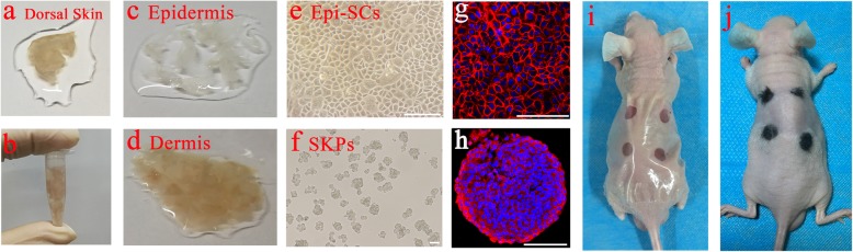
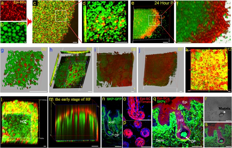
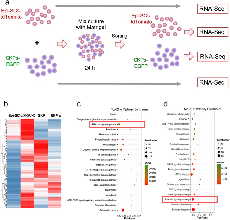
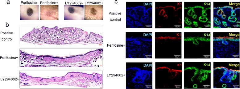
Verification
Claims the paper makes
The claims and statistics of this paper haven’t been checked yet.
Run the check — free, no account needed. It reads the open-access text and checks the sources it cites.
Claims other papers make about this one
Each is checked against this paper’s own text: does it say what the citing paper says it does?
The source backs part of the claim, not all of it — or backs it for a different population, condition or comparison.
In the citing paper
“which is consistent with previous studies reporting that these genes are crucial for the early development of hair follicles and de novo regeneration of hair follicles (44, 45).”
From Reprogramming of three-dimensional microenvironments for in vitro hair follicle induction (2022) · cites this paper as [45]
Experimental — accuracy not yet measured
Self-check
Percentages, sums and reported differences re-computed from the paper’s own numbers.
The self-check needs the paper’s full text.
Citations
Cites
Multilineage potential of adult human mesenchymal stem cells
Pittenger, M. F., Mackay, A. M., Beck, S. C., Jaiswal, R. K., Douglas, R., Mosca, J. D., Moorman, M. A., Simonetti, D. W., Craig, S., & Marshak, D. R. (1999) · Science · doi:10.1126/science.284.5411.143 · Cited by 21,052
Langer, R., & Vacanti, J. P. (1993) · Science · doi:10.1126/science.8493529 · Cited by 9,893
Cellular survival: a play in three Akts
Datta, S. R., Brunet, A., & Greenberg, M. E. (1999) · Genes & Development · doi:10.1101/gad.13.22.2905 · Cited by 4,216
Blume-Jensen, P., & Hunter, T. (2001) · Nature · doi:10.1038/35077225 · Cited by 3,738
Mesenchymal Stem Cells Enhance Wound Healing Through Differentiation and Angiogenesis
Wu, Y., Chen, L., Scott, P. G., & Tredget, E. E. (2007) · Stem Cells · doi:10.1634/stemcells.2007-0226 · Cited by 1,700
Self-citationChen, L., Tredget, E. E., Wu, P. Y. G., & Wu, Y. (2008) · PLoS ONE · doi:10.1371/journal.pone.0001886 · Cited by 1,610
Self-citationIsolation of multipotent adult stem cells from the dermis of mammalian skin
Toma, J. G., Akhavan, M., Fernandes, K. J. L., Barnabé‐Heider, F., Sadikot, A. F., Kaplan, D. R., & Miller, F. D. (2001) · Nature Cell Biology · doi:10.1038/ncb0901-778 · Cited by 1,593
Mangi, A. A., Noiseux, N., Kong, D., He, H., Rezvani, M., Ingwall, J. S., & Dzau, V. J. (2003) · Nature Medicine · doi:10.1038/nm912 · Cited by 1,506
Epidermal homeostasis: a balancing act of stem cells in the skin
Blanpain, C., & Fuchs, E. (2009) · Nature Reviews Molecular Cell Biology · doi:10.1038/nrm2636 · Cited by 1,270
WNT Signals Are Required for the Initiation of Hair Follicle Development
Andl, T., Reddy, S. T., Gaddapara, T., & Millar, S. E. (2002) · Developmental Cell · doi:10.1016/s1534-5807(02)00167-3 · Cited by 1,088
Cited by
Sorted by citations
Functional hair follicle regeneration: an updated review
Ji, S., Zhu, Z., Sun, X., & Fu, X. (2021) · Signal Transduction and Targeted Therapy · doi:10.1038/s41392-020-00441-y · Cited by 265
The PI3K/Akt Pathway: Emerging Roles in Skin Homeostasis and a Group of Non-Malignant Skin Disorders
Teng, Y., Fan, Y., Ma, J., Lü, W., Liu, N., Chen, Y.-F., Pan, W., & Tao, X. (2021) · Cells · doi:10.3390/cells10051219 · Cited by 189
Self-citationRegulation of signaling pathways in hair follicle stem cells
Wang, X., Liu, Y., He, J., Wang, J., Chen, X., & Yang, R. (2022) · Burns & Trauma · doi:10.1093/burnst/tkac022 · Cited by 125
Self-citationKim, M. J., Seong, K., Kim, D. S., Jeong, J. S., Kim, S. Y., Lee, S. S., Yang, S. Y., & An, B. (2022) · Acta Biomaterialia · doi:10.1016/j.actbio.2022.02.011 · Cited by 117
Jin, H., Zou, Z., Chang, H., Shen, Q., Liu, L., & Xing, D. (2021) · Stem Cell Reports · doi:10.1016/j.stemcr.2021.04.015 · Cited by 74
Metadata
- Authors
- Chen, Y., Fan, Z., Wang, X., Mo, M., Zeng, S. B., Xu, R.-H., Wang, X., & Wu, Y.
- Affiliations
- Tsinghua Shenzhen International Graduate School (CN)
- Sun Yat-sen University (CN)
- Tsinghua–Berkeley Shenzhen Institute (CN)
- University of Macau (MO)
- Corresponding author
- Wang, Xusheng
- Wu, Yaojiong
- Venue
- Stem Cell Research & Therapy
- Published
- 2020-04-03
- Volume
- 11
- Issue
- 1
- Pages
- 144-144
- ISSN
- 1757-6512 (ISSN-L)
- PMC
- PMC7118821
- OpenAlex
- W3014739274
- Cited by
- 130
- References
- 55
- Citations by year
- 2026: 21 · 2025: 24 · 2024: 30 · 2023: 16 · 2022: 24 · 2021: 12 · 2020: 3
- FWCI
- 19.06
- Type
- journal article
- Language
- en
- Open access
- gold
- doi.org
- Locations
- Stem Cell Research & Therapy (publishedVersion, cc-by)
- PubMed Central (publishedVersion, cc-by)
- PubMed (publishedVersion)
- DOAJ (DOAJ: Directory of Open Access Journals) (submittedVersion, cc-by-sa)
- Funders
- Natural Science Foundation of China (No. 31571429, 31961160702)
- National Key R&D Program of China (2017YFC1103304)
- Shenzhen Science and Technology Innovation Committee (JCYJ20170818093935581, JCYJ20180307123901314)
- State Key Laboratory of Chemical Oncogenomics fund
- Topics
- Health Sciences › Medicine › Dermatology › Hair Growth and Disorders
- Life Sciences › Biochemistry, Genetics and Molecular Biology › Cell Biology › Skin and Cellular Biology Research
- Health Sciences › Dentistry › General Dentistry › dental development and anomalies
- Keywords
- PI3K/Akt signaling, epidermal stem cells, skin-derived precursors, hair follicle morphogenesis, cell migration, wound healing
- MeSH terms
- Animals, Cells, Cultured, Mice, Inbred C57BL, Mice, Nude, Signal Transduction, Hair Follicle, Phosphatidylinositol 3-Kinases, Proto-Oncogene Proteins c-akt, Mice
About these checks
Registry, retraction, Cites and statistics results are computed, not judged, with one exception: a printed reference that no index lists is read once by a model before it counts as a citation error. Claims are extracted by a model and kept only when their quote appears verbatim in the text; each cited source is then checked, with a model’s help, for whether it says what the claim relies on. Those verdicts can be wrong — read the claim’s quote and the cited source before relying on one.
Statistics and arithmetic are recomputed, not judged.
Confidence bands are shown only for fields calibrated on our annotated test set. None is calibrated yet, so no band is shown.
What counts as a claim ›How we measure confidence ›How we check this ›
Are you an author, or do you see a problem with this page? Request a review
