

The DOI matches its Crossref record; 161 of the 161 references it cites were found in a registry or an index.
Skeletal muscle wasting and renewal: a pivotal role of myokine IL-6
Open accessAbstract
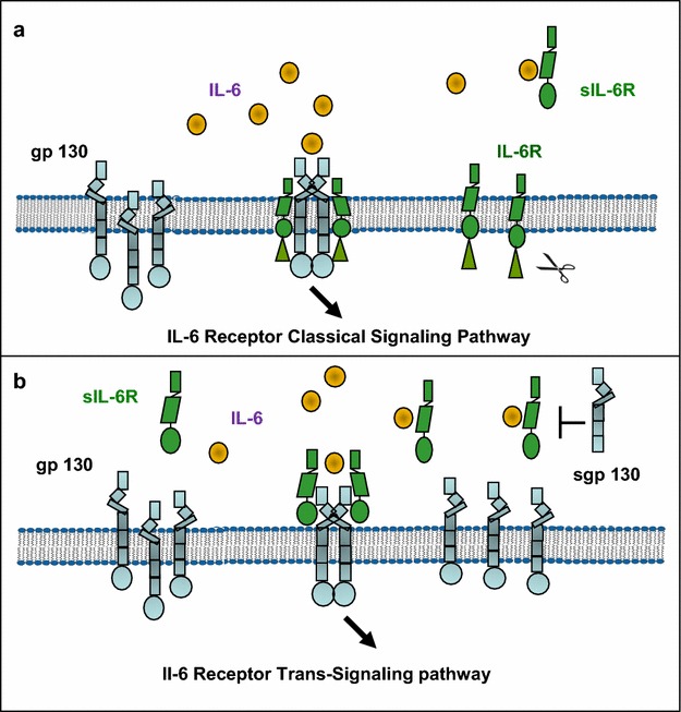
Adult skeletal tissue is composed of heterogeneous population of cells that constantly self-renew by means of a controlled process of activation and proliferation of tissue-resident stem cells named satellite cells. Many growth factors, cytokines and myokines produced by skeletal muscle cells play critical roles in local regulation of the inflammatory process and skeletal muscle regeneration during different pathological conditions. IL-6 is a pleiotropic cytokine released in large amount during infection, autoimmunity and cancer. Low levels of IL-6 can promote activation of satellite cells and myotube regeneration while chronically elevated production promote skeletal muscle wasting. These distinct effects may be explained by a crosstalk of the IL-6/IL-6 receptor and gp130 trans-signaling pathway that oppose to regenerative and anti-inflammatory of the classical IL-6 receptor signaling pathway. Here we discuss on potential therapeutic strategies using monoclonal antibodies to IL-6R for the treatment of skeletal muscle wasting and cachexia. We also highlight on the IL-6/JAK/STAT and FGF/p38αβ MAPK signaling pathways in satellite cell activation and the use of protein kinase inhibitors for tailoring and optimizing satellite cell proliferation during the skeletal muscle renewal. Future investigations on the roles of the IL-6 classical and trans-signaling pathways in both immune and non-immune cells in skeletal muscle tissue will provide new basis for therapeutic approaches to reverse atrophy and degeneration of skeletal muscles in cancer and inflammatory diseases.
Overview
The claims and statistics of this paper haven’t been checked yet.
Run the check — free, no account needed. It reads the open-access text and checks the sources it cites.
Integrity signals
Registry
Cites
Statistics
Figures
Verification
Claims the paper makes
The claims and statistics of this paper haven’t been checked yet.
Run the check — free, no account needed. It reads the open-access text and checks the sources it cites.
Claims other papers make about this one
Each is checked against this paper’s own text: does it say what the citing paper says it does?
The source backs part of the claim, not all of it — or backs it for a different population, condition or comparison.
In the citing paper
“Like TNF-α, IL-6 is considered as a “double-edged sword” with proinflammatory (catabolic) and anti-inflammatory (anabolic) effects (see the review in [46]) according to local immune cells and the cytokines’ microenvironment.”
From Main Pathogenic Mechanisms and Recent Advances in COPD Peripheral Skeletal Muscle Wasting (2023) · cites this paper as [46]
Experimental — accuracy not yet measured
Self-check
Percentages, sums and reported differences re-computed from the paper’s own numbers.
The self-check needs the paper’s full text.
Citations
Cites
1 retracted
Myostatin is a novel tumoral factor that induces cancer cachexia
Lokireddy, S., Wijesoma, I. W., Bonala, S., Wei, M., Sze, S. K., McFarlane, C., Kambadur, R., & Sharma, M. (2012) · Biochemical Journal · doi:10.1042/bj20112024 · Cited by 96
RetractedCalcium's Role in Mechanotransduction during Muscle Development
Damm, T. B., & Egli, M. (2014) · Cellular Physiology and Biochemistry · doi:10.1159/000356667 · Cited by 14,552
Definition and classification of cancer cachexia: an international consensus
Fearon, K., Strasser, F., Anker, S. D., Bosaeus, I. G., Bruera, E. D., Fainsinger, R. L., Jatoi, A., Loprinzi, C. L., Macdonald, N., Mantovani, G., et al. (2011) · The Lancet Oncology · doi:10.1016/s1470-2045(10)70218-7 · Cited by 6,020
Regulation of skeletal muscle mass in mice by a new TGF-p superfamily member
McPherron, A. C., Lawler, A. M., & Lee, S.-J. (1997) · Nature · doi:10.1038/387083a0 · Cited by 4,013
Principles of interleukin (IL)-6-type cytokine signalling and its regulation
HEINRICH, P. C., BEHRMANN, I., HAAN, S., HERMANNS, H. M., MÜLLER-NEWEN, G., & SCHAPER, F. (2003) · Biochemical Journal · doi:10.1042/bj20030407 · Cited by 3,272
The pro- and anti-inflammatory properties of the cytokine interleukin-6
Scheller, J., Chalaris, A., Schmidt-Arras, D., & Rose-John, S. (2011) · Biochimica et Biophysica Acta (BBA) - Molecular Cell Research · doi:10.1016/j.bbamcr.2011.01.034 · Cited by 3,212
Sandri, M., Sandri, C., Gilbert, A., Skurk, C., Calabria, E., Picard, A., Walsh, K., Schiaffino, S., Lecker, S. H., & Goldberg, A. L. (2004) · Cell · doi:10.1016/s0092-8674(04)00400-3 · Cited by 3,012
Muscles, exercise and obesity: skeletal muscle as a secretory organ
Pedersen, B. K., & Febbraio, M. A. (2012) · Nature Reviews Endocrinology · doi:10.1038/nrendo.2012.49 · Cited by 2,909
Bodine, S. C., Stitt, T. N., Gonzalez, M., Kline, W. O., Stover, G. L., Bauerlein, R., Zlotchenko, E., Scrimgeour, A., Lawrence, J. C., Glass, D. J., et al. (2001) · Nature Cell Biology · doi:10.1038/ncb1101-1014 · Cited by 2,752
IL-6 as a keystone cytokine in health and disease
Hunter, C. A., & Jones, S. A. (2015) · Nature Immunology · doi:10.1038/ni.3153 · Cited by 2,511
Cited by
Sorted by citations
Items 201–244 of 244
Potential Regenerative Capacity of physiological serum, L-arginine and IGF-1 on Skeletal Muscle
Alkalamawy, N. (2018) · SVU- International Journal of Veterinary Sciences · doi:10.21608/svu.2018.17935
Varghese, D. A., Christy, A. J., Pratheeba, B., Saraswathy, P., & Ravi, D. S. (2019) · International Journal of Innovative Research in Medical Science · doi:10.23958/ijirms/vol04-i09/742
WITHDRAWN: This article has been withdrawn
Spiegeleer, A. D., Elewaut, D. S., Noortgate, N. J. V. D., Janssens, Y., Debunne, N., Langenhove, S. V., Govindarajan, S., Spiegeleer, B. M. J. D., & Wynendaele, E. (2019) · Biochimica et Biophysica Acta (BBA) - Molecular Basis of Disease · doi:10.1016/j.bbadis.2019.165585
RetractedSugden, C. (2019) · SUNScholar (Stellenbosch University)
Saraf‐Bank, S., Navab, F. S., Rouhani, M. H., Hajhashemy, Z., & Mashhadi, N. S. (2021) · Research Square · doi:10.21203/rs.3.rs-1176764/v1
Kutty, P. K., & Kutty, M. K. (2021) · Springer eBooks · doi:10.1007/978-981-33-6408-0_1
Exercício físico e sistema purinérgico
Falqueto, H., Júnior, J. L. R., Cardoso, A. M., & Manfredi, L. H. (2021) · Editora UFFS eBooks · doi:10.7476/9786586545494.0004
van de Worp, W. R. P. H. (2022) · Research Publications (Maastricht University) · doi:10.26481/dis.20221214ww
Yuan, Q., Yuan, L., Zhang, L., Liu, L., Zhou, S., Zhang, T., Ding, C., & Guan, W. (2023) · Research Square · doi:10.21203/rs.3.rs-2894242/v1
Role of IL-6 and IGF-1 in sarcopenia
Riviati, N., Legiran, L., Indrajaya, T., & Saleh, I. (2023) · Jurnal Aisyah Jurnal Ilmu Kesehatan · doi:10.30604/jika.v8i1.1625
The role of genes and myokines of developing sarcopenia: A review
Valeeva, F. V., Rodygina, Z. A., & Yilmaz, T. S. (2024) · Consilium Medicum · doi:10.26442/20751753.2024.4.202735
An Assessment of Stress Patterns among Cadets of Nigerian Defence Academy, Kaduna, Nigeria
Garba, J., Okorie, A., & Aigboje, H. M. (2022) · NIU journal of social sciences. · doi:10.58709/niujss.v8i2.1463
Campanari, D. D. (2024) · Digital Library of Theses and Dissertations (Universidade de São Paulo) · doi:10.11606/t.17.2024.tde-13052024-155734
Neto, E. M. D. M. (2016) · Digital Library of Theses and Dissertations (Universidade de São Paulo) · doi:10.11606/t.42.2016.tde-16052017-135255
Effects of Medium Cut‐Off Dialyzers on Fat Tissue and Muscle Mass Indices
Bildacı, Y. D., Eşme, M., Korucu, B., Erbay, E., Yigitbasi, B., Dilek, B., Senturk, Z. S., Çavdar, C., & Değer, S. M. (2025) · Artificial Organs · doi:10.1111/aor.14997
Xue, C. (2025) · American journal of clinical and experimental immunology. · doi:10.62347/vbur5104
Yin, M., Ma, J., Li, R., Ma, W. J., Zhou, M., & Song, X. (2025) · Archives of Gerontology and Geriatrics · doi:10.1016/j.archger.2025.105971
Fundamentals of Muscle Damage and Repair of Skeletal Muscle
Kamal, M. J., Johnson, A., Wageh, M., Snijders, T., & Parise, G. (2025) · Springer eBooks · doi:10.1007/978-3-031-44270-4_2
Николаевна, Щ. Т. (2025) · Сибирский научный медицинский журнал · doi:10.18699/ssmj20250406
Amjad, I., Niazi, I. K., Kumari, N., Ghani, U., Rashid, U., Duarte, F. C. K., Fortuna, F., Iglesias, S. L., González, D., Sumich, A. L., et al. (2025) · PLoS ONE · doi:10.1371/journal.pone.0338730
Zhuang, S., Russell, A., Guo, Y., Xu, Y., & Xiao, W. (2023) · Figshare · doi:10.6084/m9.figshare.22617479
Chen, B., Li, S., Lin, S., & Dong, H. (2024) · Figshare · doi:10.6084/m9.figshare.25834254
Chen, B., Li, S., Lin, S., & Dong, H. (2024) · Figshare · doi:10.6084/m9.figshare.26720883
Chen, B., Li, S., Lin, S., & Dong, H. (2024) · Figshare · doi:10.6084/m9.figshare.25834115
Jiang, W., Huang, S., Ma, S., Gong, Y., Fu, Z., Zhou, L., Hu, W., Mao, G., Ma, Z., Yang, L., et al. (2021) · Figshare · doi:10.6084/m9.figshare.13710529
Chen, B., Li, S., Lin, S., & Dong, H. (2024) · Figshare · doi:10.6084/m9.figshare.26720886
Teixeira, L. A. D. C., Soares, L. A., Oliveira, M. C. D. M., Mourão, M. F. D. S., Nobre, J. N. P., Figueiredo, P. H. S., Costa, H. S., Parentoni, A. N., Mendonça, V. A., Taïar, R., et al. (2025) · Scientific Reports · doi:10.1038/s41598-025-33291-1
Manganese Exposure Is Associated with Reduced Grip Strength: Evidence from Humans and Mice
Fang, P., Tang, C., Wei, S., Lu, W., Liu, S., Ma, X., Zhang, L., Liang, G., Wang, J., Peng, Y., et al. (2025) · Toxics · doi:10.3390/toxics14010031
Shin, J. Y., Park, J. H., Lim, J. H., Cho, B. O., Kang, J. S., & Jang, S. I. (2026) · Food Science and Biotechnology · doi:10.1007/s10068-026-02122-y
Identification of new levers to fight muscle atrophy : benefit of the hibernating brown bear model
Cussonneau, L. (2022) · theses.fr (ABES) · doi:10.70675/1a11a81bzf829z4ba2zba9az6f3f4841c773
Impact of moderate treadmill training on cytokine levels in rats: influence of age and sex
Santo, R. F. D. E., Zhang, Y., Ro, J. Y., & Silva, J. T. D. (2026) · Frontiers in Aging · doi:10.3389/fragi.2026.1794896
Narloch, W., Jaskulak, M., Antoniak-Pietrynczak, K., & Zorena, K. (2026) · Diagnostics · doi:10.3390/diagnostics16101459
Lee, J., Shin, S., Park, J., Kim, Y., Choi, H.-Y., Bu, Y., Cho, J.-H., Choi, Y., & Lee, K. (2026) · Journal of Ethnopharmacology · doi:10.1016/j.jep.2026.121835
Lu, Z., Wang, L., Chen, S., Huo, Z., Tong, N., Li, N., & Xu, H. (2026) · Food Bioscience · doi:10.1016/j.fbio.2026.109743
Ren, Y., Zheng, C., Wang, M., & Wang, Y. (2026) · Frontiers in Endocrinology · doi:10.3389/fendo.2026.1794009
Maretti, E., Recchia, B., Votta, A., Guarnaccia, E., Cesare, F. D., Cannella, G., Rustichelli, C., Seliktar, D., Fuoco, C., Gargioli, C., et al. (2026) · Journal of Controlled Release · doi:10.1016/j.jconrel.2026.115332
IL-6 rs1800795 Polymorphism in Youth Professional Football Players and Non-Athletic Controls
Aslan, B. T., Ziya, R., Polat, T., Yılmaz, Ö. Ö., Ekmekçi, R., & Ulucan, K. (2026) · journal of new results in science · doi:10.54187/jnrs.1953435
Lee, W., 양성재, Yu, H., Kim, Y., Cho, S., Kim, H. Y., Kim, U., Kong, H., & Kim, S. B. (2026) · International Journal of Molecular Sciences · doi:10.3390/ijms27177894
CORDOVA, C. O., Morais-Junior, G. S., Moraes, C. F., Camargos, E. F., Louzada, L. L., & Nóbrega, O. D. T. (2026) · Journal of Gerontology and Geriatrics · doi:10.3390/jgg74030030
Srpčič, A., Miš, K., Gantar, B. Ž. B., Dolinar, K., Mjaaseth, U. N., Rustan, A. C., Kase, E. T., Lakota, K., Pirkmajer, K. P., & Pirkmajer, S. (2026) · American Journal of Physiology-Cell Physiology · doi:10.1152/ajpcell.00264.2026
Metadata
- Authors
- Belizário, J. E., Fontes-Oliveira, C. C., Borges, J. P., Kashiabara, J. A., & Vannier, E.
- Affiliations
- Universidade de São Paulo (BR)
- Tufts Medical Center (US)
- Corresponding author
- Vannier, Edouard
- Venue
- SpringerPlus
- Published
- 2016-05-13
- Volume
- 5
- Issue
- 1
- Pages
- 619-619
- ISSN
- 2193-1801 (ISSN-L)
- PMC
- PMC4870483
- OpenAlex
- W2388738573
- Cited by
- 244
- References
- 161
- Citations by year
- 2026: 22 · 2025: 28 · 2024: 36 · 2023: 32 · 2022: 32 · 2021: 23 · 2020: 23 · 2019: 21 · 2018: 18 · 2017: 8 · 2016: 1
- FWCI
- 1.45
- Type
- journal article
- Language
- en
- Open access
- diamond
- doi.org
- Locations
- SpringerPlus (publishedVersion, cc-by)
- Europe PMC (PubMed Central) (publishedVersion, cc-by)
- PubMed Central (publishedVersion, cc-by)
- PubMed (publishedVersion)
- Lund University Publications (Lund University) (submittedVersion)
- Funders
- Fundação de Amparo à Pesquisa do Estado de São Paulo (2001/0100-7)
- Conselho Nacional de Desenvolvimento Científico e Tecnológico (486048/2011)
- Topics
- Life Sciences › Biochemistry, Genetics and Molecular Biology › Molecular Biology › Muscle Physiology and Disorders
- Life Sciences › Immunology and Microbiology › Immunology › Cytokine Signaling Pathways and Interactions
- Health Sciences › Medicine › Physiology (medical) › Exercise and Physiological Responses
- Keywords
- skeletal muscle atrophy, cachexia, interleukin-6, myokines, satellite cells, muscle regeneration, JAK/STAT signaling, protein kinase inhibitors, muscle atrophy, muscle degeneration, cancer cachexia, inflammatory diseases
About these checks
Registry, retraction, Cites and statistics results are computed, not judged, with one exception: a printed reference that no index lists is read once by a model before it counts as a citation error. Claims are extracted by a model and kept only when their quote appears verbatim in the text; each cited source is then checked, with a model’s help, for whether it says what the claim relies on. Those verdicts can be wrong — read the claim’s quote and the cited source before relying on one.
Statistics and arithmetic are recomputed, not judged.
Confidence bands are shown only for fields calibrated on our annotated test set. None is calibrated yet, so no band is shown.
What counts as a claim ›How we measure confidence ›How we check this ›
Are you an author, or do you see a problem with this page? Request a review
