

The DOI matches its Crossref record; 45 of the 45 references it cites were found in a registry or an index.
RNA binding proteins co-localize with small tau inclusions in tauopathy
Open accessAbstract
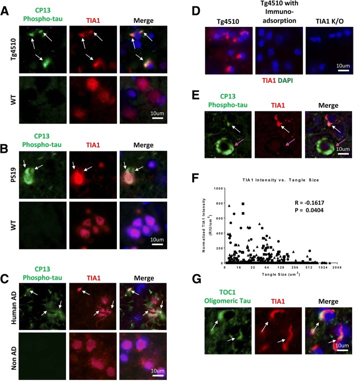
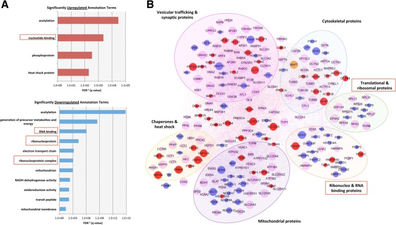
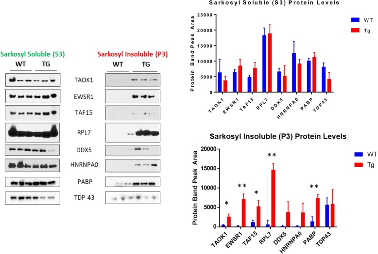
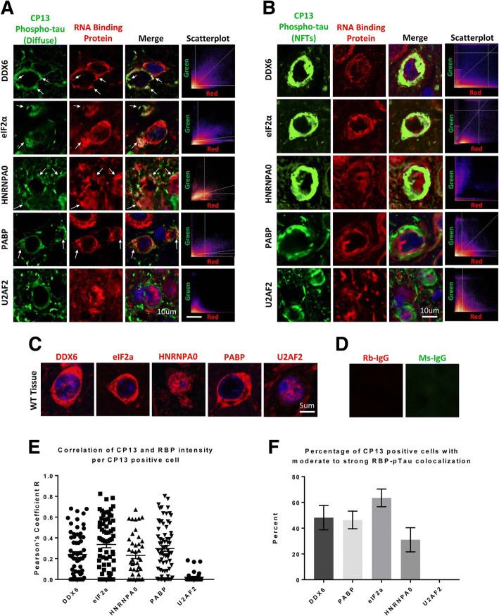
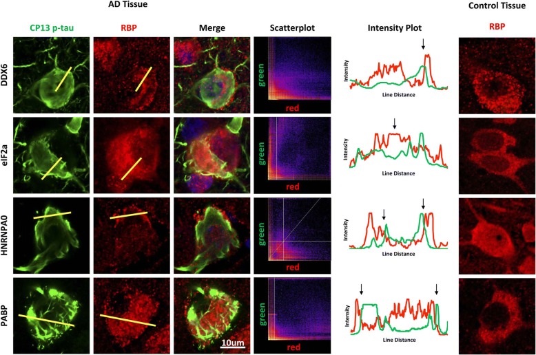
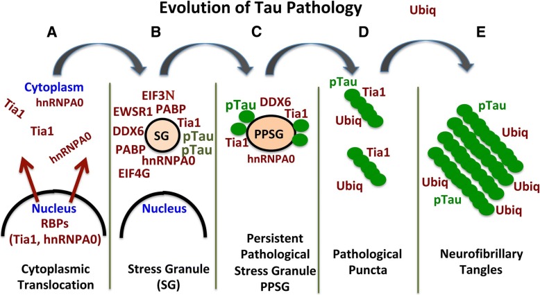
The development of insoluble, intracellular neurofibrillary tangles composed of the microtubule-associated protein tau is a defining feature of tauopathies, including Alzheimer's disease (AD). Accumulating evidence suggests that tau pathology co-localizes with RNA binding proteins (RBPs) that are known markers for stress granules (SGs). Here we used proteomics to determine how the network of tau binding proteins changes with disease in the rTg4510 mouse, and then followed up with immunohistochemistry to identify RNA binding proteins that co-localize with tau pathology. The tau interactome networks revealed striking disease-related changes in interactions between tau and a multiple RBPs, and biochemical fractionation studies demonstrated that many of these proteins including hnRNPA0, EWSR1, PABP and RPL7 form insoluble aggregates as tau pathology develops. Immunohistochemical analysis of mouse and human brain tissues suggest a model of evolving pathological interaction, in which RBPs co-localize with pathological phospho-tau but occur adjacent to larger pathological tau inclusions. We suggest a model in which tau initially interacts with RBPs in small complexes, but evolves into isolated aggregated inclusions as tau pathology matures.
Overview
The claims and statistics of this paper haven’t been checked yet.
Run the check — free, no account needed. It reads the open-access text and checks the sources it cites.
Integrity signals
Registry
Cites
Statistics
Figures
Verification
Claims the paper makes
The claims and statistics of this paper haven’t been checked yet.
Run the check — free, no account needed. It reads the open-access text and checks the sources it cites.
Claims other papers make about this one
Each is checked against this paper’s own text: does it say what the citing paper says it does?
The source backs part of the claim, not all of it — or backs it for a different population, condition or comparison.
In the citing paper
“The cell body MAPT pathology is particularly important because this pathology colocalizes with markers of the translational stress response, such as stress granule proteins HNRNPA2B1, TIA1, EIF3n and PABP, and also neuronal death (as shown by cleaved caspase 3 and loss of NeuN positive cells) (Ash et al., 2014; Apicco et al., 2018; Maziuk et al., 2018; Apicco et al., 2019; Jiang et al., 2019, 2021”
From β-amyloid accumulation enhances microtubule associated protein tau pathology in an APPNL-G-F/MAPTP301S mouse model of Alzheimer’s disease (2024) · cites this paper as Maziuk et al., 2018
Experimental — accuracy not yet measuredIn the citing paper
“previous analyses of the AD brain detergent-insoluble proteome, which have repeatedly demonstrated the association of U1 small nuclear ribonucleoproteins (U1 snRNPs) and other RNA-binding proteins with tau tangles [133–137].”
From Systems-based proteomics to resolve the biology of Alzheimer’s disease beyond amyloid and tau (2021) · cites this paper as [137]
In the citing paper
“Interestingly, our previous studies demonstrated that many of the U1 snRNPs colocalize with neurofibrillary tangles and paired helical filaments in AD brain [24, 32–34], and that accumulation of insoluble snRNPs correlates strongly with both amyloid and Tau pathology [32–37].”
From Deep proteomic network analysis of Alzheimer’s disease brain reveals alterations in RNA binding proteins and RNA splicing associated with disease (2018) · cites this paper as [36]
Self-check
Percentages, sums and reported differences re-computed from the paper’s own numbers.
The self-check needs the paper’s full text.
Citations
Cites
Amyloid oligomers exacerbate tau pathology in a mouse model of tauopathy
Selenica, M.-L. B., Brownlow, M., Jimenez, J. P., Lee, D. C., Pena, G., Dickey, C. A., Gordon, M. N., & Morgan, D. (2013) · Neurodegenerative Diseases · doi:10.1159/000337230 · Cited by 10,359
Liquid phase condensation in cell physiology and disease
Shin, Y., & Brangwynne, C. P. (2017) · Science · doi:10.1126/science.aaf4382 · Cited by 4,143
Molliex, A., Temirov, J. P., Lee, J., Coughlin, M., Kanagaraj, A., Kim, H. J., Mittag, T., & Taylor, J. P. (2015) · Cell · doi:10.1016/j.cell.2015.09.015 · Cited by 2,690
Phase transitions in the assembly of multivalent signalling proteins
Li, P., Banjade, S., Cheng, H., Kim, S., Chen, B., Guo, L., Llaguno, M. C., Hollingsworth, J. V., King, D., Banani, S. F., et al. (2012) · Nature · doi:10.1038/nature10879 · Cited by 2,565
Tau Suppression in a Neurodegenerative Mouse Model Improves Memory Function
SantaCruz, K., Lewis, J., Spires, T., Paulson, J., Kotilinek, L., Ingelsson, M., Guimaraes, A., DeTure, M., Ramsden, M., McGowan, E., et al. (2005) · Science · doi:10.1126/science.1113694 · Cited by 2,014
Nott, T. J., Petsalaki, E., Farber, P., Jervis, D., Fussner, E., Plochowietz, A., Craggs, T. D., Bazett-Jones, D. P., Pawson, T., Forman-Kay, J. D., et al. (2015) · Molecular Cell · doi:10.1016/j.molcel.2015.01.013 · Cited by 1,803
Formation and Maturation of Phase-Separated Liquid Droplets by RNA-Binding Proteins
Lin, Y., Protter, D. S. W., Rosen, M. K., & Parker, R. (2015) · Molecular Cell · doi:10.1016/j.molcel.2015.08.018 · Cited by 1,593
Tau protein liquid–liquid phase separation can initiate tau aggregation
Wegmann, S., Eftekharzadeh, B., Tepper, K., Zoltowska, K. M., Bennett, R. E., Dujardin, S., Laskowski, P. R., MacKenzie, D., Kamath, T., Commins, C., et al. (2018) · The EMBO Journal · doi:10.15252/embj.201798049 · Cited by 1,102
Liu‐Yesucevitz, L., Bilgutay, A. N., Zhang, Y., Vanderwyde, T., Citro, A., Mehta, T., Zaarur, N., McKee, A. C., Bowser, R. P., Sherman, M. Y., et al. (2010) · PLoS ONE · doi:10.1371/journal.pone.0013250 · Cited by 637
Self-citationTherapeutic reduction of ataxin-2 extends lifespan and reduces pathology in TDP-43 mice
Becker, L. A., Huang, B., Bieri, G., Ma, R., Knowles, D. A., Jafar-Nejad, P., Messing, J., Kim, H. J., Soriano, A., Auburger, G., et al. (2017) · Nature · doi:10.1038/nature22038 · Cited by 571
Sun, Z., Diaz, Z., Fang, X., Hart, M. P., Chesi, A., Shorter, J., & Gitler, A. D. (2011) · PLoS Biology · doi:10.1371/journal.pbio.1000614 · Cited by 461
Atomic structures of low-complexity protein segments reveal kinked β sheets that assemble networks
Hughes, M. P., Sawaya, M. R., Boyer, D. R., Goldschmidt, L., Rodríguez, J. A., Cascio, D., Chong, L., Gonen, T., & Eisenberg, D. S. (2018) · Science · doi:10.1126/science.aan6398 · Cited by 451
Local Nucleation of Microtubule Bundles through Tubulin Concentration into a Condensed Tau Phase
Hernández-Vega, A., Braun, M., Scharrel, L., Jahnel, M., Wegmann, S., Hyman, B. T., Alberti, S., Diez, S., & Hyman, A. A. (2017) · Cell Reports · doi:10.1016/j.celrep.2017.08.042 · Cited by 395
A yeast functional screen predicts new candidate ALS disease genes
Couthouis, J., Hart, M. P., Shorter, J., DeJesus‐Hernandez, M., Erion, R., Oristano, R., Liu, A. X., Ramos, D. M., Jethava, N., Hosangadi, D., et al. (2011) · Proceedings of the National Academy of Sciences · doi:10.1073/pnas.1109434108 · Cited by 389
Kim, H., Raphael, A. R., LaDow, E., McGurk, L., Weber, R. A., Trojanowski, J. Q., Lee, V. M. ‐., Finkbeiner, S. M., Gitler, A. D., & Bonini, N. M. (2013) · Nature Genetics · doi:10.1038/ng.2853 · Cited by 383
Mechanistic insights into mammalian stress granule dynamics
Panas, M. D., Ivanov, P. V., & Anderson, P. (2016) · The Journal of Cell Biology · doi:10.1083/jcb.201609081 · Cited by 374
Interaction of tau with the RNA-Binding Protein TIA1 Regulates tau Pathophysiology and Toxicity
Vanderweyde, T. E., Apicco, D. J., Youmans-Kidder, K., Ash, P. E. A., Cook, C. N., da Rocha, E. L., Jansen‐West, K. R., Frame, A. A., Citro, A., Leszyk, J., et al. (2016) · Cell Reports · doi:10.1016/j.celrep.2016.04.045 · Cited by 346
Self-citationU1 small nuclear ribonucleoprotein complex and RNA splicing alterations in Alzheimer’s disease
Bai, B., Hales, C. M., Chen, P.-C., Gozal, Y., Dammer, E. B., Fritz, J. J., Wang, X., Xia, Q., Duong, D. M., Street, C., et al. (2013) · Proceedings of the National Academy of Sciences · doi:10.1073/pnas.1310249110 · Cited by 335
RNA stores tau reversibly in complex coacervates
Zhang, X., Lin, Y., Eschmann, N. A., Zhou, H., Rauch, J. N., Hernández, I., Guzman, E., Kosik, K. S., & Han, S. (2017) · PLoS Biology · doi:10.1371/journal.pbio.2002183 · Cited by 312
TDP-43 gains function due to perturbed autoregulation in a Tardbp knock-in mouse model of ALS-FTD
White, M. A., Kim, E., Duffy, A., Adalbert, R., Phillips, B. U., Peters, O. M., Stephenson, J., Yang, S., Massenzio, F., Lin, Z., et al. (2018) · Nature Neuroscience · doi:10.1038/s41593-018-0113-5 · Cited by 289
Lost after translation: missorting of Tau protein and consequences for Alzheimer disease
Zempel, H., & Mandelkow�, E. (2014) · Trends in Neurosciences · doi:10.1016/j.tins.2014.08.004 · Cited by 284
Evaluating the role of the FUS/TLS-related gene EWSR1 in amyotrophic lateral sclerosis
Couthouis, J., Hart, M. P., Erion, R., King, O. D., Diaz, Z., Nakaya, T., Ibrahim, F., Kim, H.-J., Mojsilovic‐Petrovic, J., Panossian, S., et al. (2012) · Human Molecular Genetics · doi:10.1093/hmg/dds116 · Cited by 284
Reducing the RNA binding protein TIA1 protects against tau-mediated neurodegeneration in vivo
Apicco, D. J., Ash, P. E. A., Maziuk, B., LeBlang, C., Medalla, M., Al Abdullatif, A., Ferragud, A., Botelho, E., Ballance, H. I., Dhawan, U., et al. (2018) · Nature Neuroscience · doi:10.1038/s41593-017-0022-z · Cited by 281 · cited 6×
“The association of phosphorylated tau with the stress granule protein TIA1 raises the possibility that other RBPs and ribosomal proteins might also interact with tau [1, 25, 26, 37].”
BackgroundInfluentialSelf-citationContrasting Pathology of the Stress Granule Proteins TIA-1 and G3BP in Tauopathies
Vanderweyde, T. E., Yu, W. H., Varnum, M. M., Liu‐Yesucevitz, L., Citro, A., Ikezu, T., Duff, K., & Wolozin, B. L. (2012) · Journal of Neuroscience · doi:10.1523/jneurosci.1592-12.2012 · Cited by 235
Self-citationKanaan, N. M., Pigino, G. F., Brady, S. T., Lazarov, O., Binder, L. I., & Morfini, G. A. (2012) · Experimental Neurology · doi:10.1016/j.expneurol.2012.06.003 · Cited by 215
Meier, S., Bell, M., Lyons, D. N., Rodriguez-Rivera, J., Ingram, A., Fontaine, S. N., Mechas, E., Chen, J., Wolozin, B., LeVine, H., et al. (2016) · The Journal of Neuroscience · doi:10.1523/jneurosci.3029-15.2016 · Cited by 190
Self-citationWelander distal myopathy is caused by a mutation in the RNA‐binding protein TIA1
Hackman, P., Sarparanta, J., Lehtinen, S., Vihola, A., Evilä, A., Jonson, P. H., Luque, H., Kere, J., Screen, M., Chinnery, P. F., et al. (2012) · Annals of Neurology · doi:10.1002/ana.23831 · Cited by 159
Dysregulation of RNA Binding Protein Aggregation in Neurodegenerative Disorders
Maziuk, B. F., Ballance, H. I., & Wolozin, B. L. (2017) · Frontiers in Molecular Neuroscience · doi:10.3389/fnmol.2017.00089 · Cited by 147
Self-citationGunawardana, C. G., Mehrabian, M., Wang, X., Mueller, I., Lubambo, I., Jonkman, J. E. N., Wang, H., & Schmitt‐Ulms, G. (2015) · Molecular & Cellular Proteomics · doi:10.1074/mcp.m115.050724 · Cited by 127
Dysregulation of autophagy and stress granule-related proteins in stress-driven Tau pathology
Silva, J. M., Rodrigues, S., Sampaio‐Marques, B., Gomes, P., Neves‐Carvalho, A., Dioli, C., Soares‐Cunha, C., Mazuik, B. F., Takashima, A., Ludovico, P., et al. (2018) · Cell Death and Differentiation · doi:10.1038/s41418-018-0217-1 · Cited by 123
Self-citationAltered Tau Isoform Ratio Caused by Loss of FUS and SFPQ Function Leads to FTLD-like Phenotypes
Ishigaki, S., Fujioka, Y., Okada, Y., Riku, Y., Udagawa, T., Honda, D., Yokoi, S., Endo, K., Ikenaka, K., Takagi, S., et al. (2017) · Cell Reports · doi:10.1016/j.celrep.2017.01.013 · Cited by 118
Sharp, T. H., Koster, A. J., & Gros, P. (2016) · Cell Reports · doi:10.1016/j.celrep.2016.03.002 · Cited by 115
The role of ubiquitin proteasomal system and autophagy-lysosome pathway in Alzheimer’s disease
Zhang, Y., Chen, X., Zhao, Y., Ponnusamy, M., & Liu, Y. (2017) · Reviews in the Neurosciences · doi:10.1515/revneuro-2017-0013 · Cited by 99
Aggregates of Small Nuclear Ribonucleic Acids ( snRNAs ) in Alzheimer's Disease
Hales, C. M., Dammer, E. B., Diner, I., Yi, H., Seyfried, N. T., Gearing, M., Glass, J. D., Montine, T. J., Levey, A. I., & Lah, J. J. (2014) · Brain Pathology · doi:10.1111/bpa.12133 · Cited by 89
Gu, J., Chen, F., Iqbal, K., Gong, C., Wang, X., & Liu, F. (2017) · Journal of Biological Chemistry · doi:10.1074/jbc.m117.783498 · Cited by 88
Thompson, A. D., Scaglione, K. M., Prensner, J. R., Gillies, A. T., Chinnaiyan, A. M., Paulson, H. L., Jinwal, U. K., Dickey, C. A., & Gestwicki, J. E. (2012) · ACS Chemical Biology · doi:10.1021/cb3002599 · Cited by 84
Meier, S. E., Bell, M. C., Lyons, D. N., Ingram, A., Chen, J., Gensel, J. C., Zhu, H., Nelson, P. T., & Abisambra, J. F. (2015) · Journal of Alzheimer s Disease · doi:10.3233/jad-150298 · Cited by 84
Self-citationHypo- and Hyper-Assembly Diseases of RNA–Protein Complexes
Shukla, S., & Parker, R. (2016) · Trends in Molecular Medicine · doi:10.1016/j.molmed.2016.05.005 · Cited by 64
Fujita, K., Ito, H., Nakano, S., Kinoshita, Y., Wate, R., & Kusaka, H. (2008) · Acta Neuropathologica · doi:10.1007/s00401-008-0415-x · Cited by 63
Begcevic, I., Kosanam, H., Martínez‐Morillo, E., Dimitromanolakis, A., Diamandis, P., Kuzmanov, U., Hazrati, L., & Diamandis, E. P. (2013) · Clinical Proteomics · doi:10.1186/1559-0275-10-5 · Cited by 60
Hales, C. M., Seyfried, N. T., Dammer, E. B., Duong, D., Yi, H., Gearing, M., Troncoso, J. C., Mufson, E. J., Thambisetty, M., Levey, A. I., et al. (2014) · Molecular Neurodegeneration · doi:10.1186/1750-1326-9-15 · Cited by 58
Clinical and neuropathological features of ALS/FTD with TIA1 mutations
Hirsch‐Reinshagen, V., Pottier, C. P., Nicholson, A. M., Baker, M. C., Hsiung, G. R., Krieger, C., Sengdy, P., Boylan, K., Dickson, D. W., Mesulam, M. M., et al. (2017) · Acta Neuropathologica Communications · doi:10.1186/s40478-017-0493-x · Cited by 56
Sun, Y., Ip, P., & Chakrabartty, A. (2017) · Journal of Visualized Experiments · doi:10.3791/56188 · Cited by 48
Exposure of the Amino Terminus of Tau Is a Pathological Event in Multiple Tauopathies.
Combs, B., & Kanaan, N. (2017) · American Journal of Pathology · doi:10.1016/j.ajpath.2017.01.019 · Cited by 36
Ikezu, T., Chen, C.-D., DeLeo, A. M., Zeldich, E., Fallin, M. D., Kanaan, N. M., Lunetta, K. L., Abraham, C. R., Logue, M. W., & Farrer, L. A. (2018) · Journal of Neuroimmune Pharmacology · doi:10.1007/s11481-018-9781-x · Cited by 22
Cited by
Sorted by citations
Liquid–Liquid Phase Separation in Disease
Alberti, S., & Dormann, D. (2019) · Annual Review of Genetics · doi:10.1146/annurev-genet-112618-043527 · Cited by 871
Stress granules and neurodegeneration
Wolozin, B., & Ivanov, P. (2019) · Nature Reviews Neuroscience · doi:10.1038/s41583-019-0222-5 · Cited by 810
Self-citationLiquid-liquid phase separation induces pathogenic tau conformations in vitro
Kanaan, N. M., Hamel, C., Grabinski, T., & Combs, B. (2020) · Nature Communications · doi:10.1038/s41467-020-16580-3 · Cited by 397
Hofweber, M., & Dormann, D. (2018) · Journal of Biological Chemistry · doi:10.1074/jbc.tm118.001189 · Cited by 398
Zhao, J., Fu, Y., Yamazaki, Y., Ren, Y., Davis, M. D., Liu, C.-C., Lu, W., Wang, X., Chen, K., Cherukuri, Y., et al. (2020) · Nature Communications · doi:10.1038/s41467-020-19264-0 · Cited by 374
Metadata
- Authors
- Maziuk, B. F., Apicco, D. J., Cruz, A. L., Jiang, L., Ash, P. E. A., da Rocha, E. L., Zhang, C., Yu, W. H., Leszyk, J., Abisambra, J. F., Li, H., & Wolozin, B.
- Affiliations
- Boston University (US)
- Mayo Clinic (US)
- Columbia University Irving Medical Center (US)
- University of Massachusetts Chan Medical School (US)
- University of Kentucky (US)
- Corresponding author
- Wolozin, Benjamin
- ORCID
- Venue
- Acta Neuropathologica Communications
- Published
- 2018-08-01
- Volume
- 6
- Issue
- 1
- Pages
- 71-71
- ISSN
- 2051-5960 (ISSN-L)
- PMC
- PMC6069705
- OpenAlex
- W2887193453
- Cited by
- 151
- References
- 45
- Citations by year
- 2026: 18 · 2025: 17 · 2024: 14 · 2023: 20 · 2022: 18 · 2021: 17 · 2020: 25 · 2019: 17 · 2018: 5
- FWCI
- 10.98
- Type
- journal article
- Language
- en
- Open access
- gold
- doi.org
- Locations
- Acta Neuropathologica Communications (publishedVersion, cc-by)
- Europe PMC (PubMed Central) (publishedVersion, other-oa)
- PubMed Central (publishedVersion, cc-by)
- PubMed (publishedVersion)
- eScholarship@UMassChan (submittedVersion, cc-by)
- UKnowledge (University of Kentucky) (submittedVersion, cc-by)
- DOAJ (DOAJ: Directory of Open Access Journals) (submittedVersion, cc-by-sa)
- Funders
- National Institute on Aging (AG050471, AG056318, AG028383)
- National Institute of Neurological Disorders and Stroke (NS089544, NS106751, NS091329)
- National Institute of Environmental Health Sciences (ES020395)
- BrightFocus Foundation
- Alzheimer Association
- Cure Alzheimer's Fund
- National Institute of Arthritis and Musculoskeletal and Skin Diseases (MD009205)
- U.S. Department of Defense (AZ140097)
- Topics
- Life Sciences › Neuroscience › Cellular and Molecular Neuroscience › Alzheimer's disease research and treatments
- Health Sciences › Medicine › Psychiatry and Mental health › Dementia and Cognitive Impairment Research
- Health Sciences › Medicine › Clinical Neurology › Amyotrophic Lateral Sclerosis Research
- Keywords
- tauopathy, tau protein, RNA-binding proteins, stress granules, neurofibrillary tangles, EWSR1, poly(A)-binding protein, protein aggregation, phosphorylated tau, Alzheimer's disease, protein colocalization
- MeSH terms
- T-Cell Intracellular Antigen-1, Aged, Aged, 80 and over, Animals, Brain, Inclusion Bodies, Endodeoxyribonucleases, Humans, Male, Mice, Transgenic, Middle Aged, Phosphorylation, Mass Spectrometry, RNA-Binding Proteins, Neurofibrillary Tangles, tau Proteins, Cell Cycle Proteins, Tauopathies, Proteomics, Immunoprecipitation, Mice, Protein Interaction Maps, Protein Aggregation, Pathological
About these checks
Registry, retraction, Cites and statistics results are computed, not judged, with one exception: a printed reference that no index lists is read once by a model before it counts as a citation error. Claims are extracted by a model and kept only when their quote appears verbatim in the text; each cited source is then checked, with a model’s help, for whether it says what the claim relies on. Those verdicts can be wrong — read the claim’s quote and the cited source before relying on one.
Statistics and arithmetic are recomputed, not judged.
Confidence bands are shown only for fields calibrated on our annotated test set. None is calibrated yet, so no band is shown.
What counts as a claim ›How we measure confidence ›How we check this ›
Are you an author, or do you see a problem with this page? Request a review
