

The DOI matches its Crossref record; 4 of the 4 references it cites were found in a registry or an index.
Antibodies to MOG in adults with inflammatory demyelinating disease of the CNS
Open accessAbstract
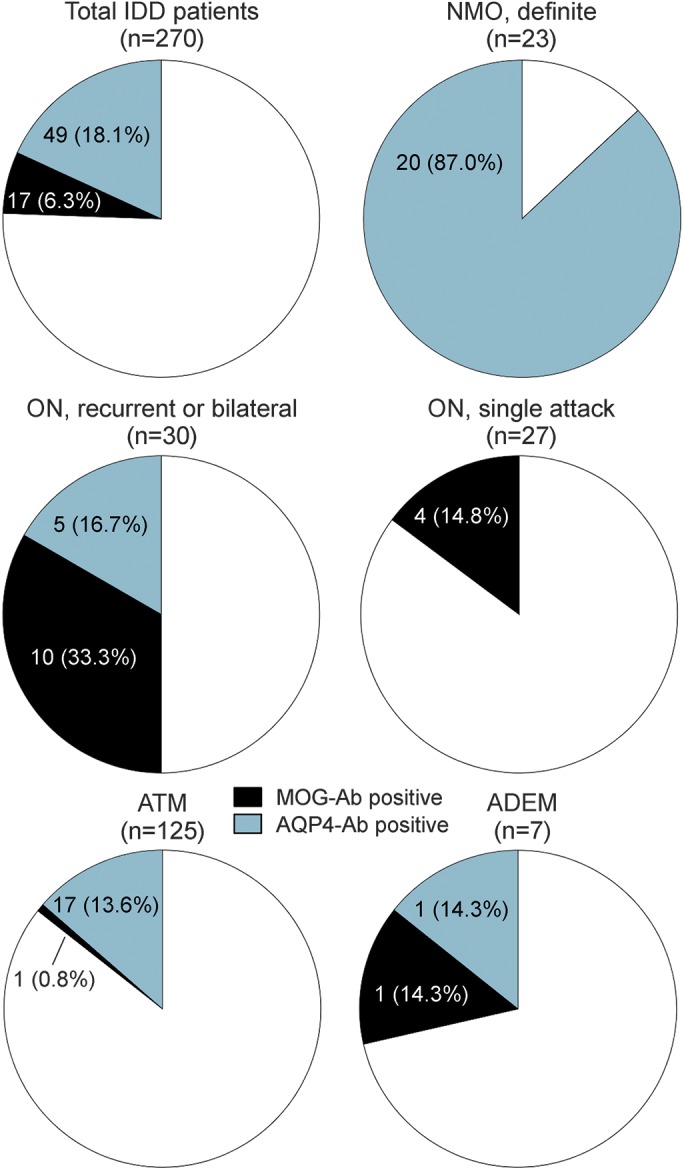
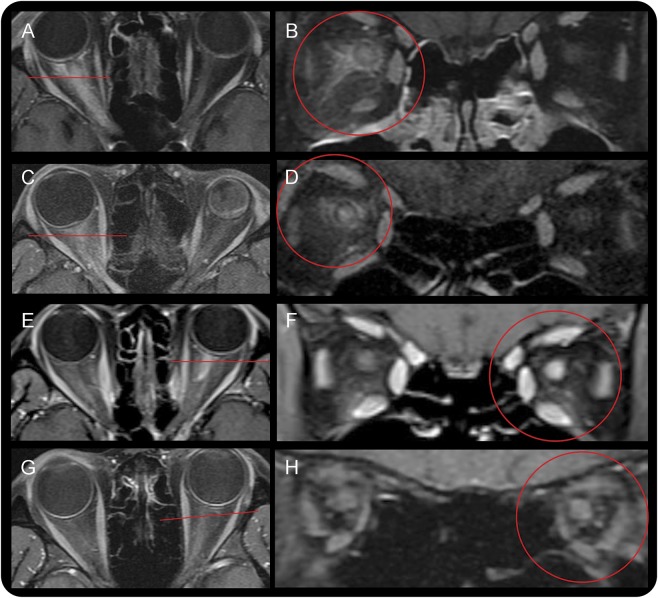
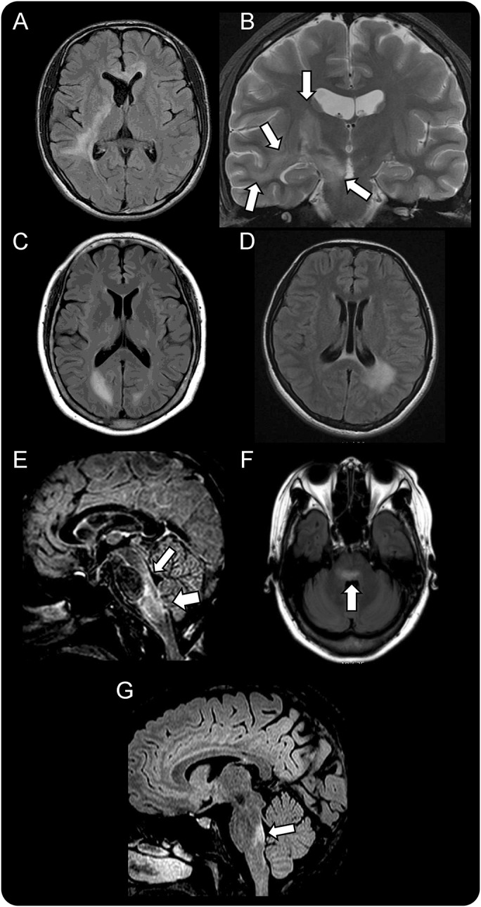
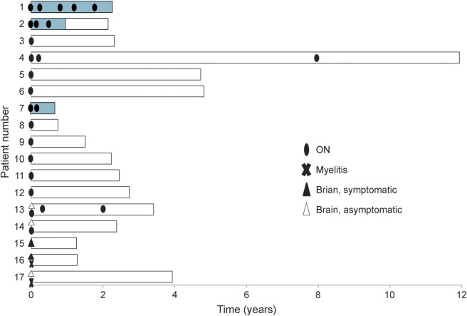
OBJECTIVE: To evaluate the clinical relevance of myelin oligodendrocyte glycoprotein antibody (MOG-Ab) in a cohort of adults with inflammatory demyelinating disease (IDD) of the CNS. METHODS: Live cell-based assays for MOG-Ab (IgG1 subset) and antibody to aquaporin-4 (AQP4-Ab) were performed in a cohort of 270 adult patients with IDD and 72 controls. Patients were first grouped by positive antibody result as MOG-Ab or AQP4-Ab, and the remainder were grouped by published diagnostic criteria. RESULTS: Seventeen patients with IDD (6.3%) had MOG-Abs and 49 patients (18.1%) had AQP4-Abs; none had both antibodies. The MOG-Ab patients predominantly manifested with isolated symptoms of optic neuritis (83%). One-third of these patients experienced relapses, which involved only the optic nerve, and all relapsed within 1 year of disease onset. At onset, MRI in the MOG-Ab group uniquely demonstrated perineural enhancement, extending to the soft tissues around the optic nerves (33%). Although about 30% of MOG-Ab patients had brain MRI lesions, they had fewer periventricular lesions than the 26 patients with relapsing-remitting multiple sclerosis (MS); none of these lesions were ovoid or perpendicular to the ventricle. Moreover, MOG-Ab patients did not meet the diagnostic criteria for definite neuromyelitis optica (NMO) and had less spinal cord involvement than the AQP4-Ab group. Four patients (23.5%) had poor visual outcomes (<0.2) or paraplegia. CONCLUSIONS: MOG-Abs may be a disease-specific biomarker in adult patients with IDD who have a disease distinct from NMO or MS. The radiologic as well as clinical manifestations of MOG-Ab patients can be useful in their differential diagnosis.
Overview
The claims and statistics of this paper haven’t been checked yet.
Run the check — free, no account needed. It reads the open-access text and checks the sources it cites.
Integrity signals
Registry
Cites
Statistics
Figures
Verification
Claims the paper makes
The claims and statistics of this paper haven’t been checked yet.
Run the check — free, no account needed. It reads the open-access text and checks the sources it cites.
Claims other papers make about this one
Each is checked against this paper’s own text: does it say what the citing paper says it does?
The source backs part of the claim, not all of it — or backs it for a different population, condition or comparison.
In the citing paper
“MOG-IgG is also found in some patients with ADEM (particularly in children), and in ADEM followed by optic neuritis, and isolated or recurrent optic neuritis.58,59”
From Diagnosis of multiple sclerosis: progress and challenges (2017) · cites this paper as [58]
Experimental — accuracy not yet measured
Self-check
Percentages, sums and reported differences re-computed from the paper’s own numbers.
The self-check needs the paper’s full text.
Citations
Cites
Rating neurologic impairment in multiple sclerosis: An expanded disability status scale (EDSS)
Kurtzke, J. F. (1983) · Neurology · doi:10.1212/wnl.33.11.1444 · Cited by 15,011
Diagnostic criteria for multiple sclerosis: 2010 revisions to the McDonald criteria
Polman, C. H., Reingold, S. C., Banwell, B., Clanet, M., Cohen, J. A., Filippi, M., Fujihara, K., Havrdova, E., Hutchinson, M., Kappos, L., et al. (2011) · Annals of Neurology · doi:10.1002/ana.22366 · Cited by 9,802
Distinction between MOG antibody-positive and AQP4 antibody-positive NMO spectrum disorders
Sato, D. K., Callegaro, D., Lana-Peixoto, M. A., Waters, P. J., Jorge, F. M. D. H., Takahashi, T., Nakashima, I., Apostolos-Pereira, S. L., Talim, N., Simm, R. F., et al. (2014) · Neurology · doi:10.1212/wnl.0000000000000101 · Cited by 850
A consensus protocol for the standardization of cerebrospinal fluid collection and biobanking
Teunissen, C. E., Petzold, A., Bennett, J. L., Berven, F. S., Brundin, L., Comabella, M., Franciotta, D., Frederiksen, J. L., Fleming, J. O., Furlan, R., et al. (2009) · Neurology · doi:10.1212/wnl.0b013e3181c47cc2 · Cited by 845
Research works printed but not linked: 19
- 10.1212/01.wnl.0000216139.44259.74 · DOI
- 10.1001/archneur.64.10.1426 · DOI
- Shams P, Plant G. Optic neuritis: a review. Int MS J 2009;16:82–89.
- 10.1212/WNL.59.4.499 · DOI
- 10.1016/S0140-6736(04)17551-X · DOI · Paper page
- 10.1016/S1474-4422(07)70216-8 · DOI
- 10.1177/1352458512472748 · DOI
- 10.1038/nrneurol.2014.141 · DOI · Paper page
- 10.1001/jamaneurol.2013.5857 · DOI · Paper page
- 10.1038/nrneurol.2013.118 · DOI
- 10.1212/NXI.0000000000000089 · DOI
- 10.1016/S1474-4422(05)70071-5 · DOI · Paper page
- 10.1056/NEJM199202273260901 · DOI
- 10.1186/1471-2377-14-93 · DOI
- Horowitz AL, Kaplan RD, Grewe G, White RT, Salberg LM. The ovoid lesion: a new MR observation in patients with multiple sclerosis. AJNR Am J Neuroradiol 1989;10:303–305.
- Ketelslegers IA Van Pelt DE Bryde S et al.. Anti-MOG antibodies plead against MS diagnosis in an Acquired Demyelinating Syndromes cohort. Mult Scler Epub 2015 Feb 6. · DOI
- 10.1002/ana.21916 · DOI
- 10.1177/1352458510389220 · DOI
- 10.1055/s-2007-1019130 · DOI
Cited by
Sorted by citations
Lassmann, H. (2018) · Cold Spring Harbor Perspectives in Medicine · doi:10.1101/cshperspect.a028936 · Cited by 937
Diagnosis of multiple sclerosis: progress and challenges
Brownlee, W. J., Hardy, T. A., Fazekas, F., & Miller, D. H. (2017) · The Lancet · doi:10.1016/s0140-6736(16)30959-x · Cited by 701
Autoantibodies to MOG in a distinct subgroup of adult multiple sclerosis
Spadaro, M., Gerdes, L. A., Krumbholz, M., Ertl-Wagner, B., Thaler, F. S., Schuh, E., Metz, I., Blaschek, A., Dick, A., Brück, W., et al. (2016) · Neurology Neuroimmunology & Neuroinflammation · doi:10.1212/nxi.0000000000000257 · Cited by 206
Metadata
- Authors
- Kim, S.-M., Woodhall, M. R., Kim, J.-S., Kim, S.-J., Park, K. S., Vincent, A., Lee, K.-W., & Waters, P.
- Affiliations
- Seoul National University (KR)
- From the Department of Neurology (S.-M.K., J.-S.K., K.S.P., K.-W.L.) and Ophthalmology (S.-J.K.), Seoul National University College of Medicine, Seoul, Republic of Korea; Nuffield Department of Clinical Neurosciences (M.R.W., A.V., P.W.), John Radcliffe Hospital, Oxford, United Kingdom; and Department of Neurology (K.S.P.), Seoul National University Bundang Hospital, Gyeonggi, Republic of Korea.
- Corresponding author
- Kim, Sung-Min
- Waters, Patrick
- ORCID
- Venue
- Neurology Neuroimmunology & Neuroinflammation
- Published
- 2015-12
- Volume
- 2
- Issue
- 6
- Pages
- e163-e163
- ISSN
- 2332-7812 (ISSN-L)
- PMC
- PMC4608758
- OpenAlex
- W2321839851
- Cited by
- 232
- References
- 4
- Citations by year
- 2026: 4 · 2025: 5 · 2024: 12 · 2023: 12 · 2022: 22 · 2021: 22 · 2020: 38 · 2019: 27 · 2018: 41 · 2017: 28 · 2016: 19 · 2015: 1
- FWCI
- 23.56
- Type
- journal article
- Language
- en
- Open access
- gold
- doi.org
- Licence
- cc-by-nc-nd
- Locations
- Neurology Neuroimmunology & Neuroinflammation (publishedVersion, cc-by-nc-nd)
- Europe PMC (PubMed Central) (publishedVersion, cc-by-nc-nd)
- PubMed Central (publishedVersion, cc-by-nc-nd)
- PubMed (publishedVersion)
- Topics
- Health Sciences › Medicine › Clinical Neurology › Multiple Sclerosis Research Studies
- Health Sciences › Medicine › Ophthalmology › Ocular Diseases and Behçet’s Syndrome
- Health Sciences › Medicine › Clinical Neurology › Peripheral Neuropathies and Disorders
- Keywords
- myelin oligodendrocyte glycoprotein antibodies, aquaporin-4 antibodies, inflammatory demyelinating diseases, optic neuritis, neuromyelitis optica, relapsing-remitting multiple sclerosis, MRI lesions, disease-specific biomarkers, differential diagnosis, relapse, visual outcome
About these checks
Registry, retraction, Cites and statistics results are computed, not judged, with one exception: a printed reference that no index lists is read once by a model before it counts as a citation error. Claims are extracted by a model and kept only when their quote appears verbatim in the text; each cited source is then checked, with a model’s help, for whether it says what the claim relies on. Those verdicts can be wrong — read the claim’s quote and the cited source before relying on one.
Statistics and arithmetic are recomputed, not judged.
Confidence bands are shown only for fields calibrated on our annotated test set. None is calibrated yet, so no band is shown.
What counts as a claim ›How we measure confidence ›How we check this ›
Are you an author, or do you see a problem with this page? Request a review
