

The DOI matches its Crossref record; 42 of the 42 references it cites were found in a registry or an index.
Loss of TBK1 is a frequent cause of frontotemporal dementia in a Belgian cohort
Open accessAbstract
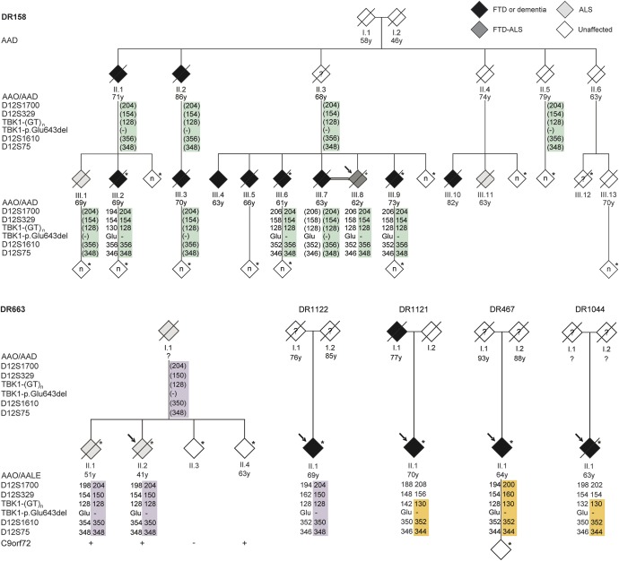
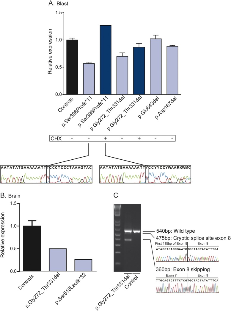
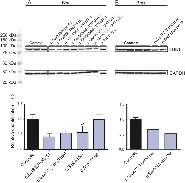
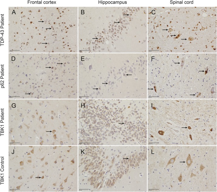
OBJECTIVE: To assess the genetic contribution of TBK1, a gene implicated in amyotrophic lateral sclerosis (ALS), frontotemporal dementia (FTD), and FTD-ALS, in Belgian FTD and ALS patient cohorts containing a significant part of genetically unresolved patients. METHODS: We sequenced TBK1 in a hospital-based cohort of 482 unrelated patients with FTD and FTD-ALS and 147 patients with ALS and an extended Belgian FTD-ALS family DR158. We followed up mutation carriers by segregation studies, transcript and protein expression analysis, and immunohistochemistry. RESULTS: We identified 11 patients carrying a loss-of-function (LOF) mutation resulting in an overall mutation frequency of 1.7% (11/629), 1.1% in patients with FTD (5/460), 3.4% in patients with ALS (5/147), and 4.5% in patients with FTD-ALS (1/22). We found 1 LOF mutation, p.Glu643del, in 6 unrelated patients segregating with disease in family DR158. Of 2 mutation carriers, brain and spinal cord was characterized by TDP-43-positive pathology. The LOF mutations including the p.Glu643del mutation led to loss of transcript or protein in blood and brain. CONCLUSIONS: TBK1 LOF mutations are the third most frequent cause of clinical FTD in the Belgian clinically based patient cohort, after C9orf72 and GRN, and the second most common cause of clinical ALS after C9orf72. These findings reinforce that FTD and ALS belong to the same disease continuum.
Overview
The claims and statistics of this paper haven’t been checked yet.
Run the check — free, no account needed. It reads the open-access text and checks the sources it cites.
Integrity signals
Registry
Cites
Statistics
Figures
Verification
Claims the paper makes
The claims and statistics of this paper haven’t been checked yet.
Run the check — free, no account needed. It reads the open-access text and checks the sources it cites.
Claims other papers make about this one
Each is checked against this paper’s own text: does it say what the citing paper says it does?
The cited source says what the claim attributes to it.
In the citing paper
“Supporting the link between ALS and FTD, TBK1 mutations have also been identified in FTD patients, and in fact occur more frequently in patients with combined ALS-FTD than in those with ALS alone [207–209].”
From Autophagy and ALS: mechanistic insights and therapeutic implications (2022) · cites this paper as [207]
Self-check
Percentages, sums and reported differences re-computed from the paper’s own numbers.
The self-check needs the paper’s full text.
Citations
Cites
Repertoires of Autophagy in the Pathogenesis of Ocular Diseases
Li, Y., Jiang, Q. L., Cao, G.-F., Yao, J., & Yan, B. (2015) · Cellular Physiology and Biochemistry · doi:10.1159/000373980 · Cited by 16,351
Autophagy fights disease through cellular self-digestion
Mizushima, N., Levine, B., Cuervo, A. M., & Klionsky, D. J. (2008) · Nature · doi:10.1038/nature06639 · Cited by 6,452
El Escorial revisited: revised criteria for the diagnosis of amyotrophic lateral sclerosis
Brooks, B. R., Miller, R. G., Swash, M., & Munsat, T. L. (2000) · Amyotrophic Lateral Sclerosis and Other Motor Neuron Disorders · doi:10.1080/146608200300079536 · Cited by 5,433
Frontotemporal lobar degeneration: A consensus on clinical diagnostic criteria
Neary, D., Snowden, J. S., Gustafson, L., Passant, U., Stuss, D., Black, S., Freedman, M., Kertesz, A., Robert, P. H., Albert, M., et al. (1998) · Neurology · doi:10.1212/wnl.51.6.1546 · Cited by 5,047
DeJesus-Hernandez, M., Mackenzie, I. R., Boeve, B. F., Boxer, A. L., Baker, M., Rutherford, N. J., Nicholson, A. M., Finch, N. A., Flynn, H., Adamson, J., et al. (2011) · Neuron · doi:10.1016/j.neuron.2011.09.011 · Cited by 4,936
A Hexanucleotide Repeat Expansion in C9ORF72 Is the Cause of Chromosome 9p21-Linked ALS-FTD
Renton, A. E., Majounie, E., Waite, A., Simón-Sánchez, J., Rollinson, S., Gibbs, J. R., Schymick, J. C., Laaksovirta, H., van Swieten, J. C., Myllykangas, L., et al. (2011) · Neuron · doi:10.1016/j.neuron.2011.09.010 · Cited by 4,409
Systematic RNA interference reveals that oncogenic KRAS-driven cancers require TBK1
Barbie, D. A., Tamayo, P., Boehm, J. S., Kim, S. Y., Moody, S. E., Dunn, I. F., Schinzel, A. C., Sandy, P., Meylan, E., Scholl, C., et al. (2009) · Nature · doi:10.1038/nature08460 · Cited by 3,922
Watts, G. D. J., Wymer, J., Kovach, M. J., Mehta, S. G., Mumm, S., Darvish, D., Pestronk, A., Whyte, M. P., & Kimonis, V. E. (2004) · Nature Genetics · doi:10.1038/ng1332 · Cited by 1,391
Phosphorylation of the Autophagy Receptor Optineurin Restricts Salmonella Growth
Wild, P., Farhan, H., McEwan, D. G., Wagner, S., Rogov, V. V., Brady, N. R., Richter, B., Korac, J., Waidmann, O., Choudhary, C., et al. (2011) · Science · doi:10.1126/science.1205405 · Cited by 1,298
Mutations of optineurin in amyotrophic lateral sclerosis
Maruyama, H., Morino, H., Ito, H., Izumi, Y., Kato, H., Watanabe, Y., Kinoshita, Y., Kamada, M., Nodera, H., Suzuki, H., et al. (2010) · Nature · doi:10.1038/nature08971 · Cited by 1,265
Cited by
Sorted by citations
TBK1: a new player in ALS linking autophagy and neuroinflammation
Oakes, J. A., Davies, M. C., & Collins, M. O. (2017) · Molecular Brain · doi:10.1186/s13041-017-0287-x · Cited by 319
Human TBK1: A Gatekeeper of Neuroinflammation
Ahmad, L., Zhang, S.-Y., Casanova, J.-L., & Sancho-Shimizu, V. (2016) · Trends in Molecular Medicine · doi:10.1016/j.molmed.2016.04.006 · Cited by 208
Autophagy and ALS: mechanistic insights and therapeutic implications
Chua, J. P., De Calbiac, H., Kabashi, E., & Barmada, S. J. (2022) · Autophagy · doi:10.1080/15548627.2021.1926656 · Cited by 189
Review: neuropathology of non-tau frontotemporal lobar degeneration
Neumann, M., & Mackenzie, I. R. A. (2019) · Neuropathology and Applied Neurobiology · doi:10.1111/nan.12526 · Cited by 144
Van Mossevelde, S., van der Zee, J., Gijselinck, I., Engelborghs, S., Sieben, A., Van Langenhove, T., De Bleecker, J., Baets, J., Vandenbulcke, M., Van Laere, K., et al. (2016) · Brain · doi:10.1093/brain/awv358 · Cited by 98
Self-citationGossye, H., Van Broeckhoven, C., & Engelborghs, S. (2019) · Frontiers in Neuroscience · doi:10.3389/fnins.2019.00757 · Cited by 34
Self-citation
Metadata
- Authors
- Gijselinck, I., Van Mossevelde, S., van der Zee, J., Sieben, A., Philtjens, S., Heeman, B., Engelborghs, S., Vandenbulcke, M., De Baets, G., Bäumer, V., Cuijt, I., Van den Broeck, M., Peeters, K., Mattheijssens, M., Rousseau, F., Vandenberghe, R., De Jonghe, P., Cras, P., De Deyn, P. P., Martin, J.-J., Cruts, M., Van Broeckhoven, C., BELNEU Consortium, Nuytten, D., Van Langenhove, T., Smets, K., Baets, J., Robberecht, W., Van Damme, P., De Bleecker, J., Santens, P., Dermaut, B., Deryck, O., Bergmans, B., Delbeck, J., Versijpt, J., Michotte, A., Willems, C., Ivanoiu, A., & Salmon, E.
- Affiliations
- University Medical Center Groningen (NL)
- From the Department of Molecular Genetics (I.G., S.V.M., J.v.d.Z., A.S., S.P., B.H., V.B., I.C., M.V.d.B., K.P., M.M., P.D.J., M.C., C.V.B.), VIB, Antwerp; Institute Born-Bunge (I.G., S.V.M., J.v.d.Z., A.S., S.P., B.H., S.E., V.B., I.C., M.V.d.B., K.P., M.M., P.D.J., P.C., P.P.D.D., J.-J.M., M.C., C.V.B.), University of Antwerp; the Department of Neurology (A.S.), University Hospital Ghent and University of Ghent; the Department of Neurology and Memory Clinic (S.E., P.P.D.D.), Hospital Network...
- Corresponding author
- Cruts, Marc
- ORCID
- Venue
- Neurology
- Published
- 2015-12-15
- Volume
- 85
- Issue
- 24
- Pages
- 2116-2125
- ISSN
- 0028-3878 (ISSN-L)
- 1526-632X
- PMC
- PMC4691687
- OpenAlex
- W2210071362
- Cited by
- 174
- References
- 42
- Citations by year
- 2026: 4 · 2025: 8 · 2024: 10 · 2023: 11 · 2022: 13 · 2021: 24 · 2020: 22 · 2019: 19 · 2018: 23 · 2017: 19 · 2016: 18 · 2015: 3
- FWCI
- 14.81
- Type
- journal article
- Language
- en
- Open access
- hybrid
- doi.org
- Licence
- cc-by-nc-nd
- Locations
- Neurology (publishedVersion, cc-by-nc-nd)
- Europe PMC (PubMed Central) (publishedVersion, cc-by-nc-nd)
- PubMed Central (publishedVersion, cc-by-nc-nd)
- PubMed (publishedVersion)
- Ghent University Academic Bibliography (Ghent University) (submittedVersion, cc-by)
- Lirias (submittedVersion, other-oa)
- Ghent University Academic Bibliography (Ghent University) (submittedVersion)
- Digital Access to Libraries (submittedVersion)
- University of Groningen research database (University of Groningen / Centre for Information Technology) (submittedVersion, cc-by-nc-nd)
- VUBIR (Vrije Universiteit Brussel) (submittedVersion)
- Funders
- MetLife Foundation
- Belgian Federal Science Policy Office
- Fonds Wetenschappelijk Onderzoek
- Universiteit Antwerpen
- Vlaamse regering
- Instituut Born-Bunge
- Fondation pour la Recherche sur Alzheimer
- Topics
- Health Sciences › Medicine › Clinical Neurology › Amyotrophic Lateral Sclerosis Research
- Life Sciences › Neuroscience › Cellular and Molecular Neuroscience › Alzheimer's disease research and treatments
- Health Sciences › Medicine › Psychiatry and Mental health › Dementia and Cognitive Impairment Research
- Keywords
- TBK1, frontotemporal dementia, amyotrophic lateral sclerosis, loss-of-function mutations, TDP-43 pathology, genetic susceptibility, family segregation
- MeSH terms
- Adult, Aged, Aged, 80 and over, Belgium, Female, Humans, Male, Middle Aged, Mutation, Pedigree, Cohort Studies, Protein Serine-Threonine Kinases, Frontotemporal Dementia
About these checks
Registry, retraction, Cites and statistics results are computed, not judged, with one exception: a printed reference that no index lists is read once by a model before it counts as a citation error. Claims are extracted by a model and kept only when their quote appears verbatim in the text; each cited source is then checked, with a model’s help, for whether it says what the claim relies on. Those verdicts can be wrong — read the claim’s quote and the cited source before relying on one.
Statistics and arithmetic are recomputed, not judged.
Confidence bands are shown only for fields calibrated on our annotated test set. None is calibrated yet, so no band is shown.
What counts as a claim ›How we measure confidence ›How we check this ›
Are you an author, or do you see a problem with this page? Request a review
