

The DOI matches its Crossref record; 8 of the 8 references it cites were found in a registry or an index.
C. elegans midbodies are released, phagocytosed and undergo LC3-dependent degradation independent of macroautophagy
Abstract
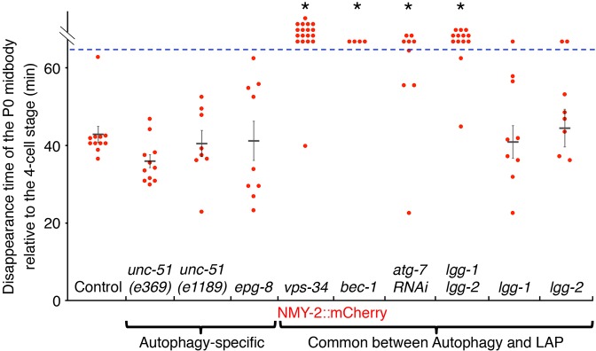
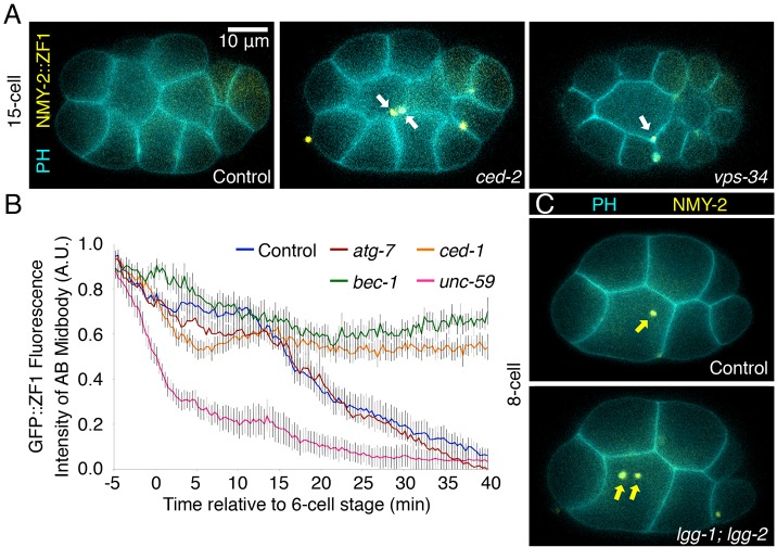
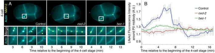
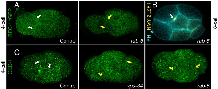
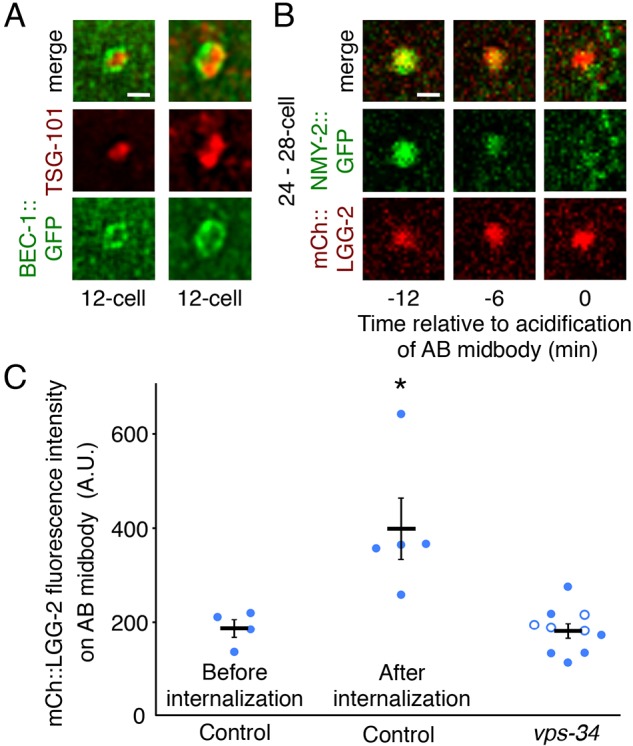
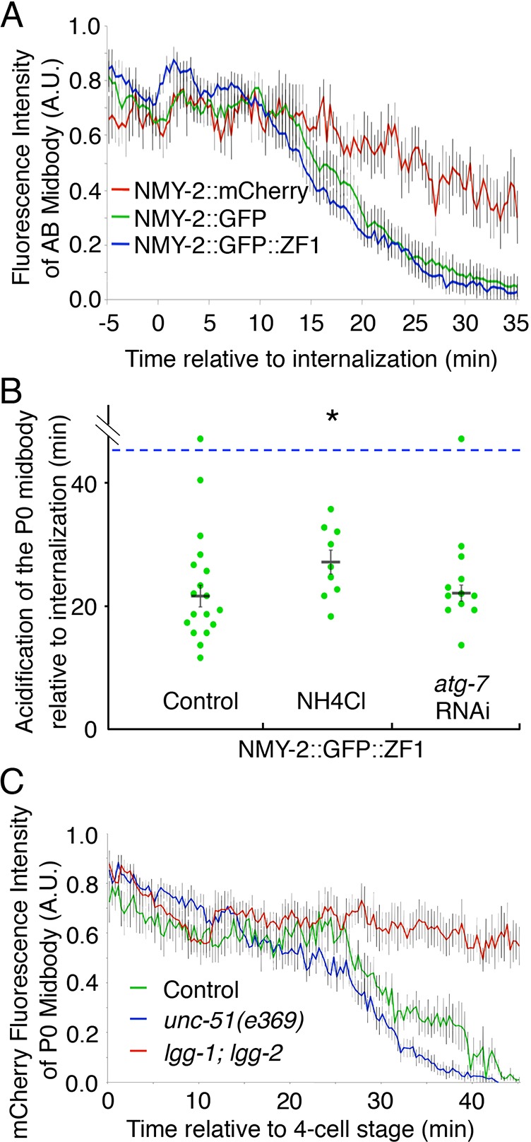
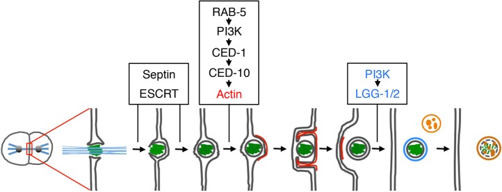
In animals, the midbody coordinates the end of cytokinesis when daughter cells separate through abscission. The midbody was thought to be sequestered by macroautophagy, but recent evidence suggests that midbodies are primarily released and phagocytosed. It was unknown, however, whether autophagy proteins play a role in midbody phagosome degradation. Using a protein degradation assay, we show that midbodies are released in Caenorhabditis elegans. Released midbodies are known to be internalized by actin-driven phagocytosis, which we show requires the RAB-5 GTPase to localize the class III phosphoinositide 3-kinase (PI3K) complex at the cortex. Autophagy-associated proteins, including the Beclin 1 homolog BEC-1 and the Atg8/LC3-family members LGG-1 and LGG-2, localize around the midbody phagosome and are required for midbody degradation. In contrast, proteins required specifically for macroautophagy, such as UNC-51 and EPG-8 (homologous to ULK1/Atg1 and Atg14, respectively) are not required for midbody degradation. These data suggest that the C. elegans midbody is degraded by LC3-associated phagocytosis (LAP), not macroautophagy. Our findings reconcile the two prevailing models on the role of phagocytic and autophagy proteins, establishing a new non-canonical role for autophagy proteins in midbody degradation.
Overview
The claims and statistics of this paper haven’t been checked yet.
Run the check — free, no account needed. It reads the open-access text and checks the sources it cites.
Integrity signals
Registry
Cites
Statistics
Figures
Verification
Claims the paper makes
The claims and statistics of this paper haven’t been checked yet.
Run the check — free, no account needed. It reads the open-access text and checks the sources it cites.
Claims other papers make about this one
Each is checked against this paper’s own text: does it say what the citing paper says it does?
In the citing paper
“TORC1 activity does not necessarily preclude autophagy induction because there are TOR-independent mechanisms that induce autophagy both in mammals and yeast [757-761]”
From Guidelines for the use and interpretation of assays for monitoring autophagy (4th edition) 1 (2021) · cites this paper as [758]
Self-check
Percentages, sums and reported differences re-computed from the paper’s own numbers.
The self-check needs the paper’s full text.
Citations
Cites
3 retracted
Martinez, J., Malireddi, R. K. S., Lu, Q., Cunha, L. D., Pelletier, S., Gingras, S., Orchard, R., Guan, J.-L., Tan, H., Peng, J., et al. (2015) · Nature Cell Biology · doi:10.1038/ncb3192 · Cited by 758 · cited 2×
“In contrast, we find proteins specifically required for autophagy, such as UNC-51 and EPG-8 (homologous to ULK1 and ATG14, respectively) (Martinez et al., 2011, 2015; Yang and Zhang, 2011), are not required for degradation of the midbody phagosome.”
BackgroundRetractedMartinez, J., Almendinger, J., Oberst, A., Ness, R., Dillon, C. P., Fitzgerald, P., Hengartner, M. O., & Green, D. R. (2011) · Proceedings of the National Academy of Sciences · doi:10.1073/pnas.1113421108 · Cited by 655 · cited 4×
“Cell corpses are also wrapped in their plasma membrane before phagocytosis (Florey et al., 2011; Martinez et al., 2011).”
BackgroundInfluentialRetractedMartinez, J., Cunha, L. D., Park, S., Yang, M., Lu, Q., Orchard, R., Li, Q.-Z., Yan, M., Janke, L., Guy, C., et al. (2016) · Nature · doi:10.1038/nature17950 · Cited by 381
RetractedToll-like receptor signalling in macrophages links the autophagy pathway to phagocytosis
Sanjuan, M. A., Dillon, C. P., Tait, S. W. G., Moshiach, S., Dorsey, F., Connell, S., Komatsu, M., Tanaka, K., Cleveland, J. L., Withoff, S., et al. (2007) · Nature · doi:10.1038/nature06421 · Cited by 1,347
Florey, O., Kim, S. E., Sandoval, C. P., Haynes, C. M., & Overholtzer, M. (2011) · Nature Cell Biology · doi:10.1038/ncb2363 · Cited by 459
Lucin, K. M., O’Brien, C. E., Bieri, G., Czirr, E., Mosher, K. I., Abbey, R. J., Mastroeni, D. F., Rogers, J., Spencer, B., Masliah, E., et al. (2013) · Neuron · doi:10.1016/j.neuron.2013.06.046 · Cited by 374
Noncanonical autophagy promotes the visual cycle
Kim, J.-Y., Zhao, H., Martinez, J., Doggett, T. A., Kolesnikov, A. V., Tang, P. H., Ablonczy, Z., Chan, C.-C., Zhou, Z., Green, D. R., et al. (2013) · Cell · doi:10.1016/j.cell.2013.06.012 · Cited by 347
A pathway for phagosome maturation during engulfment of apoptotic cells
Kinchen, J. M., Doukoumetzidis, K., Almendinger, J., Stergiou, L., Tosello-Trampont, A., Sifri, C. D., Hengartner, M. O., & Ravichandran, K. S. (2008) · Nature Cell Biology · doi:10.1038/ncb1718 · Cited by 284
Research works printed but not linked: 52
- Al Rawi, 2011, Postfertilization autophagy of sperm organelles prevents paternal mitochondrial DNA transmission, Science, 334, 1144, 10.1126/science.1211878 · DOI · Paper page
- Anderson, 2008, Polarization of the C. elegans embryo by RhoGAP-mediated exclusion of PAR-6 from cell contacts, Science, 320, 1771, 10.1126/science.1156063 · DOI
- Arai, 2015, Lipidome of midbody released from neural stem and progenitor cells during mammalian cortical neurogenesis, Front. Cell. Neurosci., 9, 325, 10.3389/fncel.2015.00325 · DOI
- Artal-Sanz, 2006, Lysosomal biogenesis and function is critical for necrotic cell death in Caenorhabditis elegans, J. Cell Biol., 173, 231, 10.1083/jcb.200511103 · DOI
- Audhya, 2005, A complex containing the Sm protein CAR-1 and the RNA helicase CGH-1 is required for embryonic cytokinesis in Caenorhabditis elegans, J. Cell Biol., 171, 267, 10.1083/jcb.200506124 · DOI
- Brenner, 1974, The genetics of Caenorhabditis elegans, Genetics, 77, 71, 10.1093/genetics/77.1.71 · DOI
- Carlsson, 2015, Membrane dynamics in autophagosome biogenesis, J. Cell Sci., 128, 193, 10.1242/jcs.141036 · DOI
- Chai, 2012, Apoptotic regulators promote cytokinetic midbody degradation in C. elegans, J. Cell Biol., 199, 1047, 10.1083/jcb.201209050 · DOI
- Chen, 2010, Retromer is required for apoptotic cell clearance by phagocytic receptor recycling, Science, 327, 1261, 10.1126/science.1184840 · DOI
- Crowell, 2014, Engulfment of the midbody remnant after cytokinesis in mammalian cells, J. Cell Sci., 127, 3840, 10.1242/jcs.154732 · DOI
- Derenzo, 2003, Exclusion of germ plasm proteins from somatic lineages by cullin-dependent degradation, Nature, 424, 685, 10.1038/nature01887 · DOI · Paper page
- Dubreuil, 2007, Midbody and primary cilium of neural progenitors release extracellular membrane particles enriched in the stem cell marker prominin-1, J. Cell Biol., 176, 483, 10.1083/jcb.200608137 · DOI
- Elia, 2011, Dynamics of endosomal sorting complex required for transport (ESCRT) machinery during cytokinesis and its role in abscission, Proc. Natl. Acad. Sci. USA, 108, 4846, 10.1073/pnas.1102714108 · DOI
- Ettinger, 2011, Proliferating versus differentiating stem and cancer cells exhibit distinct midbody-release behaviour, Nat. Commun., 2, 503, 10.1038/ncomms1511 · DOI
- Fraser, 2000, Functional genomic analysis of C. elegans chromosome I by systematic RNA interference, Nature, 408, 325, 10.1038/35042517 · DOI
- Funderburk, 2010, The Beclin 1-VPS34 complex--at the crossroads of autophagy and beyond, Trends Cell Biol., 20, 355, 10.1016/j.tcb.2010.03.002 · DOI
- Green, 2008, Expression and imaging of fluorescent proteins in the C. elegans gonad and early embryo, Methods Cell Biol., 85, 179, 10.1016/s0091-679x(08)85009-1 · DOI
- Green, 2011, A high-resolution C. elegans essential gene network based on phenotypic profiling of a complex tissue, Cell, 145, 470, 10.1016/j.cell.2011.03.037 · DOI
- Green, 2012, Cytokinesis in animal cells, Annu. Rev. Cell Dev. Biol., 28, 29, 10.1146/annurev-cellbio-101011-155718 · DOI
- Green, 2013, The midbody ring scaffolds the abscission machinery in the absence of midbody microtubules, J. Cell Biol., 203, 505, 10.1083/jcb.201306036 · DOI
- Guizetti, 2011, Cortical constriction during abscission involves helices of ESCRT-III-dependent filaments, Science, 331, 1616, 10.1126/science.1201847 · DOI
- Huang, 2013, Autophagy genes function in apoptotic cell corpse clearance during C. elegans embryonic development, Autophagy, 9, 138, 10.4161/auto.22352 · DOI
- Isakson, 2013, TRAF6 mediates ubiquitination of KIF23/MKLP1 and is required for midbody ring degradation by selective autophagy, Autophagy, 9, 1955, 10.4161/auto.26085 · DOI
- Kamath, 2003, Genome-wide RNAi screening in Caenorhabditis elegans, Methods, 30, 313, 10.1016/S1046-2023(03)00050-1 · DOI
- Kneen, 1998, Green fluorescent protein as a noninvasive intracellular pH indicator, Biophys. J., 74, 1591, 10.1016/S0006-3495(98)77870-1 · DOI
- Konishi, 2012, Involvement of Beclin 1 in engulfment of apoptotic cells, J. Biol. Chem., 287, 13919, 10.1074/jbc.M112.348375 · DOI
- Kuo, 2011, Midbody accumulation through evasion of autophagy contributes to cellular reprogramming and tumorigenicity, Nat. Cell Biol., 13, 1214, 10.1038/ncb2332 · DOI
- Levine, 2015, Beclin orthologs: integrative hubs of cell signaling, membrane trafficking, and physiology, Trends Cell Biol., 25, 533, 10.1016/j.tcb.2015.05.004 · DOI
- Lu, 2012, Two PI 3-kinases and one PI 3-phosphatase together establish the cyclic waves of phagosomal PtdIns(3)P critical for the degradation of apoptotic cells, PLoS Biol., 10, e1001245, 10.1371/journal.pbio.1001245 · DOI
- Manil-Segalen, 2014, The C. elegans LC3 acts downstream of GABARAP to degrade autophagosomes by interacting with the HOPS subunit VPS39, Dev. Cell, 28, 43, 10.1016/j.devcel.2013.11.022 · DOI · Paper page
- Morais-De-Sa, 2013, Connecting polarized cytokinesis to epithelial architecture, Cell Cycle, 12, 3583, 10.4161/cc.26910 · DOI
- Nance, 2003, C. elegans PAR-3 and PAR-6 are required for apicobasal asymmetries associated with cell adhesion and gastrulation, Development, 130, 5339, 10.1242/dev.00735 · DOI
- Ogura, 1994, Caenorhabditis elegans unc-51 gene required for axonal elongation encodes a novel serine/threonine kinase, Genes Dev., 8, 2389, 10.1101/gad.8.20.2389 · DOI
- Ou, 2014, Stereotyped distribution of midbody remnants in early C. elegans embryos requires cell death genes and is dispensable for development, Cell Res., 24, 251, 10.1038/cr.2013.140 · DOI
- Pohl, 2009, Midbody ring disposal by autophagy is a post-abscission event of cytokinesis, Nat. Cell Biol., 11, 65, 10.1038/ncb1813 · DOI
- Praitis, 2001, Creation of low-copy integrated transgenic lines in Caenorhabditis elegans, Genetics, 157, 1217, 10.1093/genetics/157.3.1217 · DOI
- Ravikumar, 2008, Rab5 modulates aggregation and toxicity of mutant huntingtin through macroautophagy in cell and fly models of Huntington disease, J. Cell Sci., 121, 1649, 10.1242/jcs.025726 · DOI
- Reese, 2000, Asymmetric segregation of PIE-1 in C. elegans is mediated by two complementary mechanisms that act through separate PIE-1 protein domains, Mol. Cell, 6, 445, 10.1016/S1097-2765(00)00043-5 · DOI
- Russell, 2013, ULK1 induces autophagy by phosphorylating Beclin-1 and activating VPS34 lipid kinase, Nat. Cell Biol., 15, 741, 10.1038/ncb2757 · DOI · Paper page
- Salzmann, 2014, Centrosome-dependent asymmetric inheritance of the midbody ring in Drosophila germline stem cell division, Mol. Biol. Cell, 25, 267, 10.1091/mbc.E13-09-0541 · DOI
- Sarov, 2012, A genome-scale resource for in vivo tag-based protein function exploration in C. elegans, Cell, 150, 855, 10.1016/j.cell.2012.08.001 · DOI
- Sato, 2011, Degradation of paternal mitochondria by fertilization-triggered autophagy in C. elegans embryos, Science, 334, 1141, 10.1126/science.1210333 · DOI
- Schiel, 2013, Endocytic transport and cytokinesis: from regulation of the cytoskeleton to midbody inheritance, Trends Cell Biol., 23, 319, 10.1016/j.tcb.2013.02.003 · DOI
- Shaner, 2004, Improved monomeric red, orange and yellow fluorescent proteins derived from Discosoma sp. red fluorescent protein, Nat. Biotechnol., 22, 1567, 10.1038/nbt1037 · DOI · Paper page
- Shelton, 1999, The nonmuscle myosin regulatory light chain gene mlc-4 is required for cytokinesis, anterior-posterior polarity, and body morphology during Caenorhabditis elegans embryogenesis, J. Cell Biol., 146, 439, 10.1083/jcb.146.2.439 · DOI
- Sijen, 2001, On the role of RNA amplification in dsRNA-triggered gene silencing, Cell, 107, 465, 10.1016/S0092-8674(01)00576-1 · DOI
- Singh, 2014, Coupling of rotational cortical flow, asymmetric midbody positioning, and spindle rotation mediates dorsoventral axis formation in C. elegans, Dev. Cell, 28, 253, 10.1016/j.devcel.2014.01.002 · DOI
- Sonnichsen, 2005, Full-genome RNAi profiling of early embryogenesis in Caenorhabditis elegans, Nature, 434, 462, 10.1038/nature03353 · DOI
- Tian, 2010, C. elegans screen identifies autophagy genes specific to multicellular organisms, Cell, 141, 1042, 10.1016/j.cell.2010.04.034 · DOI · Paper page
- Wehman, 2011, The P4-ATPase TAT-5 inhibits the budding of extracellular vesicles in C. elegans embryos, Curr. Biol., 21, 1951, 10.1016/j.cub.2011.10.040 · DOI
- Yang, 2011, The coiled-coil domain protein EPG-8 plays an essential role in the autophagy pathway in C. elegans, Autophagy, 7, 159, 10.4161/auto.7.2.14223 · DOI
- Zhang, 2009, SEPA-1 mediates the specific recognition and degradation of P granule components by autophagy in C. elegans, Cell, 136, 308, 10.1016/j.cell.2008.12.022 · DOI
Cited by
Sorted by citations
Guidelines for the use and interpretation of assays for monitoring autophagy (4th edition) 1
Klionsky, D. J., Abdel-Aziz, A. K., Abdelfatah, S., Abdellatif, M., Abdoli, A., Abel, S., Abeliovich, H., Abildgaard, M. H., Abudu, Y. P., Acevedo-Arozena, A., et al. (2021) · Autophagy · doi:10.1080/15548627.2020.1797280 · Cited by 2,897
Self-citation
Metadata
- Authors
- Fazeli, G., Trinkwalder, M., Irmisch, L., & Wehman, A. M.
- Affiliations
- Rudolf Virchow Center for Experimental Biomedicine, University of Würzburg , Würzburg 97080 , Germany
- ORCID
- Wehman, Ann Marie
- Venue
- Journal of Cell Science
- Publisher
- The Company of Biologists
- Published
- 2016-10-15
- Volume
- 129
- Issue
- 20
- Pages
- 3721-3731
- ISSN
- 1477-9137
- 0021-9533
- PMC
- PMC5087666
- Cited by
- 50
- References
- 8
- Language
- en
About these checks
Registry, retraction, Cites and statistics results are computed, not judged, with one exception: a printed reference that no index lists is read once by a model before it counts as a citation error. Claims are extracted by a model and kept only when their quote appears verbatim in the text; each cited source is then checked, with a model’s help, for whether it says what the claim relies on. Those verdicts can be wrong — read the claim’s quote and the cited source before relying on one.
Statistics and arithmetic are recomputed, not judged.
Confidence bands are shown only for fields calibrated on our annotated test set. None is calibrated yet, so no band is shown.
What counts as a claim ›How we measure confidence ›How we check this ›
Are you an author, or do you see a problem with this page? Request a review
