Cited by 135

The DOI matches its Crossref record; 3 of the 3 references it cites were found in a registry or an index.

Proteolytic Origin of the Soluble Human IL-6R In Vivo and a Decisive Role of N-Glycosylation

Riethmueller, Steffen, Somasundaram, Prasath, Ehlers, Johanna C., Hung, Chien-Wen, Flynn, Charlotte M., Lokau, Juliane, Agthe, Maria, Düsterhöft, Stefan, Zhu, Yijue, Grötzinger, Joachim, Lorenzen, Inken, Koudelka, Tomas, Yamamoto, Kosuke, Pickhinke, Ute, Wichert, Rielana, Becker-Pauly, Christoph, Rädisch, Marisa, Albrecht, Alexander, Hessefort, Markus, Stahnke, Dominik, Unverzagt, Carlo, Rose-John, Stefan, Tholey, Andreas, Garbers, Christoph·2017·PLOS Biology

Overview

The claims and statistics of this paper haven’t been checked yet.

Run the check — free, no account needed. It reads the open-access text and checks the sources it cites.

A few free checks a day · papers already verified open instantly · results are public

Integrity signals

Registry

Crossref · 10.1371/journal.pbio.2000080

Cites

3 of 3 references verified0 citation errors · 0 not checkable

Statistics

Figures

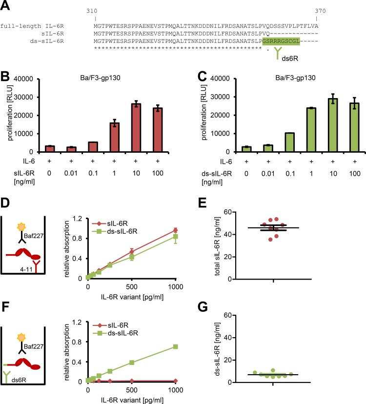
Fig 1 The majority of sIL-6R in human serum is not generated by alternative mRNA splicing. (A) Multiple sequence alignment of amino acids Met-310 to Ala-370 of the full-length human IL-6R with the sIL-6R (Met-310 to Gln-357) and amino acids Met-310 to Leu-365 of the differentially spliced sIL-6R (ds-sIL-6R). The ten unique C-terminal amino-acid residues of the sIL-6R, which represent the epitope against which the antibody ds6R was raised, are colored in green. (B, C) Proliferation assay of Ba/F3-gp130 cells in response to 10 ng/ml IL-6 and increasing amounts (0–100 ng/ml) of either (B) recombinant sIL-6R or (C) recombinant ds-sIL-6R. Cell viability was assessed after 48 h. Data shown are the mean ± the standard deviation (SD) of one representative experiment with three biological replicates. (D) Schematic representation of the sandwich ELISA that recognizes all forms of the sIL-6R (left) and proof-of-principle experiment with recombinant proteins (mean ± SD, n = 3). (E) Determination of the total sIL-6R levels in the serum of eight healthy donors. Values for the mean ± the standard error of the mean (SEM) are indicated. (F) Schematic representation of the sandwich ELISA that recognizes only the ds-sIL-6R (left) and proof-of-principle experiment with recombinant proteins (mean ± SD, n = 3). (G) Determination of the ds-sIL-6R levels in the serum of the same eight healthy donors as in (E). The values for the mean ± SEM are indicated.
Fig 1 The majority of sIL-6R in human serum is not generated by alternative mRNA splicing. (A) Multiple sequence alignment of amino acids Met-310 to Ala-370 of the full-length human IL-6R with the sIL-6R (Met-310 to Gln-357) and amino acids Met-310 to Leu-365 of the differentially spliced sIL-6R (ds-sIL-6R). The ten unique C-terminal amino-acid residues of the sIL-6R, which represent the epitope against which the antibody ds6R was raised, are colored in green. (B, C) Proliferation assay of Ba/F3-gp130 cells in response to 10 ng/ml IL-6 and increasing amounts (0–100 ng/ml) of either (B) recombinant sIL-6R or (C) recombinant ds-sIL-6R. Cell viability was assessed after 48 h. Data shown are the mean ± the standard deviation (SD) of one representative experiment with three biological replicates. (D) Schematic representation of the sandwich ELISA that recognizes all forms of the sIL-6R (left) and proof-of-principle experiment with recombinant proteins (mean ± SD, n = 3). (E) Determination of the total sIL-6R levels in the serum of eight healthy donors. Values for the mean ± the standard error of the mean (SEM) are indicated. (F) Schematic representation of the sandwich ELISA that recognizes only the ds-sIL-6R (left) and proof-of-principle experiment with recombinant proteins (mean ± SD, n = 3). (G) Determination of the ds-sIL-6R levels in the serum of the same eight healthy donors as in (E). The values for the mean ± SEM are indicated.CC BY · PubMed Central
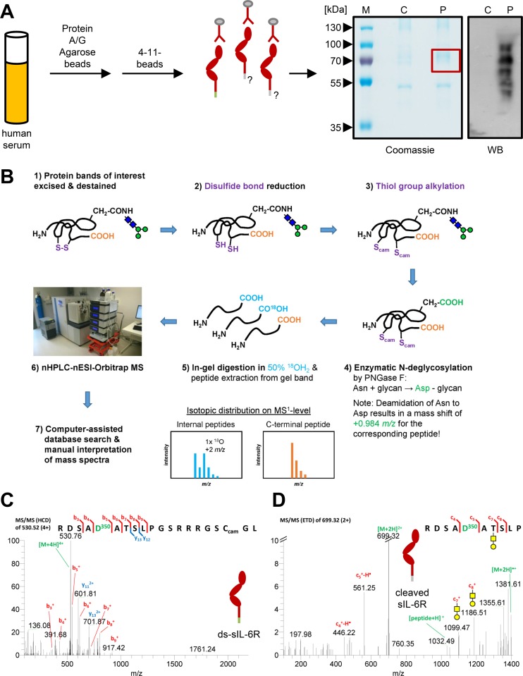
Fig 2 Identification of the ds-sIL-6R and a novel protease-derived sIL-6R from human serum via mass spectrometry (MS). (A) Schematic illustration of the procedure to precipitate total sIL-6R from human serum. A representative coomassie-stained sodium dodecyl sulfate (SDS) gel and a western blot of the precipitated protein are shown on the right. The SDS gels were run under nonreducing conditions (see Materials and Methods for details), and the region of the SDS gel excised for MS is indicated with a red box. The precipitated sIL-6R is detected with an antibody that specifically recognizes the ectodomain of the human IL-6R (4–11). (B) Proteomics workflow including disulfide bond reduction, thiol alkylation, enzymatic N-deglycosylation, proteolysis in presence of 50% H218O, LC-MS/MS analysis, and data interpretation. (C) MS/MS spectrum (higher-energy collisional dissociation [HCD]) of the C-terminal peptide of the ds-sIL-6R identified via database searching. The N-glycan site Asn-350, which is modified to an Asp-350 because of PNGase F treatment, is shown in green. (D) MS/MS spectrum (electron-transfer dissociation [ETD]) of the C-terminal peptide of the protease-derived sIL-6R identified via database searching. The N-glycan site Asn-350, which is modified to an Asp-350 because of PNGase F treatment, is shown in green. One of the identified O-glycan structures on Thr-352 is shown.
Fig 2 Identification of the ds-sIL-6R and a novel protease-derived sIL-6R from human serum via mass spectrometry (MS). (A) Schematic illustration of the procedure to precipitate total sIL-6R from human serum. A representative coomassie-stained sodium dodecyl sulfate (SDS) gel and a western blot of the precipitated protein are shown on the right. The SDS gels were run under nonreducing conditions (see Materials and Methods for details), and the region of the SDS gel excised for MS is indicated with a red box. The precipitated sIL-6R is detected with an antibody that specifically recognizes the ectodomain of the human IL-6R (4–11). (B) Proteomics workflow including disulfide bond reduction, thiol alkylation, enzymatic N-deglycosylation, proteolysis in presence of 50% H218O, LC-MS/MS analysis, and data interpretation. (C) MS/MS spectrum (higher-energy collisional dissociation [HCD]) of the C-terminal peptide of the ds-sIL-6R identified via database searching. The N-glycan site Asn-350, which is modified to an Asp-350 because of PNGase F treatment, is shown in green. (D) MS/MS spectrum (electron-transfer dissociation [ETD]) of the C-terminal peptide of the protease-derived sIL-6R identified via database searching. The N-glycan site Asn-350, which is modified to an Asp-350 because of PNGase F treatment, is shown in green. One of the identified O-glycan structures on Thr-352 is shown.CC BY · PubMed Central
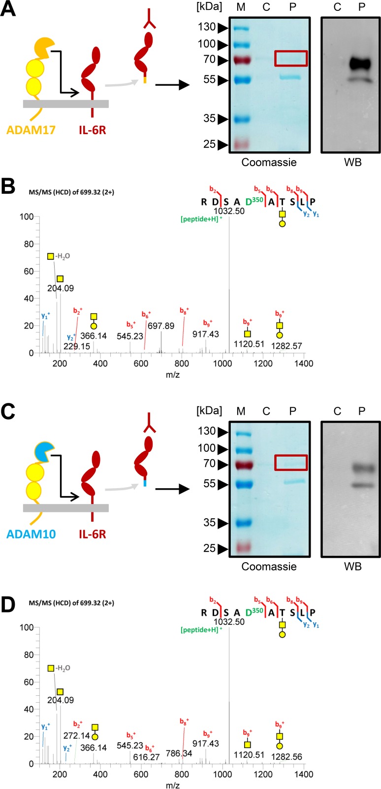
Fig 3 ADAM17 and ADAM10 cleave the IL-6R between Pro-355 and Val-356 in vitro. (A) A schematic illustration, a representative coomassie-stained SDS gel, and a western blot of the precipitated protein after PMA-induced ADAM17-mediated sIL-6R generation are shown. The SDS gels were run under reducing conditions (see Materials and Methods for details), and the region of the SDS gel excised for MS is indicated with a red box. The precipitated sIL-6R is detected with an antibody that specifically recognizes the ectodomain of the human IL-6R (4–11). (B) MS/MS spectra (HCD) of the C-terminal peptide of the sIL-6R identified by manual spectra interpretation. One of the identified O-glycan structures on Thr-352 is shown. (C) A schematic illustration, a representative coomassie-stained SDS gel, and a western blot of the precipitated protein after ionomycin-induced ADAM10-mediated sIL-6R generation are shown. The SDS gels and the western blot were performed as described under panel (A). (D) MS/MS spectra (HCD) of the C-terminal peptide of the sIL-6R identified by manual spectra interpretation. One of the identified O-glycan structures on Thr-352 is shown.
Fig 3 ADAM17 and ADAM10 cleave the IL-6R between Pro-355 and Val-356 in vitro. (A) A schematic illustration, a representative coomassie-stained SDS gel, and a western blot of the precipitated protein after PMA-induced ADAM17-mediated sIL-6R generation are shown. The SDS gels were run under reducing conditions (see Materials and Methods for details), and the region of the SDS gel excised for MS is indicated with a red box. The precipitated sIL-6R is detected with an antibody that specifically recognizes the ectodomain of the human IL-6R (4–11). (B) MS/MS spectra (HCD) of the C-terminal peptide of the sIL-6R identified by manual spectra interpretation. One of the identified O-glycan structures on Thr-352 is shown. (C) A schematic illustration, a representative coomassie-stained SDS gel, and a western blot of the precipitated protein after ionomycin-induced ADAM10-mediated sIL-6R generation are shown. The SDS gels and the western blot were performed as described under panel (A). (D) MS/MS spectra (HCD) of the C-terminal peptide of the sIL-6R identified by manual spectra interpretation. One of the identified O-glycan structures on Thr-352 is shown.CC BY · PubMed Central
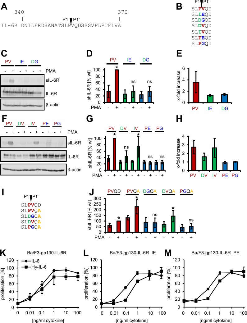
Fig 4 Mutation of Val-356 is sufficient to block proteolysis of the IL-6R and the Asp358Ala variant. (A) Amino-acid residues from Asp-340 to Ala-370 of the human IL-6R. The identified cleavage site between Pro-355 (P1) and Val-356 (P1′) is indicated. (B) Overview of the six different IL-6R cleavage site mutants. Mutations are colored in either green or blue; the amino-acid residues of the wild-type cleavage site are shown in red. (C) HEK293 cells were transiently transfected with expression plasmids encoding the wild-type IL-6R (PV) or the double mutants (IE, DG) as indicated. Cells were either treated with 100 nM PMA for 2 h or DMSO as vehicle control. sIL-6R was precipitated from the supernatant with concanavalin A-covered sepharose beads, and the cells were lysed. Both were analyzed via western blot, and β-actin served as the loading control. One out of three experiments with similar outcomes is shown. (D, E) The experiment was performed as described under (C), but sIL-6R generation was analyzed via ELISA. In (D), the amount of sIL-6R generated after PMA stimulation of the wild-type IL-6R (PV) was set to 100%, and all other values were calculated accordingly. In (E), the amount of sIL-6R without stimulation was considered as constitutive shedding and set to 1, and the increase of sIL-6R was calculated. Data shown are the mean ± SD from at least three independent experiments (*p < 0.05, ns = no significant difference). (F–H) HEK293 cells were transiently transfected with expression plasmids encoding the wild-type IL-6R (PV) or the single mutants (DV, IV, PE, PG) as indicated. The experiments were performed as described in (C) to (E). (I) Overview of the four different IL-6R cleavage site mutants of the IL-6R Asp358Ala variant. Mutations are colored in either green or blue, the amino-acid residues of the wild-type cleavage site are shown in red, and the Asp358Ala single nucleotide polymorphism (SNP) is colored in orange. (J) ADAM17-mediated proteolysis of the IL-6R variants depicted in (I) were analyzed as described in (D). (K–M) Equal numbers of Ba/F3-gp130 cells were incubated for 48 h with increasing amounts (0–100 ng/ml) of either IL-6 or Hyper-IL-6. The stably transduced IL-6R variant is indicated above the diagram. One representative experiment out of three performed is shown (mean ± SD, biological triplicates).
Fig 4 Mutation of Val-356 is sufficient to block proteolysis of the IL-6R and the Asp358Ala variant. (A) Amino-acid residues from Asp-340 to Ala-370 of the human IL-6R. The identified cleavage site between Pro-355 (P1) and Val-356 (P1′) is indicated. (B) Overview of the six different IL-6R cleavage site mutants. Mutations are colored in either green or blue; the amino-acid residues of the wild-type cleavage site are shown in red. (C) HEK293 cells were transiently transfected with expression plasmids encoding the wild-type IL-6R (PV) or the double mutants (IE, DG) as indicated. Cells were either treated with 100 nM PMA for 2 h or DMSO as vehicle control. sIL-6R was precipitated from the supernatant with concanavalin A-covered sepharose beads, and the cells were lysed. Both were analyzed via western blot, and β-actin served as the loading control. One out of three experiments with similar outcomes is shown. (D, E) The experiment was performed as described under (C), but sIL-6R generation was analyzed via ELISA. In (D), the amount of sIL-6R generated after PMA stimulation of the wild-type IL-6R (PV) was set to 100%, and all other values were calculated accordingly. In (E), the amount of sIL-6R without stimulation was considered as constitutive shedding and set to 1, and the increase of sIL-6R was calculated. Data shown are the mean ± SD from at least three independent experiments (*p < 0.05, ns = no significant difference). (F–H) HEK293 cells were transiently transfected with expression plasmids encoding the wild-type IL-6R (PV) or the single mutants (DV, IV, PE, PG) as indicated. The experiments were performed as described in (C) to (E). (I) Overview of the four different IL-6R cleavage site mutants of the IL-6R Asp358Ala variant. Mutations are colored in either green or blue, the amino-acid residues of the wild-type cleavage site are shown in red, and the Asp358Ala single nucleotide polymorphism (SNP) is colored in orange. (J) ADAM17-mediated proteolysis of the IL-6R variants depicted in (I) were analyzed as described in (D). (K–M) Equal numbers of Ba/F3-gp130 cells were incubated for 48 h with increasing amounts (0–100 ng/ml) of either IL-6 or Hyper-IL-6. The stably transduced IL-6R variant is indicated above the diagram. One representative experiment out of three performed is shown (mean ± SD, biological triplicates).CC BY · PubMed Central
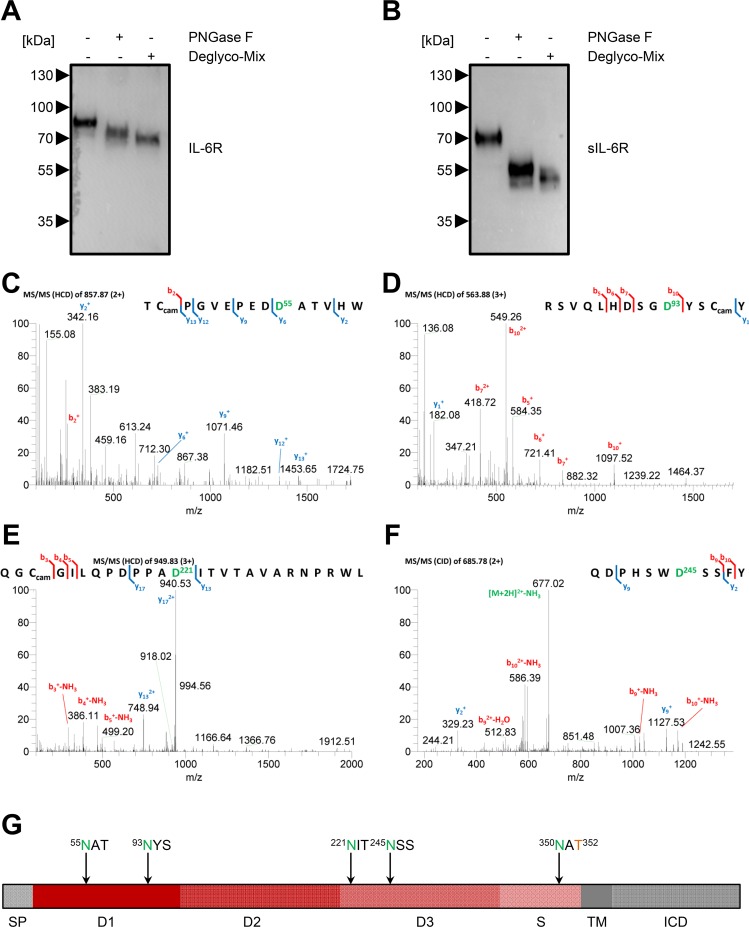
Fig 5 Determination of the N-glycosylation sites on the sIL-6R in vitro and in vivo via MS. (A) HEK293 cells were transiently transfected with an expression plasmid encoding IL-6R, and cells were lysed 48 h later. Lysates were treated with either PNGase F or Deglyco-Mix or were left untreated. Proteins were resolved on a 10% SDS gel and analyzed by western blot. One representative experiment out of two performed is shown. (B) HEK293 cells were transiently transfected with an expression plasmid encoding IL-6R, and cells were stimulated after 48 h with 100 nM PMA for 2 h. The sIL-6R was precipitated from the cell supernatant and afterwards treated and analyzed as described under panel (A). (C–F) MS/MS spectra (HCD) of the different peptides of the sIL-6R that contained N-linked glycans. The four glycosylation sites (Asn-55, Asn-93, Asn-221, and Asn-245) were modified to aspartic acid residues due to PNGase F treatment in the presence of H218O-containing buffer and are shown in green. The use of Asn-350 as an N-glycosylation site was already shown in Fig 2C and 2D. Peptides were identified via database searching. (G) Schematic overview of the different domains of the IL-6R and the localization of the five N-glycosylation sites (green) and one O-glycosylation site (orange). D, domain; ICD, intracellular domain; S, stalk region; SP, signal peptide; TM, transmembrane region.
Fig 5 Determination of the N-glycosylation sites on the sIL-6R in vitro and in vivo via MS. (A) HEK293 cells were transiently transfected with an expression plasmid encoding IL-6R, and cells were lysed 48 h later. Lysates were treated with either PNGase F or Deglyco-Mix or were left untreated. Proteins were resolved on a 10% SDS gel and analyzed by western blot. One representative experiment out of two performed is shown. (B) HEK293 cells were transiently transfected with an expression plasmid encoding IL-6R, and cells were stimulated after 48 h with 100 nM PMA for 2 h. The sIL-6R was precipitated from the cell supernatant and afterwards treated and analyzed as described under panel (A). (C–F) MS/MS spectra (HCD) of the different peptides of the sIL-6R that contained N-linked glycans. The four glycosylation sites (Asn-55, Asn-93, Asn-221, and Asn-245) were modified to aspartic acid residues due to PNGase F treatment in the presence of H218O-containing buffer and are shown in green. The use of Asn-350 as an N-glycosylation site was already shown in Fig 2C and 2D. Peptides were identified via database searching. (G) Schematic overview of the different domains of the IL-6R and the localization of the five N-glycosylation sites (green) and one O-glycosylation site (orange). D, domain; ICD, intracellular domain; S, stalk region; SP, signal peptide; TM, transmembrane region.CC BY · PubMed Central
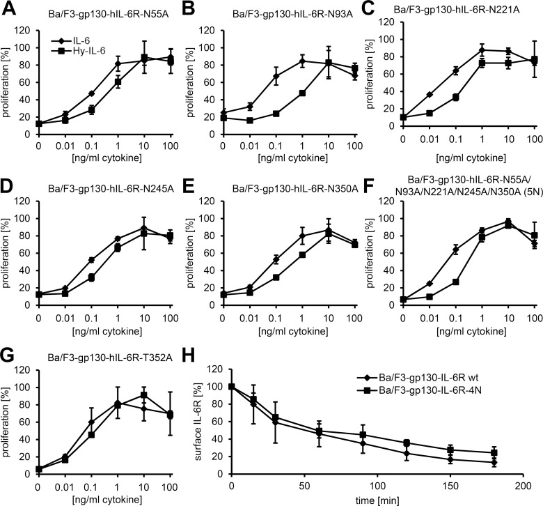
Fig 6 N- and O-linked glycosylation are dispensable for intracellular transport and signaling of the IL-6R. (A–G) Equal numbers of Ba/F3-gp130 cells were incubated for 48 h with increasing amounts (0–100 ng/ml) of either IL-6 or Hyper-IL-6. The stably transduced IL-6R variant is indicated above the diagram. One representative experiment out of three performed is shown (mean ± SD, biological triplicates). (H) Equal numbers of Ba/F3-gp130-hIL-6R and Ba/F3-gp130-hIL-6R-4N cells were labeled with an anti-IL-6R antibody and incubated at 37°C for the indicated time periods. The remaining cell-surface expression of the IL-6R was analyzed via flow cytometry. Data shown are the mean ± SD from three independent experiments.
Fig 6 N- and O-linked glycosylation are dispensable for intracellular transport and signaling of the IL-6R. (A–G) Equal numbers of Ba/F3-gp130 cells were incubated for 48 h with increasing amounts (0–100 ng/ml) of either IL-6 or Hyper-IL-6. The stably transduced IL-6R variant is indicated above the diagram. One representative experiment out of three performed is shown (mean ± SD, biological triplicates). (H) Equal numbers of Ba/F3-gp130-hIL-6R and Ba/F3-gp130-hIL-6R-4N cells were labeled with an anti-IL-6R antibody and incubated at 37°C for the indicated time periods. The remaining cell-surface expression of the IL-6R was analyzed via flow cytometry. Data shown are the mean ± SD from three independent experiments.CC BY · PubMed Central
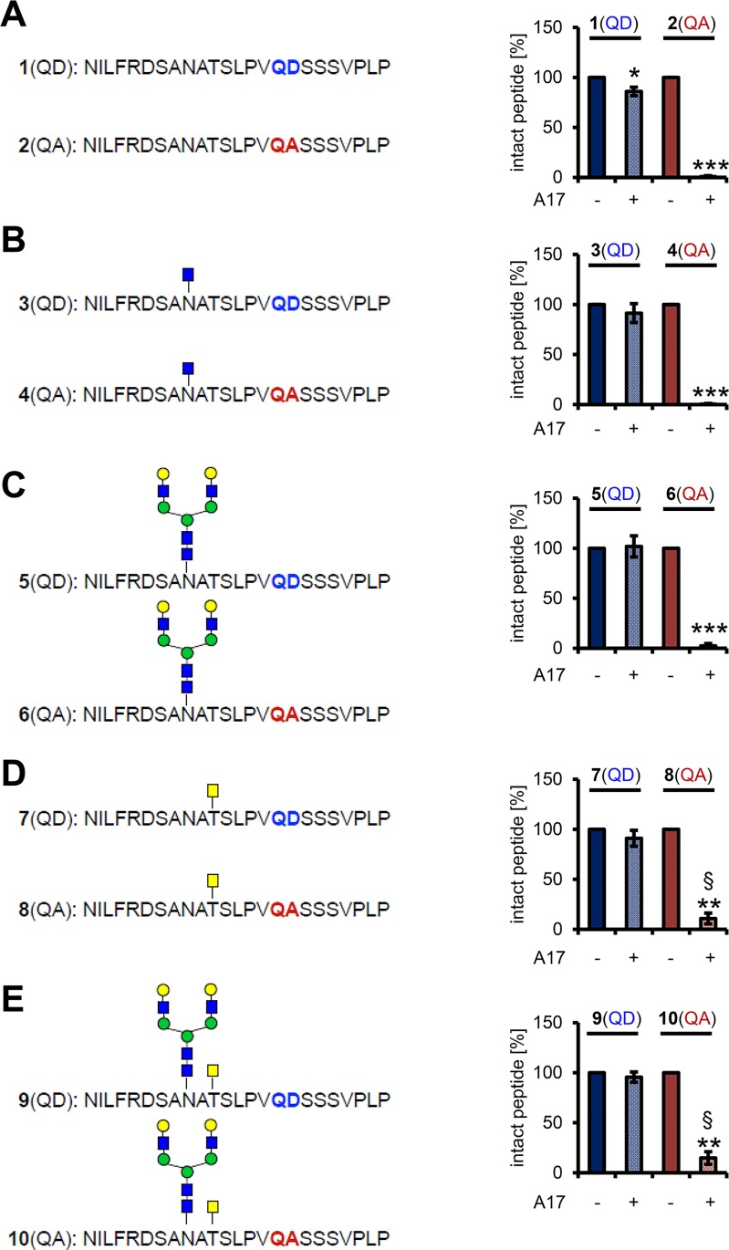
Fig 7 The N- and O-glycosylated sequon adjacent to the cleavage site is of minor importance for IL-6R cleavage. (A) Amino-acid sequence of the two unglycosylated peptides 1(QD) and 2(QA). Peptides were incubated either with or without recombinant ADAM17 for 16 h at 37°C, and the amount of cleaved peptide was analyzed by reversed-phase (RP) chromatography. (B) Amino-acid sequence of the two glycopeptides 3(QD) and 4(QA), which have a GlcNAc attached to the amino-acid residue corresponding to Asn-350 of the IL-6R. Cleavage was determined as described in panel (A). (C) Amino-acid sequence of the two glycopeptides 5(QD) and 6(QA), which have a biantennary N-glycan attached to the amino-acid residue corresponding to Asn-350 of the IL-6R. Cleavage was determined as described in panel (A). (D) Amino-acid sequence of the two glycopeptides 7(QD) and 8(QA), which have a GalNAc attached to the amino-acid residue corresponding toThr-352 of the IL-6R. Cleavage was determined as described in panel (A). (E) Amino-acid sequence of the two glycopeptides 9(QD) and 10(QA), which have a biantennary N-glycan attached to the amino-acid residue corresponding to Asn-350 and a GalNAc attached to the amino-acid residue corresponding toThr-352 of the IL-6R. Cleavage was determined as described in panel (A). All data shown are the mean ± SD from three independent experiments (*p < 0.05, **p < 0.01, ***p < 0.001, compared to the untreated peptide; §p < 0.05, compared to the cleaved nonglycosylated peptide).
Fig 7 The N- and O-glycosylated sequon adjacent to the cleavage site is of minor importance for IL-6R cleavage. (A) Amino-acid sequence of the two unglycosylated peptides 1(QD) and 2(QA). Peptides were incubated either with or without recombinant ADAM17 for 16 h at 37°C, and the amount of cleaved peptide was analyzed by reversed-phase (RP) chromatography. (B) Amino-acid sequence of the two glycopeptides 3(QD) and 4(QA), which have a GlcNAc attached to the amino-acid residue corresponding to Asn-350 of the IL-6R. Cleavage was determined as described in panel (A). (C) Amino-acid sequence of the two glycopeptides 5(QD) and 6(QA), which have a biantennary N-glycan attached to the amino-acid residue corresponding to Asn-350 of the IL-6R. Cleavage was determined as described in panel (A). (D) Amino-acid sequence of the two glycopeptides 7(QD) and 8(QA), which have a GalNAc attached to the amino-acid residue corresponding toThr-352 of the IL-6R. Cleavage was determined as described in panel (A). (E) Amino-acid sequence of the two glycopeptides 9(QD) and 10(QA), which have a biantennary N-glycan attached to the amino-acid residue corresponding to Asn-350 and a GalNAc attached to the amino-acid residue corresponding toThr-352 of the IL-6R. Cleavage was determined as described in panel (A). All data shown are the mean ± SD from three independent experiments (*p < 0.05, **p < 0.01, ***p < 0.001, compared to the untreated peptide; §p < 0.05, compared to the cleaved nonglycosylated peptide).CC BY · PubMed Central
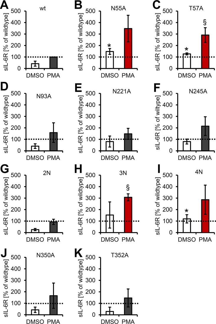
Fig 8 The Asn-55 N-glycan in domain D1 is a protease-regulatory exosite. (A–K) PMA-mediated ectodomain shedding of IL-6R variants lacking single or multiple N-glycans. Experiments were performed with stably transduced Ba/F3-gp130 cell lines. The IL-6R variant is indicated above the respective diagram. Data shown are the mean ± SD from three independent experiments, which were compared to wild-type IL-6R (*p < 0.05, compared to DMSO-treated Ba/F3-gp130-IL-6R cells; §p < 0.05, compared to PMA-stimulated Ba/F3-gp130-IL-6R cells).
Fig 8 The Asn-55 N-glycan in domain D1 is a protease-regulatory exosite. (A–K) PMA-mediated ectodomain shedding of IL-6R variants lacking single or multiple N-glycans. Experiments were performed with stably transduced Ba/F3-gp130 cell lines. The IL-6R variant is indicated above the respective diagram. Data shown are the mean ± SD from three independent experiments, which were compared to wild-type IL-6R (*p < 0.05, compared to DMSO-treated Ba/F3-gp130-IL-6R cells; §p < 0.05, compared to PMA-stimulated Ba/F3-gp130-IL-6R cells).CC BY · PubMed Central
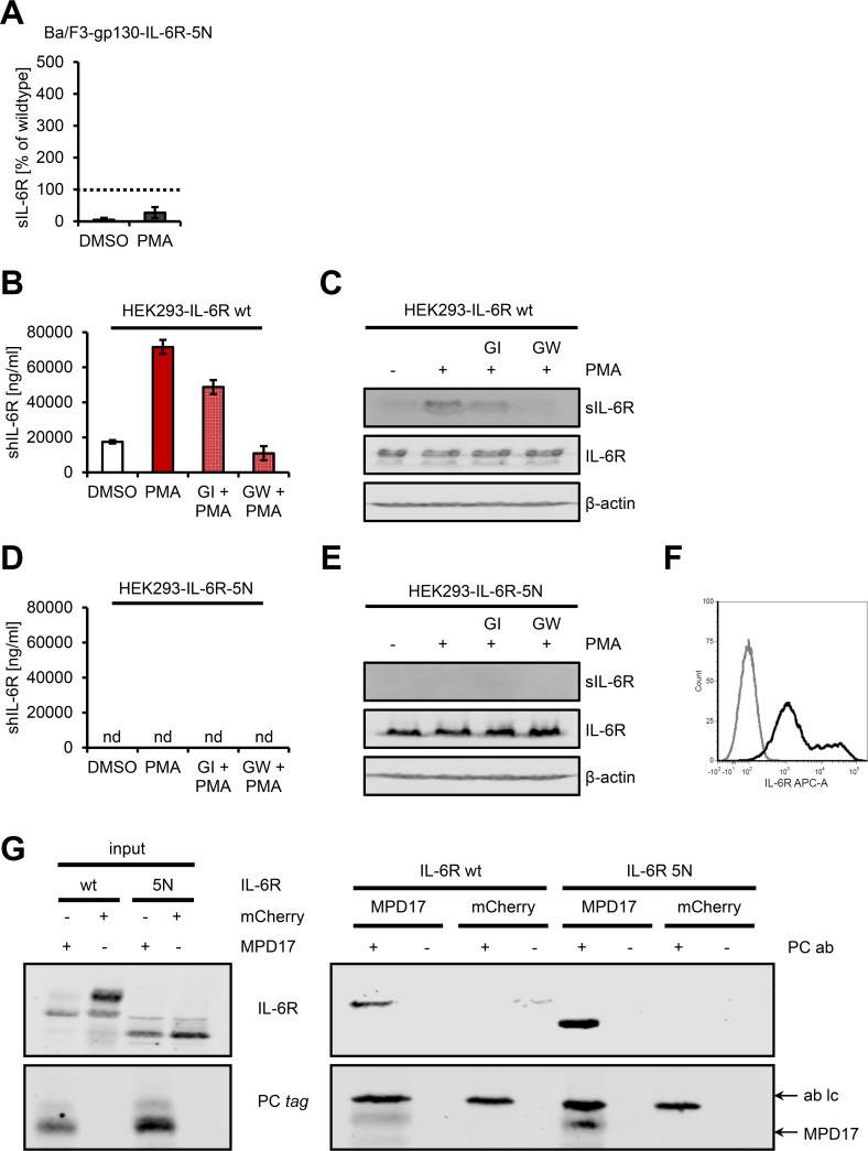
Fig 9 An IL-6R devoid of all N-glycans is defective in ADAM17-mediated proteolysis. (A) PMA-mediated ectodomain shedding of IL-6R-5N with a stably transduced Ba/F3-gp130 cell line. Data shown are the mean ± SD from three independent experiments compared to wild-type IL-6R. (B) HEK293 cells were transiently transfected with a cDNA encoding wild-type IL-6R. Cells were pretreated with the ADAM10-specific inhibitor GI or the combined ADAM10/ADAM17-specific inhibitor GW for 30 min and then stimulated with 100 nM PMA for 2 h as indicated. The amount of sIL-6R in the cell supernatant was determined by ELISA. One out of three experiments with similar outcomes is shown (mean ± SD, n = 3). (C) The experiment was performed as described under (B), but sIL-6R was precipitated from cell supernatant and analyzed by western blot. Furthermore, the cells were lysed, and IL-6R expression in the lysates was also determined by western blot. β-actin served as loading control. One experiment out of three with similar outcomes is shown. (D, E) The experiment was performed as described in (C) and (D), but HEK293 cells were transiently transfected with a cDNA encoding IL-6R-5N. nd, not detected. (F) Cell-surface expression of the IL-6R-5N variant on transiently transfected HEK293 cells analyzed via flow cytometry (black histogram). The control staining is shown in gray. (G) HEK293 cells were transiently transfected with expression plasmids encoding wild-type IL-6R or IL-6R-5N in combination with either an expression plasmid encoding mCherry or MPD17-CANDIS. After 48 h, cells were lysed and the MPD17-CANDIS precipitated via its protein C (PC) tag. The expression of all proteins (input) and the interaction of the IL-6R variants with MPD17-CANDIS were analyzed via western blot. One experiment out of two with similar outcomes is shown.
Fig 9 An IL-6R devoid of all N-glycans is defective in ADAM17-mediated proteolysis. (A) PMA-mediated ectodomain shedding of IL-6R-5N with a stably transduced Ba/F3-gp130 cell line. Data shown are the mean ± SD from three independent experiments compared to wild-type IL-6R. (B) HEK293 cells were transiently transfected with a cDNA encoding wild-type IL-6R. Cells were pretreated with the ADAM10-specific inhibitor GI or the combined ADAM10/ADAM17-specific inhibitor GW for 30 min and then stimulated with 100 nM PMA for 2 h as indicated. The amount of sIL-6R in the cell supernatant was determined by ELISA. One out of three experiments with similar outcomes is shown (mean ± SD, n = 3). (C) The experiment was performed as described under (B), but sIL-6R was precipitated from cell supernatant and analyzed by western blot. Furthermore, the cells were lysed, and IL-6R expression in the lysates was also determined by western blot. β-actin served as loading control. One experiment out of three with similar outcomes is shown. (D, E) The experiment was performed as described in (C) and (D), but HEK293 cells were transiently transfected with a cDNA encoding IL-6R-5N. nd, not detected. (F) Cell-surface expression of the IL-6R-5N variant on transiently transfected HEK293 cells analyzed via flow cytometry (black histogram). The control staining is shown in gray. (G) HEK293 cells were transiently transfected with expression plasmids encoding wild-type IL-6R or IL-6R-5N in combination with either an expression plasmid encoding mCherry or MPD17-CANDIS. After 48 h, cells were lysed and the MPD17-CANDIS precipitated via its protein C (PC) tag. The expression of all proteins (input) and the interaction of the IL-6R variants with MPD17-CANDIS were analyzed via western blot. One experiment out of two with similar outcomes is shown.CC BY · PubMed Central

Verification

Claims the paper makes

The claims and statistics of this paper haven’t been checked yet.

Run the check — free, no account needed. It reads the open-access text and checks the sources it cites.

A few free checks a day · papers already verified open instantly · results are public

Claims other papers make about this one

Each is checked against this paper’s own text: does it say what the citing paper says it does?

Self-check

Percentages, sums and reported differences re-computed from the paper’s own numbers.

The self-check needs the paper’s full text.

Citations

Cites

Show all 3 references

Research works printed but not linked: 60

  1. J Scheller, 2014, Interleukin-6: From basic biology to selective blockade of pro-inflammatory activities, Semin Immunol, 26, 2, 10.1016/j.smim.2013.11.002 · DOI · Paper page
  2. J Wolf, 2014, Interleukin-6 and its receptors: a highly regulated and dynamic system, Cytokine, 70, 11, 10.1016/j.cyto.2014.05.024 · DOI · Paper page
  3. C Garbers, 2012, Plasticity and cross-talk of Interleukin 6-type cytokines, Cytokine Growth Factor Rev, 23, 85, 10.1016/j.cytogfr.2012.04.001 · DOI
  4. C Garbers, 2015, The IL-6/gp130/STAT3 signaling axis: recent advances towards specific inhibition, Curr Opin Immunol, 34, 75, 10.1016/j.coi.2015.02.008 · DOI
  5. C Garbers, 2013, Interleukin-6 and interleukin-11: same same but different, Biol Chem, 394, 1145, 10.1515/hsz-2013-0166 · DOI
  6. J Scheller, 2011, The pro- and anti-inflammatory properties of the cytokine interleukin-6, Biochim Biophys Acta, 1813, 878, 10.1016/j.bbamcr.2011.01.034 · DOI · Paper page
  7. P Baran, 2013, Minimal interleukin (IL-)6 receptor stalk composition for IL-6R shedding and IL-6 classic signaling, J Biol Chem, 288, 14756, 10.1074/jbc.M113.466169 · DOI
  8. N Mitra, 2006, N-linked oligosaccharides as outfitters for glycoprotein folding, form and function, Trends Biochem Sci, 31, 156, 10.1016/j.tibs.2006.01.003 · DOI · Paper page
  9. GH Waetzig, 2010, N-linked glycosylation is essential for the stability but not the signaling function of the interleukin-6 signal transducer glycoprotein 130, J Biol Chem, 285, 1781, 10.1074/jbc.M109.075952 · DOI
  10. LJ Slieker, 1985, Post-translational processing of the epidermal growth factor receptor. Glycosylation-dependent acquisition of ligand-binding capacity, J Biol Chem, 260, 687, 10.1016/S0021-9258(20)71149-X · DOI
  11. DXH Ding, 1995, N-glycosylation of the human granulocyte-macrophage colony-stimulating factor receptor α subunit is essential for ligand binding and signal transduction, J Biol Chem, 270, 24580, 10.1074/jbc.270.41.24580 · DOI
  12. H Zhou, 1999, Characterization of recombinant human CXCR4 in insect cells: role of extracellular domains and N-glycosylation in ligand binding, Arch Biochem Biophys, 369, 267, 10.1006/abbi.1999.1368 · DOI
  13. AR Cole, 1999, Disulfide bond structure and N-glycosylation sites of the extracellular domain of the human interleukin-6 receptor, J Biol Chem, 274, 7207, 10.1074/jbc.274.11.7207 · DOI
  14. J Müllberg, 1994, The soluble human IL-6 receptor. Mutational characterization of the proteolytic cleavage site, J Immunol, 152, 4958, 10.4049/jimmunol.152.10.4958 · DOI
  15. SA Jones, 2011, Therapeutic strategies for the clinical blockade of IL-6/gp130 signaling, J Clin Invest, 121, 3375, 10.1172/JCI57158 · DOI
  16. S Horiuchi, 1994, Soluble interleukin-6 receptors released from T cell or granulocyte/macrophage cell lines and human peripheral blood mononuclear cells are generated through an alternative splicing mechanism, Eur J Immunol, 24, 1945, 10.1002/eji.1830240837 · DOI
  17. G Müller-Newen, 1996, Purification and Characterization of the Soluble Interleukin-6 Receptor from Human Plasma and Identification of An Isoform Generated through Alternative Splicing, Eur J Biochem, 236, 837, 10.1111/j.1432-1033.1996.00837.x · DOI
  18. S Horiuchi, 1998, High-level production of alternatively spliced soluble interleukin-6 receptor in serum of patients with adult T-cell leukaemia/HTLV-I-associated myelopathy, Immunology, 95, 360, 10.1046/j.1365-2567.1998.00622.x · DOI
  19. M Leggate, 2010, The response of interleukin-6 and soluble interleukin-6 receptor isoforms following intermittent high intensity and continuous moderate intensity cycling, Cell Stress Chaperones, 15, 827, 10.1007/s12192-010-0192-z · DOI
  20. C Garbers, 2014, The Interleukin-6 Receptor Asp358Ala Single Nucleotide Polymorphism rs2228145 Confers Increased Proteolytic Conversion Rates by ADAM Proteases, Biochim Biophys Acta, 1842, 1485, 10.1016/j.bbadis.2014.05.018 · DOI · Paper page
  21. N Schumacher, 2015, Shedding of Endogenous Interleukin-6 Receptor (IL-6R) Is Governed by A Disintegrin and Metalloproteinase (ADAM) Proteases while a Full-length IL-6R Isoform Localizes to Circulating Microvesicles, J Biol Chem, 290, 26059, 10.1074/jbc.M115.649509 · DOI
  22. J Tucher, 2014, LC-MS based cleavage site profiling of the proteases ADAM10 and ADAM17 using proteome-derived peptide libraries, J Proteome Res, 13, 2205, 10.1021/pr401135u · DOI
  23. ND Rawlings, 2013, MEROPS: the database of proteolytic enzymes, their substrates and inhibitors, Nucleic Acids Res, 42, D503, 10.1093/nar/gkt953 · DOI
  24. CK Goth, 2015, A systematic study of modulation of ADAM-mediated ectodomain shedding by site-specific O-glycosylation, Proc Natl Acad Sci U S A, 112, 14623, 10.1073/pnas.1511175112 · DOI · Paper page
  25. S Düsterhöft, 2014, A disintegrin and metalloprotease 17 dynamic interaction sequence, the sweet tooth for the human interleukin 6 receptor, J Biol Chem, 289, 16336, 10.1074/jbc.M114.557322 · DOI
  26. C Garbers, 2011, Species specificity of ADAM10 and ADAM17 proteins in interleukin-6 (IL-6) trans-signaling and novel role of ADAM10 in inducible IL-6 receptor shedding, J Biol Chem, 286, 14804, 10.1074/jbc.M111.229393 · DOI · Paper page
  27. K Althoff, 2001, Recognition sequences and structural elements contribute to shedding susceptibility of membrane proteins, Biochem J, 353, 663, 10.1042/bj3530663 · DOI
  28. D Novick, 1989, Soluble cytokine receptors are present in normal human urine, J Exp Med, 170, 1409, 10.1084/jem.170.4.1409 · DOI
  29. M Honda, 1992, Human soluble IL-6 receptor: its detection and enhanced release by HIV infection, J Immunol, 148, 2175, 10.4049/jimmunol.148.7.2175 · DOI
  30. A Chalaris, 2007, Apoptosis is a natural stimulus of IL6R shedding and contributes to the pro-inflammatory trans-signaling function of neutrophils, Blood, 110, 1748, 10.1182/blood-2007-01-067918 · DOI
  31. M Schnölzer, 1996, Protease-catalyzed incorporation of 18O into peptide fragments and its application for protein sequencing by electrospray and matrix-assisted laser desorption/ionization mass spectrometry, Electrophoresis, 17, 945, 10.1002/elps.1150170517 · DOI
  32. V Matthews, 2003, Cellular cholesterol depletion triggers shedding of the human interleukin-6 receptor by ADAM10 and ADAM17 (TACE), J Biol Chem, 278, 38829, 10.1074/jbc.M210584200 · DOI · Paper page
  33. J Lokau, 2016, Proteolytic Cleavage Governs Interleukin-11, Trans-signaling. Cell Rep, 14, 1761
  34. U Bank, 1999, Selective proteolytic cleavage of IL-2 receptor and IL-6 receptor ligand binding chains by neutrophil-derived serine proteases at foci of inflammation, J Interferon Cytokine Res, 19, 1277, 10.1089/107999099312957 · DOI
  35. S Jones, 1998, Shedding of the soluble IL-6 receptor is triggered by Ca2+ mobilization, while basal release is predominantly the product of differential mRNA splicing in THP-1 cells, Eur J Immunol, 28, 3514, 10.1002/(SICI)1521-4141(199811)28:11<3514::AID-IMMU3514>3.0.CO;2-T · DOI · Paper page
  36. S Rafiq, 2007, A common variant of the interleukin 6 receptor (IL-6r) gene increases IL-6r and IL-6 levels, without other inflammatory effects, Genes Immun, 8, 552, 10.1038/sj.gene.6364414 · DOI
  37. J Galicia, 2004, Polymorphisms in the IL-6 receptor (IL-6R) gene: strong evidence that serum levels of soluble IL-6R are genetically influenced, Genes Immun, 5, 513, 10.1038/sj.gene.6364120 · DOI
  38. MJ Mohan, 2002, The tumor necrosis factor-alpha converting enzyme (TACE): a unique metalloproteinase with highly defined substrate selectivity, Biochemistry, 41, 9462, 10.1021/bi0260132 · DOI
  39. I Lorenzen, 2012, The membrane-proximal domain of A Disintegrin and Metalloprotease 17 (ADAM17) is responsible for recognition of the interleukin-6 receptor and interleukin-1 receptor II, FEBS Lett, 586, 1093, 10.1016/j.febslet.2012.03.012 · DOI
  40. D Aderka, 1991, Increased serum levels of soluble receptors for tumor necrosis factor in cancer patients, Cancer Res, 51, 5602
  41. S Siebert, 2015, Cytokines as therapeutic targets in rheumatoid arthritis and other inflammatory diseases, Pharmacological Rev, 67, 280, 10.1124/pr.114.009639 · DOI
  42. K Stone, 2013, Interleukin-6 Receptor Polymorphism Is Prevalent in HIV-negative Castleman Disease and Is Associated with Increased Soluble Interleukin-6 Receptor Levels, PLoS ONE, 8, e54610, 10.1371/journal.pone.0054610 · DOI
  43. R Takakura-Yamamoto, 1996, O-glycosylated species of natural human tumor-necrosis factor-alpha, Eur J Biochem, 235, 431, 10.1111/j.1432-1033.1996.00431.x · DOI
  44. TS Bringman, 1987, Different transforming growth factor-alpha species are derived from a glycosylated and palmitoylated transmembrane precursor, Cell, 48, 429, 10.1016/0092-8674(87)90194-2 · DOI
  45. D Minond, 2012, Discovery of Novel Inhibitors of a Disintegrin and Metalloprotease 17 (ADAM17) Using Glycosylated and Non-glycosylated Substrates, J Biol Chem, 287, 36473, 10.1074/jbc.M112.389114 · DOI
  46. M Fischer, 1997, I. A bioactive designer cytokine for human hematopoietic progenitor cell expansion, Nat Biotechnol, 15, 142, 10.1038/nbt0297-142 · DOI
  47. P Vollmer, 1999, A role for the immunoglobulin-like domain of the human IL-6 receptor. Intracellular protein transport and shedding, FEBS J, 263, 438, 10.1046/j.1432-1327.1999.00511.x · DOI
  48. MJ Boulanger, 2003, Hexameric structure and assembly of the interleukin-6/IL-6 alpha-receptor/gp130 complex, Science, 300, 2101, 10.1126/science.1083901 · DOI
  49. AJ Gearing, 1994, Processing of tumour necrosis factor-alpha precursor by metalloproteinases, Nature, 370, 555, 10.1038/370555a0 · DOI
  50. M Kovaleva, 2006, Abrogation of viral interleukin-6 (vIL-6)-induced signaling by intracellular retention and neutralization of vIL-6 with an anti-vIL-6 single-chain antibody selected by phage display, J Virol, 80, 8510, 10.1128/JVI.00420-06 · DOI
  51. M Fischer, 1997, A bioactive designer cytokine for human hematopoietic progenitor cell expansion, Nat Biotech, 15, 145, 10.1038/nbt0297-142 · DOI
  52. A Schroers, 2005, Dynamics of the gp130 cytokine complex: a model for assembly on the cellular membrane, Protein Sci, 14, 783, 10.1110/ps.041117105 · DOI
  53. A Mackiewicz, 1992, Complex of soluble human IL-6-receptor/IL-6 up-regulates expression of acute-phase proteins, J Immunol, 149, 2021, 10.4049/jimmunol.149.6.2021 · DOI
  54. R Ketteler, 2002, Enhanced transgene expression in primitive hematopoietic progenitor cells and embryonic stem cells efficiently transduced by optimized retroviral hybrid vectors, Gene Ther, 9, 477, 10.1038/sj.gt.3301653 · DOI
  55. C Garbers, 2011, Inhibition of classic signaling is a novel function of soluble glycoprotein 130 (sgp130), which is controlled by the ratio of interleukin 6 and soluble interleukin 6 receptor, J Biol Chem, 286, 42959, 10.1074/jbc.M111.295758 · DOI
  56. J Suthaus, 2010, Forced homo- and heterodimerization of all gp130-type receptor complexes leads to constitutive ligand-independent signaling and cytokine-independent growth, Mol Biol Cell, 21, 2797, 10.1091/mbc.E10-03-0240 · DOI
  57. P Somasundaram, 2016, C-Terminal Charge-Reversal Derivatization and Parallel Use of Multiple Proteases Facilitates Identification of Protein C-Termini by C-Terminomics, J Proteome Res, 15, 1369, 10.1021/acs.jproteome.6b00146 · DOI
  58. Z Wu, 2010, Chemoenzymatic synthesis of glycosylphosphatidylinositol-anchored glycopeptides, Chem Commun, 46, 5773, 10.1039/c0cc00828a · DOI
  59. V Ullmann, 2012, Convergent solid-phase synthesis of N-glycopeptides facilitated by pseudoprolines at consensus-sequence Ser/Thr residues, Angew Chem Int Ed Engl, 51, 11566, 10.1002/anie.201204272 · DOI
  60. R Chen, 2010, Study of on-resin convergent synthesis of N-linked glycopeptides containing a large high mannose N-linked oligosaccharide, J Am Chem Soc, 132, 3211, 10.1021/ja9104073 · DOI

Cited by

Sorted by citations

See all 135

Metadata

Authors
Riethmueller, S., Somasundaram, P., Ehlers, J. C., Hung, C.-W., Flynn, C. M., Lokau, J., Agthe, M., Düsterhöft, S., Zhu, Y., Grötzinger, J., Lorenzen, I., Koudelka, T., Yamamoto, K., Pickhinke, U., Wichert, R., Becker-Pauly, C., Rädisch, M., Albrecht, A., Hessefort, M., Stahnke, D., Unverzagt, C., Rose-John, S., Tholey, A., & Garbers, C.
Venue
PLOS Biology
Published
2017-01-06
Volume
15
Issue
1
Pages
e2000080
ISSN
1545-7885
Cited by
135
References
3
Language
en

About these checks

Registry, retraction, Cites and statistics results are computed, not judged, with one exception: a printed reference that no index lists is read once by a model before it counts as a citation error. Claims are extracted by a model and kept only when their quote appears verbatim in the text; each cited source is then checked, with a model’s help, for whether it says what the claim relies on. Those verdicts can be wrong — read the claim’s quote and the cited source before relying on one.

Statistics and arithmetic are recomputed, not judged.

Confidence bands are shown only for fields calibrated on our annotated test set. None is calibrated yet, so no band is shown.

What counts as a claim ›How we measure confidence ›How we check this ›

Are you an author, or do you see a problem with this page? Request a review

How to cite

Cite CoChat for the checks on this page, and the paper for its own findings.

This page
CoChat. (2026). Citation check: Proteolytic Origin of the Soluble Human IL-6R In Vivo and a Decisive Role of N-Glycosylation. https://app.cochat.ai/papers/10.1371/journal.pbio.2000080
The paper
Riethmueller, S., Somasundaram, P., Ehlers, J. C., Hung, C.-W., Flynn, C. M., Lokau, J., Agthe, M., Düsterhöft, S., Zhu, Y., Grötzinger, J., Lorenzen, I., Koudelka, T., Yamamoto, K., Pickhinke, U., Wichert, R., Becker-Pauly, C., Rädisch, M., Albrecht, A., Hessefort, M., . . . Garbers, C. (2017). Proteolytic Origin of the Soluble Human IL-6R In Vivo and a Decisive Role of N-Glycosylation. PLOS Biology. https://doi.org/10.1371/journal.pbio.2000080