

The DOI matches its Crossref record; 74 of the 74 references it cites were found in a registry or an index.
Mechanistic computational modeling of sFLT1 secretion dynamics
Open accessPublished version of a preprint
Abstract
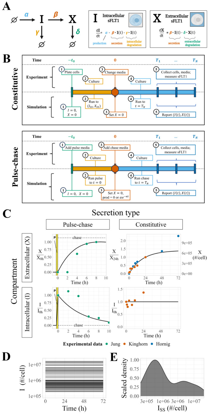
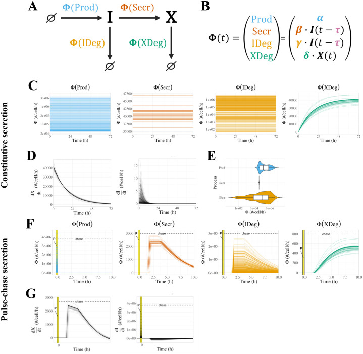
Constitutively secreted by endothelial cells, soluble FLT1 (sFLT1 or sVEGFR1) binds and sequesters extracellular vascular endothelial growth factors (VEGF), thereby reducing VEGF binding to VEGF receptor tyrosine kinases and their downstream signaling. In doing so, sFLT1 plays an important role in vascular development and in the patterning of new blood vessels in angiogenesis. Here, we develop multiple mechanistic models of sFLT1 secretion and identify a minimal mechanistic model that recapitulates key qualitative and quantitative features of temporal experimental datasets of sFLT1 secretion from multiple studies. We show that the experimental data on sFLT1 secretion is best represented by a delay differential equation (DDE) system including a maturation term, reflecting the time required between synthesis and secretion. Using optimization to identify appropriate values for the key mechanistic parameters in the model, we show that two model parameters (extracellular degradation rate constant and maturation time) are very strongly constrained by the experimental data, and that the remaining parameters are related by two strongly constrained constants. Thus, only one degree of freedom remains, and measurements of the intracellular levels of sFLT1 would fix the remaining parameters. Comparison between simulation predictions and additional experimental data of the outcomes of chemical inhibitors and genetic perturbations suggest that intermediate values of the secretion rate constant best match the simulation with experiments, which would completely constrain the model. However, some of the inhibitors tested produce results that cannot be reproduced by the model simulations, suggesting that additional mechanisms not included here are required to explain those inhibitors. Overall, the model reproduces most available experimental data and suggests targets for further quantitative investigation of the sFLT1 system.
Overview
The claims and statistics of this paper haven’t been checked yet.
Run the check — free, no account needed. It reads the open-access text and checks the sources it cites.
Integrity signals
Registry
Cites
Statistics
Figures




Verification
Claims the paper makes
The claims and statistics of this paper haven’t been checked yet.
Run the check — free, no account needed. It reads the open-access text and checks the sources it cites.
Self-check
Percentages, sums and reported differences re-computed from the paper’s own numbers.
The self-check needs the paper’s full text.
Citations
Cites
Fiji: an open-source platform for biological-image analysis
Schindelin, J., Arganda-Carreras, I., Frise, E., Kaynig, V., Longair, M., Pietzsch, T., Preibisch, S., Rueden, C., Saalfeld, S., Schmid, B., et al. (2012) · Nature Methods · doi:10.1038/nmeth.2019 · Cited by 66,890
Information Theory and an Extension of the Maximum Likelihood Principle
Akaike, H. (1998) · Springer Series in Statistics · doi:10.1007/978-1-4612-1694-0_15 · Cited by 17,781
Tissue-based map of the human proteome
Uhlén, M., Fagerberg, L., Hallström, B. M., Lindskog, C., Oksvold, P., Mardinoglu, A., Sivertsson, Å., Kampf, C., Sjöstedt, E., Asplund, A., et al. (2015) · Science · doi:10.1126/science.1260419 · Cited by 15,286
Jaffe, E. A., Nachman, R. L., Becker, C. G., & Minick, C. R. (1973) · Journal of Clinical Investigation · doi:10.1172/jci107470 · Cited by 6,856
Global quantification of mammalian gene expression control
Schwanhäusser, B., Busse, D., Li, N., Dittmar, G., Schuchhardt, J., Wolf, J., Chen, W., & Selbach, M. (2011) · Nature · doi:10.1038/nature10098 · Cited by 6,566
Bozdogan, H. (1987) · Psychometrika · doi:10.1007/bf02294361 · Cited by 4,468
Maynard, S. E., Min, J.-Y., Merchan, J., Lim, K.-H., Li, J., Mondal, S., Libermann, T. A., Morgan, J. P., Sellke, F. W., Stillman, I. E., et al. (2003) · Journal of Clinical Investigation · doi:10.1172/jci17189 · Cited by 4,101
Intracellular Aspects of the Process of Protein Synthesis
Palade, G. (1975) · Science · doi:10.1126/science.1096303 · Cited by 3,564
An Introduction to Systems Biology: Design Principles of Biological Circuits
Alon, U. (2006) · doi:10.1201/9781420011432 · Cited by 2,825
Mechanisms, regulation and functions of the unfolded protein response
Hetz, C., Zhang, K., & Kaufman, R. J. (2020) · Nature Reviews Molecular Cell Biology · doi:10.1038/s41580-020-0250-z · Cited by 2,666
Chloroquine inhibits autophagic flux by decreasing autophagosome-lysosome fusion
Mauthe, M., Orhon, I., Rocchi, C., Zhou, X., Luhr, M., Hijlkema, K.-J., Coppes, R. P., Engedal, N., Mari, M., & Reggiori, F. (2018) · Autophagy · doi:10.1080/15548627.2018.1474314 · Cited by 2,122
Glycosylation in health and disease
Reily, C., Stewart, T. J., Renfrow, M. B., & Novak, J. (2019) · Nature Reviews Nephrology · doi:10.1038/s41581-019-0129-4 · Cited by 2,075
Lippincott-Schwartz, J., Yuan, L. C., Bonifacino, J. S., & Klausner, R. D. (1989) · Cell · doi:10.1016/0092-8674(89)90685-5 · Cited by 1,687
Kendall, R. L., & Thomas, K. A. (1993) · Proceedings of the National Academy of Sciences · doi:10.1073/pnas.90.22.10705 · Cited by 1,394
Cellular Heterogeneity: Do Differences Make a Difference?
Altschuler, S. J., & Wu, L. F. (2010) · Cell · doi:10.1016/j.cell.2010.04.033 · Cited by 1,214
Mis-assembly of clathrin lattices on endosomes reveals a regulatory switch for coated pit formation.
Wang, L. H., Rothberg, K. G., & Anderson, R. G. (1993) · The Journal of cell biology · doi:10.1083/jcb.123.5.1107 · Cited by 1,177
Endocytosis and signalling: intertwining molecular networks
Sorkin, A., & von Zastrow, M. (2009) · Nature Reviews Molecular Cell Biology · doi:10.1038/nrm2748 · Cited by 1,064
Hiratsuka, S., Minowa, O., Kuno, J., Noda, T., & Shibuya, M. (1998) · Proceedings of the National Academy of Sciences · doi:10.1073/pnas.95.16.9349 · Cited by 1,062
Fujiwara, T., Oda, K., Yokota, S., Takatsuki, A., & Ikehara, Y. (1988) · Journal of Biological Chemistry · doi:10.1016/s0021-9258(19)81393-5 · Cited by 885
Misumi, Y., Misumi, Y., Miki, K., Takatsuki, A., Tamura, G., & Ikehara, Y. (1986) · Journal of Biological Chemistry · doi:10.1016/s0021-9258(18)67398-3 · Cited by 847
Endocytosis and intracellular trafficking of ErbBs
Sorkin, A., & Goh, L. K. (2009) · Experimental Cell Research · doi:10.1016/j.yexcr.2008.07.029 · Cited by 756
Kendall, R. L., Wang, G., & Thomas, K. A. (1996) · Biochemical and Biophysical Research Communications · doi:10.1006/bbrc.1996.1355 · Cited by 724
Corneal avascularity is due to soluble VEGF receptor-1
Ambati, B. K., Nozaki, M., Singh, N., Takeda, A., Jani, P. D., Suthar, T., Albuquerque, R. J. C., Richter, E., Sakurai, E., Newcomb, M. T., et al. (2006) · Nature · doi:10.1038/nature05249 · Cited by 684
Uhlén, M., Karlsson, M. J., Hober, A., Svensson, A.-S., Scheffel, J., Kotol, D., Zhong, W., Tebani, A., Strandberg, L., Edfors, F., et al. (2019) · Science Signaling · doi:10.1126/scisignal.aaz0274 · Cited by 495
Proteome Half-Life Dynamics in Living Human Cells
Eden, E., Geva-Zatorsky, N., Issaeva, I., Cohen, A., Dekel, E., Danon, T., Cohen, L., Mayo, A., & Alon, U. (2011) · Science · doi:10.1126/science.1199784 · Cited by 326
A Cell-Based Model Exhibiting Branching and Anastomosis during Tumor-Induced Angiogenesis
Bauer, A. L., Jackson, T. L., & Jiang, Y. (2007) · Biophysical Journal · doi:10.1529/biophysj.106.101501 · Cited by 297
Syntaxin 6 functions in trans-Golgi network vesicle trafficking.
Bock, J. B., Klumperman, J., Davanger, S., & Scheller, R. H. (1997) · Molecular Biology of the Cell · doi:10.1091/mbc.8.7.1261 · Cited by 288
Localization of proteins to the Golgi apparatus
Munro, S. (1998) · Trends in Cell Biology · doi:10.1016/s0962-8924(97)01197-5 · Cited by 262
Local Guidance of Emerging Vessel Sprouts Requires Soluble Flt-1
Chappell, J. C., Taylor, S. M., Ferrara, N., & Bautch, V. L. (2009) · Developmental Cell · doi:10.1016/j.devcel.2009.07.011 · Cited by 235
Self-citationRab27a enables myosin Va-dependent melanosome capture by recruiting the myosin to the organelle
Wu, X., Rao, K., Bowers, M., Copeland, N. G., Jenkins, N. A., & Hammer, J. A. (2001) · Journal of Cell Science · doi:10.1242/jcs.114.6.1091 · Cited by 233
Release and Complex Formation of Soluble VEGFR-1 from Endothelial Cells and Biological Fluids
Hornig, C., Barleon, B., Ahmad, S., Vuorela, P., Ahmed, A., & Weich, H. A. (2000) · Laboratory Investigation · doi:10.1038/labinvest.3780050 · Cited by 224
Wu, F. T. H., Stefanini, M. O., Gabhann, F. M., Kontos, C. D., Annex, B. H., & Popel, A. S. (2009) · Journal of Cellular and Molecular Medicine · doi:10.1111/j.1582-4934.2009.00941.x · Cited by 207
Central dogma rates and the trade-off between precision and economy in gene expression
Hausser, J., Mayo, A., Keren, L., & Alon, U. (2019) · Nature Communications · doi:10.1038/s41467-018-07391-8 · Cited by 188
The VEGF receptor Flt-1 spatially modulates Flk-1 signaling and blood vessel branching
Kappas, N. C., Zeng, G., Chappell, J. C., Kearney, J. B., Hazarika, S., Kallianos, K. G., Patterson, C., Annex, B. H., & Bautch, V. L. (2008) · The Journal of Cell Biology · doi:10.1083/jcb.200709114 · Cited by 183
Self-citationPerfahl, H., Byrne, H. M., Chen, T., Estrella, V. C., Alarcón, T., Lapin, A., Gatenby, R. A., Gillies, R. J., Lloyd, M. C., Maini, P. K., et al. (2011) · PLoS ONE · doi:10.1371/journal.pone.0014790 · Cited by 168
Inactivation of lysosomal function in normal cultured human fibroblasts by chloroquine
Lie, S. O., & Schofield, B. (1973) · Biochemical Pharmacology · doi:10.1016/0006-2952(73)90197-4 · Cited by 155
Huang, S. S., & Huang, J. S. (1988) · Journal of Biological Chemistry · doi:10.1016/s0021-9258(18)37798-6 · Cited by 145
Nonlinear delay differential equations and their application to modeling biological network motifs
Glass, D. S., Jin, X., & Riedel-Kruse, I. H. (2021) · Nature Communications · doi:10.1038/s41467-021-21700-8 · Cited by 139
Differential Binding Characteristics and Cellular Inhibition by Soluble VEGF Receptors 1 and 2
Röckl, W., Hecht, D., Sztajer, H., Waltenberger, J. L., Yayon, A., & Weich, H. A. (1998) · Experimental Cell Research · doi:10.1006/excr.1998.4039 · Cited by 135
Qutub, A. A., & Popel, A. S. (2009) · BMC Systems Biology · doi:10.1186/1752-0509-3-13 · Cited by 130
Syntaxin 6: The Promiscuous Behaviour of a SNARE Protein
Wendler, F., & Tooze, S. A. (2001) · Traffic · doi:10.1034/j.1600-0854.2001.20903.x · Cited by 117
Agent-based modeling of morphogenetic systems: advantages and challenges.
Glen, C. M., Kemp, M. L., & Voit, E. O. (2019) · PLoS Computational Biology · doi:10.1371/journal.pcbi.1006577 · Cited by 116
Orecchia, A., Lacal, P. M., Schietroma, C., Morea, V., Zambruno, G., & Failla, C. M. (2003) · Journal of Cell Science · doi:10.1242/jcs.00673 · Cited by 115
Manickam, V., Tiwari, A., Jung, J.-J., Bhattacharya, R., Goel, A., Mukhopadhyay, D., & Choudhury, A. (2010) · Blood · doi:10.1182/blood-2010-06-291690 · Cited by 104
VEGF-Directed Blood Vessel Patterning: From Cells to OrganismPreprint
Bautch, V. (2011) · Cold Spring Harbor Perspectives in Medicine · doi:10.1101/cshperspect.a006452 · Cited by 103
Self-citation(2008) · Experimental Cell Research · doi:10.1016/s0014-4827(08)00404-7 · Cited by 96
The Origin of Phenotypic Heterogeneity in a Clonal Cell Population In Vitro
Stockholm, D., Benchaouir, R., Picot, J., Rameau, P., Neildez‐Nguyen, T. M. A., Landini, G., Laplace‐Builhé, C., & Páldi, A. (2007) · PLoS ONE · doi:10.1371/journal.pone.0000394 · Cited by 94
Apical targeting of syntaxin 3 is essential for epithelial cell polarity
Sharma, N., Low, S. H., Misra, S., Bhattaram, P., & Weimbs, T. (2006) · The Journal of Cell Biology · doi:10.1083/jcb.200603132 · Cited by 93
Sela, S., Natanson‐Yaron, S., Zcharia, E., Vlodavsky, I., Yagel, S., & Keshet, E. (2011) · Circulation Research · doi:10.1161/circresaha.110.239665 · Cited by 88
Rahimi, N., Golde, T. E., & Meyer, R. (2009) · Cancer Research · doi:10.1158/0008-5472.can-08-2905 · Cited by 82
Park, M., & Lee, S. (1999) · Biochemical and Biophysical Research Communications · doi:10.1006/bbrc.1999.1580 · Cited by 75
Autocrine activity of soluble Flt-1 controls endothelial cell function and angiogenesis
Ahmad, S., Hewett, P. W., Al‐Ani, B., Sissaoui, S., Fujisawa, T., Cudmore, M., & Ahmed, A. (2011) · Vascular Cell · doi:10.1186/2045-824x-3-15 · Cited by 74
Du, Y., Shen, J. Z., Hsu, J. L., et al. (2013) · Oncogene · doi:10.1038/onc.2013.1 · Cited by 65
Alzahofi, N., Welz, T., Robinson, C. L., Page, E. L., Briggs, D. A., Stainthorp, A. K., Reekes, J., Elbe, D. A., Straub, F., Kallemeijn, W. W., et al. (2020) · Nature Communications · doi:10.1038/s41467-020-17212-6 · Cited by 61
VEGFR1 receptor tyrosine kinase localization to the Golgi apparatus is calcium-dependent
Mittar, S., Ulyatt, C., Howell, G. J., Bruns, A. F., Zachary, I. C., Walker, J. H., & Ponnambalam, S. (2009) · Experimental Cell Research · doi:10.1016/j.yexcr.2008.12.020 · Cited by 54
Saito, T., Takeda, N., Amiya, E., Nakao, T., Abe, H., Semba, H., Soma, K., Koyama, K., Hosoya, Y., Imai, Y., et al. (2013) · FEBS Letters · doi:10.1016/j.febslet.2013.05.038 · Cited by 54
Flt-1 (VEGFR-1) coordinates discrete stages of blood vessel formation
Chappell, J. C., Cluceru, J. G., Nesmith, J. E., Mouillesseaux, K. P., Bradley, V. B., Hartland, C. M., Hashambhoy-Ramsay, Y., Walpole, J., Peirce, S. M., Gabhann, F. M., et al. (2016) · Cardiovascular Research · doi:10.1093/cvr/cvw091 · Cited by 52
Self-citationMolecular mechanism matters: Benefits of mechanistic computational models for drug development
Clegg, L. E., & Gabhann, F. M. (2015) · Pharmacological Research · doi:10.1016/j.phrs.2015.06.002 · Cited by 50
Wu, F. T. H., Stefanini, M. O., Gabhann, F. M., Kontos, C. D., Annex, B. H., & Popel, A. S. (2010) · American Journal of Physiology-Heart and Circulatory Physiology · doi:10.1152/ajpheart.00365.2009 · Cited by 50
de Galarreta, M. R., Navarro‐Corcuera, A., Ansorena, E., García-Garzón, A., Mòdol, T., López-Zabalza, M. J., Martínez-Irujo, J. J., & Iraburu, M. J. (2016) · Biochimica et Biophysica Acta (BBA) - Molecular Cell Research · doi:10.1016/j.bbamcr.2016.05.002 · Cited by 46
Membrane fusion by VAMP3 and plasma membrane t-SNAREs
Hu, C., Hardee, D., & Minnear, F. L. (2007) · Experimental Cell Research · doi:10.1016/j.yexcr.2007.06.008 · Cited by 41
Agent-based model of angiogenesis simulates capillary sprout initiation in multicellular networks
Walpole, J., Chappell, J. C., Cluceru, J. G., Gabhann, F. M., Bautch, V. L., & Peirce, S. M. (2015) · Integrative Biology · doi:10.1039/c5ib00024f · Cited by 41
Self-citationComputational Modeling of Interacting VEGF and Soluble VEGF Receptor Concentration Gradients
Hashambhoy, Y. L., Chappell, J. C., Peirce, S. M., Bautch, V. L., & Gabhann, F. M. (2011) · Frontiers in Physiology · doi:10.3389/fphys.2011.00062 · Cited by 38
Self-citationJung, J., Tiwari, A., Inamdar, S. M., Thomas, C. P., Goel, A., & Choudhury, A. (2012) · PLoS ONE · doi:10.1371/journal.pone.0044572 · Cited by 34
A Novel Class of Fusion Polypeptides Inhibits Exocytosis
Matsushita, K., Morrell, C. N., & Lowenstein, C. J. (2004) · Molecular Pharmacology · doi:10.1124/mol.104.004275 · Cited by 34
A quantitative study of the Golgi retention of glycosyltransferases
Sun, X., Mahajan, D., Chen, B., Song, Z., & Lü, L. (2021) · Journal of Cell Science · doi:10.1242/jcs.258564 · Cited by 33
A computational analysis of in vivo VEGFR activation by multiple co-expressed ligands
Clegg, L. E., & Gabhann, F. M. (2017) · PLoS Computational Biology · doi:10.1371/journal.pcbi.1005445 · Cited by 32
Walpole, J., Gabhann, F. M., Peirce, S. M., et al. (2017) · Microcirculation · doi:10.1111/micc.12393 · Cited by 32
Trafficking dynamics of VEGFR1, VEGFR2, and NRP1 in human endothelial cells
Sarabipour, S., Kinghorn, K., Quigley, K., Kovács‐Kása, A., Annex, B. H., Bautch, V. L., & Gabhann, F. M. (2024) · PLoS Computational Biology · doi:10.1371/journal.pcbi.1011798 · Cited by 23
Self-citationWu, F. T. H., Stefanini, M. O., Gabhann, F. M., Kontos, C. D., Annex, B. H., & Popel, A. S. (2009) · Physiological Genomics · doi:10.1152/physiolgenomics.00031.2009 · Cited by 20
Silva, J. A. F., Qi, X., Grant, M. B., & Boulton, M. E. (2021) · Scientific Reports · doi:10.1038/s41598-021-96964-7 · Cited by 14
Kinghorn, K., Gill, A., Marvin, A., Li, R., Quigley, K., Singh, S., Gore, M. T., le Noble, F. A. C., Gabhann, F. M., & Bautch, V. L. (2023) · Angiogenesis · doi:10.1007/s10456-023-09893-6 · Cited by 11
Self-citationMechanistic characterization of endothelial sprouting mediated by pro‐angiogenic signaling
Song, M., & Finley, S. D. (2021) · Microcirculation · doi:10.1111/micc.12744 · Cited by 10
A computational analysis of pro-angiogenic therapies for peripheral artery disease
Clegg, L. E., & Gabhann, F. M. (2018) · Integrative Biology · doi:10.1039/c7ib00218a · Cited by 9
Metadata
- Authors
- Gill, A., Kinghorn, K., Bautch, V. L., & Mac Gabhann, F.
- Affiliations
- Johns Hopkins University (US)
- University of North Carolina at Chapel Hill (US)
- Corresponding author
- Gill, Amy
- Venue
- PLOS Computational Biology
- Publisher
- Public Library of Science (PLoS)
- Published
- 2025-08-18
- Volume
- 21
- Issue
- 8
- Pages
- e1013324
- ISSN
- 1553-734X (ISSN-L)
- 1553-7358
- PMC
- PMC12370208
- OpenAlex
- W4413314500
- Cited by
- 1
- References
- 74
- Citations by year
- 2025: 1
- FWCI
- 0.30
- Type
- journal article
- Language
- en
- Update notices
- new_version (2025-08-21)
- Open access
- gold
- doi.org
- Licence
- Locations
- PLoS Computational Biology (publishedVersion, cc-by)
- bioRxiv (Cold Spring Harbor Laboratory) (acceptedVersion, cc-by)
- PubMed Central (publishedVersion, cc-by)
- PubMed (publishedVersion)
- Carolina Digital Repository (University of North Carolina at Chapel Hill) (submittedVersion, cc-by)
- PubMed Central (submittedVersion, cc-by)
- DOAJ (DOAJ: Directory of Open Access Journals) (submittedVersion, cc-by-sa)
- UNC Libraries
- Funders
- National Institute of General Medical Sciences (R01-GM129074)
- National Heart, Lung, and Blood Institute (R01-HL101200, R35-HL139950)
- Topics
- Life Sciences › Biochemistry, Genetics and Molecular Biology › Molecular Biology › Angiogenesis and VEGF in Cancer
- Physical Sciences › Mathematics › Modeling and Simulation › Mathematical Biology Tumor Growth
- Life Sciences › Biochemistry, Genetics and Molecular Biology › Molecular Biology › Gene Regulatory Network Analysis
- Keywords
- soluble Flt-1, delay differential equations, angiogenesis, endothelial cells, parameter estimation, chemical inhibitors, gene perturbation, mechanistic modeling
About these checks
Registry, retraction, Cites and statistics results are computed, not judged, with one exception: a printed reference that no index lists is read once by a model before it counts as a citation error. Claims are extracted by a model and kept only when their quote appears verbatim in the text; each cited source is then checked, with a model’s help, for whether it says what the claim relies on. Those verdicts can be wrong — read the claim’s quote and the cited source before relying on one.
Statistics and arithmetic are recomputed, not judged.
Confidence bands are shown only for fields calibrated on our annotated test set. None is calibrated yet, so no band is shown.
What counts as a claim ›How we measure confidence ›How we check this ›
Are you an author, or do you see a problem with this page? Request a review
Data sources
Bibliographic data: Semantic Scholar, OpenAlex, Crossref.
Contains information from the Semantic Scholar Academic Graph Datasets, made available under the ODC Attribution License (ODC-BY 1.0).
Semantic Scholar’s data is described in: Kinney, R., et al. (2023). The Semantic Scholar Open Data Platform. arXiv:2301.10140.
