

The DOI matches its Crossref record; 38 of the 39 references it cites were found in a registry or an index.
The Architecture of the Adhesive Apparatus of Cultured Osteoclasts: From Podosome Formation to Sealing Zone Assembly
Open accessAbstract
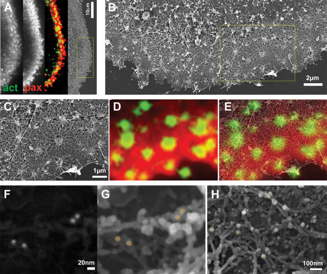
BACKGROUND: Osteoclasts are bone-degrading cells, which play a central role in physiological bone remodeling. Unbalanced osteoclast activity is largely responsible for pathological conditions such as osteoporosis. Osteoclasts develop specialized adhesion structures, the so-called podosomes, which subsequently undergo dramatic reorganization into sealing zones. These ring-like adhesion structures, which delimit the resorption site, effectively seal the cell to the substrate forming a diffusion barrier. The structural integrity of the sealing zone is essential for the cell ability to degrade bone, yet its structural organization is poorly understood. PRINCIPAL FINDINGS: Combining high-resolution scanning electron microscopy with fluorescence microscopy performed on the same sample, we mapped the molecular architecture of the osteoclast resorptive apparatus from individual podosomes to the sealing zone, at an unprecedented resolution. Podosomes are composed of an actin-bundle core, flanked by a ring containing adhesion proteins connected to the core via dome-like radial actin fibers. The sealing zone, hallmark of bone-resorbing osteoclasts, consists of a dense array of podosomes communicating through a network of actin filaments, parallel to the substrate and anchored to the adhesive plaque domain via radial actin fibers. SIGNIFICANCE: The sealing zone of osteoclasts cultured on bone is made of structural units clearly related to individual podosomes. It differs from individual or clustered podosomes in the higher density and degree of inter-connectivity of its building blocks, thus forming a unique continuous functional structure connecting the cell to its extracellular milieu. Through this continuous structure, signals reporting on the substrate condition may be transmitted to the whole cell, modulating the cell response under physiological and pathological conditions.
Overview
The claims and statistics of this paper haven’t been checked yet.
Run the check — free, no account needed. It reads the open-access text and checks the sources it cites.
Integrity signals
Registry
Cites
Statistics
Figures
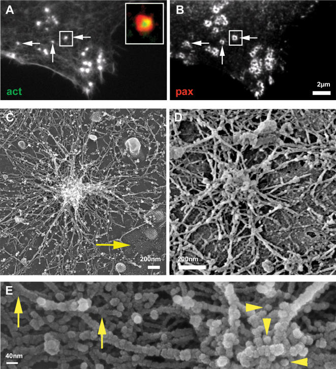
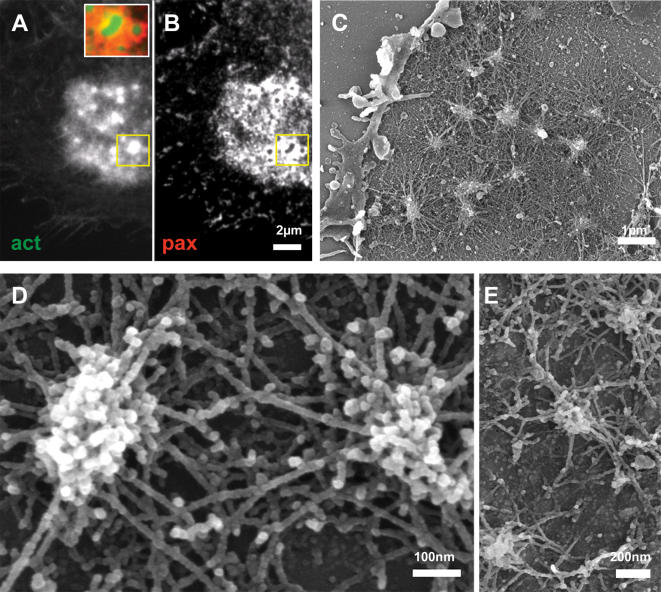
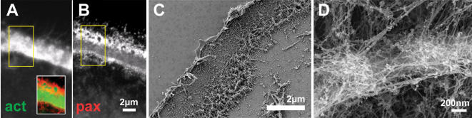
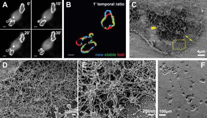
Verification
Claims the paper makes
The claims and statistics of this paper haven’t been checked yet.
Run the check — free, no account needed. It reads the open-access text and checks the sources it cites.
Claims other papers make about this one
Each is checked against this paper’s own text: does it say what the citing paper says it does?
In the citing paper
“It is important to mention that these domains are only formed when osteoclasts are in contact with extracellular mineralized matrix, in a process which α v β 3-integrin, as well as the CD44, mediates the attachment of the osteoclast podosomes to the bone surface [96–99].”
From Biology of Bone Tissue: Structure, Function, and Factors That Influence Bone Cells (2015) · cites this paper as [98]
Self-check
Percentages, sums and reported differences re-computed from the paper’s own numbers.
The self-check needs the paper’s full text.
Citations
Cites
Bone Resorption by Osteoclasts
Teitelbaum, S. L. (2000) · Science · doi:10.1126/science.289.5484.1504 · Cited by 3,962
THE MATERIAL BONE: Structure-Mechanical Function Relations
Weiner, S., & Wagner, H. D. (1998) · Annual Review of Materials Science · doi:10.1146/annurev.matsci.28.1.271 · Cited by 2,717
Therapeutic Approaches to Bone Diseases
Rodan, G. A., & Martin, T. J. (2000) · Science · doi:10.1126/science.289.5484.1508 · Cited by 1,767
Genetic regulation of osteoclast development and function
Teitelbaum, S. L., & Ross, F. P. (2003) · Nature Reviews Genetics · doi:10.1038/nrg1122 · Cited by 1,720
The cell biology of osteoclast function
Väänänen, H. K., Zhao, H., Mulari, M. T. K., & Halleen, J. M. (2000) · Journal of Cell Science · doi:10.1242/jcs.113.3.377 · Cited by 750
Podosomes: adhesion hot-spots of invasive cells
Linder, S., & Aepfelbacher, M. (2003) · Trends in Cell Biology · doi:10.1016/s0962-8924(03)00128-4 · Cited by 615
Destaing, O., Saltel, F., Géminard, J.-C., Jurdic, P., & Bard, F. (2003) · Molecular Biology of the Cell · doi:10.1091/mbc.e02-07-0389 · Cited by 547
Molecular diversity of cell-matrix adhesions
Zamir, E., Katz, B.-Z., Aota, S., Yamada, K. M., Geiger, B., & Kam, Z. (1999) · Journal of Cell Science · doi:10.1242/jcs.112.11.1655 · Cited by 459
Self-citationRemoval of Osteoclast Bone Resorption Products by Transcytosis
Salo, J., Lehenkari, P. P., Mulari, M. T. K., Metsikkö, K., & Väänänen, H. K. (1997) · Science · doi:10.1126/science.276.5310.270 · Cited by 427
Trafficking of Matrix Collagens Through Bone-Resorbing Osteoclasts
Nesbitt, S. A., & Horton, M. A. (1997) · Science · doi:10.1126/science.276.5310.266 · Cited by 378
Cited by
Sorted by citations
Items 201–285 of 285
Hic-5 regulates Src-induced invadopodia rosette formation and organization
Gulvady, A. C., Forsythe, I. J., & Turner, C. E. (2019) · Molecular Biology of the Cell · doi:10.1091/mbc.e18-10-0629 · Cited by 11
El-Farrash, R. A., Ali, R. H., & Barakat, N. M. (2019) · Seminars in Fetal and Neonatal Medicine · doi:10.1016/j.siny.2019.101077 · Cited by 12
Regulation of Osteoclast Differentiation at Multiple Stages by Protein Kinase D Family Kinases
Leightner, A. C., Meyers, C. M. G., Evans, M. D., Mansky, K. C., Gopalakrishnan, R., & Jensen, E. D. (2020) · International Journal of Molecular Sciences · doi:10.3390/ijms21031056 · Cited by 11
Arnett, T. R. (2013) · Elsevier eBooks · doi:10.1016/b978-0-12-415853-5.00008-x · Cited by 12
Das, B. K., Gogoi, J., Kannan, A., Gao, L., Xing, W., Mohan, S., & Zhao, H. (2021) · Cells · doi:10.3390/cells11010013 · Cited by 12
Biomaterial Cues for Regulation of Osteoclast Differentiation and Function in Bone Regeneration
Shariati, K., Bedar, M., Huang, K. X., Moghadam, S., Mirzaie, S., LaGuardia, J. S., Chen, W., Kang, Y., Ren, X., & Lee, J. C. (2024) · Advanced Therapeutics · doi:10.1002/adtp.202400296 · Cited by 15
Disruption of Collagen Matrix Alignment in Osteolytic Bone Metastasis Induced by Breast Cancer
Sekita, A., Matsugaki, A., & Nakano, T. (2016) · MATERIALS TRANSACTIONS · doi:10.2320/matertrans.m2016341 · Cited by 11
Rho-GEF trio regulates osteoclast differentiation and function by Rac1/Cdc42
Gu, J., Yang, Z., Yuan, L., Guo, S., Wang, D., Zhao, N., Li, M., Liu, H., Chen, W., & Ma, J. (2020) · Experimental Cell Research · doi:10.1016/j.yexcr.2020.112265 · Cited by 10
Akisaka, T., & Yoshida, A. (2019) · Cytoskeleton · doi:10.1002/cm.21543 · Cited by 10
Gramoun, A., Goto, T., Nordström, T., Rotstein, O. D., Grinstein, S., Heersche, J. N. M., & Manolson, M. F. (2010) · Journal of Cellular Biochemistry · doi:10.1002/jcb.22705 · Cited by 10
Maurin, J., Morel, A., Hassen‐Khodja, C., Vives, V., Jurdic, P., Machuca‐Gayet, I., & Blangy, A. (2018) · European Journal of Cell Biology · doi:10.1016/j.ejcb.2018.10.002 · Cited by 9
Cell-Substrate Patterns Driven by Curvature-Sensitive Actin Polymerization: Waves and Podosomes
Naoz, M., & Gov, N. S. (2020) · Cells · doi:10.3390/cells9030782 · Cited by 9
The osteoclast cytoskeleton: How does it work?
Teitelbaum, S. L., & Zou, W. (2011) · IBMS BoneKEy · doi:10.1138/20110493 · Cited by 9
Gabner, S., Haeusler, G., & Böck, P. (2016) · The Anatomical Record · doi:10.1002/ar.23537 · Cited by 8
Tong, L., Wang, Y., Wang, J., He, F., Zhai, J., Bai, J., & Zhu, G. (2020) · International Journal of Radiation Biology · doi:10.1080/09553002.2020.1798542 · Cited by 8
Akisaka, T., & Yoshida, A. (2016) · Micron · doi:10.1016/j.micron.2016.05.006 · Cited by 7
A novel regulatory role of TRAPPC9 in L‐plastin‐mediated osteoclast actin ring formation
Hussein, N. J., Mbimba, T., Al-Adlaan, A. A., Ansari, M. Y., Jaber, F. A., McDermott, S., Kasumov, T., & Safadi, F. F. (2019) · Journal of Cellular Biochemistry · doi:10.1002/jcb.29168 · Cited by 7
Anti-osteoclastic effects of C-glucosidic ellagitannins mediated by actin perturbation
Georgess, D., Spuul, P., Clainche, C. L., Nihouannen, D. L., Frémaux, I., Dakhli, T., López, D. M. D., Deffieux, D., Jurdic, P., Quideau, S., et al. (2018) · European Journal of Cell Biology · doi:10.1016/j.ejcb.2018.09.003 · Cited by 7
RPTPε promotes M2-polarized macrophage migration through ROCK2 signaling and podosome formation
Lapointe, F., Turcotte, S., Roy, J., Bissonnette, É. Y., Rola‐Pleszczynski, M., & Staňková, J. (2019) · Journal of Cell Science · doi:10.1242/jcs.234641 · Cited by 7
Paolini, A., Battafarano, G., Doria, V., Mura, F., Sennato, S., Mussi, V., Risoluti, R., Materazzi, S., Fattore, A. D., & Masotti, A. (2019) · International Journal of Nanomedicine · doi:10.2147/ijn.s224819 · Cited by 7
Lauko, D., Ohkawa, T., Mares, S. E., & Welch, M. D. (2021) · Molecular Biology of the Cell · doi:10.1091/mbc.e20-11-0705 · Cited by 7
Spectrin coordinates cell shape and signaling essential for epidermal differentiation
Soffer, A., Bhosale, A., Ghodrat, R., Peskoller, M., Matsui, T., Niessen, C. M., Luxenburg, C., & Rübsam, M. (2025) · The Journal of Cell Biology · doi:10.1083/jcb.202502071 · Cited by 6
Self-citationKlapproth, S., Richter, K., Türk, C., Bock, T., Bromberger, T., Dominik, J., Huck, K., Pfaller, K., Hess, M. W., Reichel, C. A., et al. (2021) · Journal of Cell Science · doi:10.1242/jcs.259040 · Cited by 6
Hem1 is essential for ruffled border formation in osteoclasts and efficient bone resorption
Werbenko, E., de Gorter, D. J. J., Kleimann, S., Beckmann, D., Kracke, V., Reinhardt, J., Geers, F., Paruzel, P., Hansen, U. J., Pap, T., et al. (2024) · Scientific Reports · doi:10.1038/s41598-024-58110-x · Cited by 6
The Rényi divergence enables accurate and precise cluster analysis for localization microscopy
Staszowska, A., Fox‐Roberts, P., Hirvonen, L. M., Peddie, C. J., Collinson, L., Jones, G. E., & Cox, S. (2018) · Bioinformatics · doi:10.1093/bioinformatics/bty403 · Cited by 6
Effects of varying gelatin coating concentrations on RANKL induced osteoclastogenesis
Kuo, C.-H., Chen, J., Chen, C., Huang, C. W., & Liou, Y. (2021) · Experimental Cell Research · doi:10.1016/j.yexcr.2021.112509 · Cited by 6
Castillo, L. M., Guerrero, C., & Acosta, O. (2017) · Journal of Molecular Histology · doi:10.1007/s10735-017-9717-4 · Cited by 7
Disease-related PSS1 mutant impedes the formation and function of osteoclasts
Sugahara, S., Ishino, Y., Sawada, K., Iwata, T., Shimanaka, Y., Aoki, J., Arai, H., & Kono, N. (2023) · Journal of Lipid Research · doi:10.1016/j.jlr.2023.100443 · Cited by 5
Deguchi, T., Fazeli, E., Koho, S., Pennanen, P., Alanne, M. H., Modi, M. K., Eriksson, J., Vienola, K. V., Hanninen, P. E., Peltonen, J. T. K., et al. (2019) · Journal of Physics D Applied Physics · doi:10.1088/1361-6463/ab4838 · Cited by 5
Cell Migration in Microfluidic Devices: Invadosomes Formation in Confined Environments
Chi, P., Spuul, P., Tseng, F., Genot, E. M., Chou, C., & Taloni, A. (2019) · Advances in experimental medicine and biology · doi:10.1007/978-3-030-17593-1_6 · Cited by 5
Small GTPases all over invadosomes
Rivier, P., Mubalama, M., & Destaing, O. (2021) · Small GTPases · doi:10.1080/21541248.2021.1877081 · Cited by 7
Genot, E. M. (2009) · médecine/sciences · doi:10.1051/medsci/2009252168 · Cited by 5
Integrin-mediated adhesion as self-sustained waves of enzymatic activation
Block, M. R., Destaing, O., Petropoulos, C., Planus, E., Albigès‐Rizo, C., & Fourcade, B. (2015) · Physical Review E · doi:10.1103/physreve.92.042704 · Cited by 5
Morel, A., Douat, C., Blangy, A., & Vives, V. (2024) · Frontiers in Physiology · doi:10.3389/fphys.2024.1342024 · Cited by 4
Li, Y., Wu, M., & Chen, W. (2019) · Elsevier eBooks · doi:10.1016/b978-0-12-801238-3.11165-1 · Cited by 4
Kumar, C. S. S., Valades‐Cruz, C. A., Sison, M., Vesga, A. G., Rey-Barroso, J., Curcio, V., Alemán-Castañeda, L. A., Alonso, M. A., Poincloux, R., Mavrakis, M., et al. (2026) · Nature Communications · doi:10.1038/s41467-026-70852-y · Cited by 3
Dissecting the Inorganic Nanoparticle-Driven Interferences on Adhesome Dynamics
Mulens‐Arias, V. (2021) · Journal of Nanotheranostics · doi:10.3390/jnt2030011 · Cited by 3
Kotake, S., Yago, T., Kawamoto, M., & Nanke, Y. (2011) · BioMed Research International · doi:10.1155/2012/675317 · Cited by 3
Vitamin Regulation of Osteoclast Differentiation
Odkhuu, E., Narmandakh, S., Zinamyadar, M., Bold, J., Amgalanbaatar, A., & Sundui, E. (2020) · Central Asian Journal of Medical Sciences · doi:10.24079/cajms.2020.09.002 · Cited by 3
Lymphocyte–Endothelial Interactions
Carman, C. V., & Martinelli, R. (2015) · Elsevier eBooks · doi:10.1016/b978-0-12-394447-4.30095-5 · Cited by 3
The role of T-cell leukemia translocation-associated gene (TCTA) protein in human osteoclastogenesis
Kotake, S., Yago, T., Kawamoto, M., & Nanke, Y. (2009) · Japanese Journal of Clinical Immunology · doi:10.2177/jsci.32.466 · Cited by 3
Identification of Osteoclasts in Culture
Udagawa, N., Yamashita, T., Kobayashi, Y., & Takahashi, N. (2010) · Methods in molecular biology · doi:10.1007/978-1-60761-962-8_18 · Cited by 2
Simon, P., Pompe, W., Bobeth, M., Worch, H., Kniep, R., Formánek, P., Hild, A., Wenisch, S., & Sturm, E. V. (2021) · ACS Biomaterials Science & Engineering · doi:10.1021/acsbiomaterials.0c01493 · Cited by 3
Polarized Membrane Trafficking in Development and Disease
Linklater, E. S., Jewett, C. E., & Prekeris, R. (2017) · Elsevier eBooks · doi:10.1016/b978-0-12-802438-6.00005-x · Cited by 2
Organization of Immunological Synapses and Kinapses
Fritzsche, M., & Dustin, M. L. (2018) · Elsevier eBooks · doi:10.1016/b978-0-12-803369-2.00001-2 · Cited by 2
Takito, J., & Nonaka, N. (2025) · International Journal of Molecular Sciences · doi:10.3390/ijms26104759 · Cited by 1
Vig, S., Sotelo, L., Christiansen, S. H., Monteiro‐Reis, S., Jerónimo, C., & Fernandes, M. H. (2026) · Biomaterials Advances · doi:10.1016/j.bioadv.2026.214914 · Cited by 1
Actin Organizing Proteins in Regulation of Osteoclast Function
McMichael, B. K., & Lee, B. S. (2015) · Springer eBooks · doi:10.1007/978-1-4939-2904-7_14 · Cited by 1
Variations sur le thème des podosomes, une affaire de contexte
Alonso, F., Spuul, P., Kramer, I. M., & Genot, E. M. (2018) · médecine/sciences · doi:10.1051/medsci/2018296 · Cited by 1
Bone Health: Basic and Applied Bone Biology
Miedany, Y. M. E. (2022) · Springer eBooks · doi:10.1007/978-3-030-87950-1_1 · Cited by 1
Osteoclast Biology and Mechanosensing
Pawlak, G., Vives, V., Blangy, A., Planus, E., & Albigès‐Rizo, C. (2010) · Springer eBooks · doi:10.1007/978-4-431-89757-6_14 · Cited by 1
The Urocortin System: A Novel Bone Regulatory Triad, Working In Opposition to RANKL/RANK/OPG
Lawrence, K. M., & Townsend, S. M. R. P. A. (2015) · Journal of Osteoporosis and Physical Activity · doi:10.4172/2329-9509.1000147 · Cited by 1
Lymphocyte-Endothelial Interactions
Carman, C. V., & Martinelli, R. (2016) · Elsevier eBooks · doi:10.1016/b978-0-12-821618-7.30095-5 · Cited by 1
The role of JNK2 and JNK1 in breast cancer mediated invasion and metastasis
Nasrazadani, A. (2010) · Texas ScholarWorks (Texas Digital Library)
WIP, What an Interesting Protein for leukocyte activation, migration and inflammation
Bañón‐Rodríguez, I., Franco, A. C. G., Pereda, I., & Anton, I. M. (2008) · Inmunología · doi:10.1016/s0213-9626(08)70055-6
Genetic studies of SH3PXD2B and its contributions to ocular diseases
Mao, M., Anderson, M. G., Henry, M. D., Johnson, W. A., Stamnes, M. A., Russo, A. F., & Slusarski, D. C. (2010) · doi:10.17077/etd.7b7w80l8
Integrins and Reorganization of Osteoclast Cytoskeleton under Orthodontic Force
Feng, J., Yang, X., Xu, L., Guan, Q., Wang, Z., Bai, D., & Han, X. (2015) · Journal of Advances in Medicine and Medical Research · doi:10.9734/bjmmr/2015/19544
Regulation of Podosomes in Vascular Smooth Muscle Cell Invasion of the Extracellular Matrix
Mak, A. S. (2016) · WORLD SCIENTIFIC eBooks · doi:10.1142/9789813144064_0006
Impaired Bone Matrix Alignment Induced by Breast Cancer Metastasis
Sekita, A., Matsugaki, A., & Nakano, T. (2017) · Journal of the Japan Institute of Metals and Materials · doi:10.2320/jinstmet.j2017003
Dynamin-2 regulates synaptic podosome maturation to facilitate neuromuscular junction developmentPreprint
Lin, S., Hsieh, T.-L., Liou, G., Li, T.-N., Lin, H.-C., Chang, C.-W. M., Wu, H.-Y., Yao, C., & Liu, Y. (2020) · bioRxiv (Cold Spring Harbor Laboratory) · doi:10.1101/2020.05.30.125062
Osteoclast Cytoskeleton, Podosome, Motility, Attachment, and Signaling by Receptors
Chellaiah, M. A. (2019) · Elsevier eBooks · doi:10.1016/b978-0-12-801238-3.11164-x
Osteoclast activity in ankylosing spondylitis patients before and after TNF-blocking therapy
Raposeiro, R. (2014) · Portuguese National Funding Agency for Science, Research and Technology (RCAAP Project by FCT)
Dreyer, T. J. (2020) · UpSpace Institutional Repository (University of Pretoria)
Arnett, T. R. (2020) · Elsevier eBooks · doi:10.1016/b978-0-12-813073-5.00005-8
Bone Development and Remodeling
Maes, C., & Kronenberg, H. M. (2010) · Elsevier eBooks · doi:10.1016/b978-1-4160-5583-9.00059-9
Actin nano-architecture of phagocytic podosomesPreprint
Herron, J. C., Hu, S., Watanabe, T., Nogueira, A. T., Liu, B., Kern, M., Aaron, J., Taylor, A., Pablo, M., Chew, T., et al. (2022) · bioRxiv (Cold Spring Harbor Laboratory) · doi:10.1101/2022.05.04.490675
The Role of T-Cell Leukemia Translocation-Associated Gene (TCTA) Protein in Human Osteoclastogenesis
Kotake, S., Yago, T., Kawamoto, M., & Nanke, Y. (2011) · InTech eBooks · doi:10.5772/22695
Invadosomes as “shape-shifters” of cellular maturation: insights from megakaryocytes
Gaits‐Iacovoni, F. (2025) · Frontiers in Cell and Developmental Biology · doi:10.3389/fcell.2025.1644011
Rôle de la protéine FAK (Focal Adhésion Kinase) dans les mécanismes d'invasion cellulaire
Kolli-Bouhafs, K. (2012) · theses.fr (ABES) · doi:10.70675/06ad798ez5890z49c6z9210z8f8d5a98a4f1
Touaitahuata, H. (2012) · theses.fr (ABES) · doi:10.70675/5d98385ezeff6z4f0cz84c7z28cc535f0c39
Common and specific functions of paxillin and hic-5 in invadosome formation and activity
Petropoulos, C. (2014) · theses.fr (ABES) · doi:10.70675/9edc90ddz42e8z4372zbd2az621e5c5e2786
Développement d'analogues urotensinergiques radiomarqués pour l'imagerie de tumeurs solides
Poret, B. (2018) · theses.fr (ABES) · doi:10.70675/bbffea07z6c9cz43f4zb871z7b6df9df9cd2
Impact du microenvironnement dans la composition, la plasticité et la formation des invadosomes
Henriet, E. (2017) · theses.fr (ABES) · doi:10.70675/b6bc42f3z4b88z41c0z8c31z890b1ab044bc
Hypoxia and FGF-2 priming for bone tissue engineering : an in vitro and in vivo study
Novais, A. (2019) · theses.fr (ABES) · doi:10.70675/9259747ezadcez4488zbf3fz0f2e537f5dd9
New approaches to study the architecture and dynamics of the sealing zone in osteoclasts
Portes, M. (2019) · theses.fr (ABES) · doi:10.70675/dfd35c44z8405z424az8b31z903d83d14648
ZO-1 shuttles between apical junctional complexes and podosomes by riding ERK activation waves
Hirano, S., Kondo, Y., Kitajima, A., Kinoshita, N., Otani, T., Furuse, M., Ueno, N., & Aoki, K. (2026) · Nature Communications · doi:10.1038/s41467-026-72840-8 · Cited by 3
Podosomes and Mechanical Force
Kloc, M., Thabet, S. A., Ranmale, S., & Wosik, J. (2026) · Results and problems in cell differentiation · doi:10.1007/978-3-032-19509-8_4
Metadata
- Authors
- Luxenburg, C., Geblinger, D., Klein, E., Anderson, K., Hanein, D., Geiger, B., & Addadi, L.
- Affiliations
- Weizmann Institute of Science (IL)
- Sanford Burnham Prebys Medical Discovery Institute (US)
- Corresponding author
- Addadi, Lia
- Venue
- PLoS ONE
- Publisher
- Public Library of Science (PLoS)
- Published
- 2007-01-31
- Volume
- 2
- Issue
- 1
- Pages
- e179
- ISSN
- 1932-6203 (ISSN-L)
- PMC
- PMC1779809
- OpenAlex
- W2026266643
- Cited by
- 285
- References
- 39
- Citations by year
- 2026: 4 · 2025: 4 · 2024: 4 · 2023: 6 · 2022: 7 · 2021: 11 · 2020: 25 · 2019: 20 · 2018: 17 · 2017: 14 · 2016: 19 · 2015: 19 · 2014: 21 · 2013: 19 · 2012: 17
- FWCI
- 7.43
- Type
- journal article
- Language
- en
- Open access
- gold
- doi.org
- Licence
- Locations
- PLoS ONE (publishedVersion, public-domain)
- PubMed Central (publishedVersion, public-domain)
- PubMed (publishedVersion)
- DOAJ (DOAJ: Directory of Open Access Journals) (submittedVersion, cc-by-sa)
- Figshare (submittedVersion, cc-by)
- Funders
- National Institutes of Health (5u54gm064346-10, GM-64346, U54 GM64346)
- National Institute of General Medical Sciences (U54-GM064346, GM-64346, U54 GM 64346)
- Topics
- Life Sciences › Biochemistry, Genetics and Molecular Biology › Molecular Biology › Bone Metabolism and Diseases
- Life Sciences › Biochemistry, Genetics and Molecular Biology › Cell Biology › Cellular Mechanics and Interactions
- Life Sciences › Biochemistry, Genetics and Molecular Biology › Biochemistry › Bone and Dental Protein Studies
- Keywords
- osteoclasts, podosomes, sealing zone, bone resorption, actin cytoskeleton, cell-substrate adhesion, bone remodeling, osteoporosis
- MeSH terms
- Actins, Animals, Cell Adhesion, Cells, Cultured, Cytoskeleton, Immunohistochemistry, Microscopy, Electron, Scanning, Osteoclasts, Recombinant Fusion Proteins, Cell Surface Extensions, Mice, Paxillin
About these checks
Registry, retraction, Cites and statistics results are computed, not judged, with one exception: a printed reference that no index lists is read once by a model before it counts as a citation error. Claims are extracted by a model and kept only when their quote appears verbatim in the text; each cited source is then checked, with a model’s help, for whether it says what the claim relies on. Those verdicts can be wrong — read the claim’s quote and the cited source before relying on one.
Statistics and arithmetic are recomputed, not judged.
Confidence bands are shown only for fields calibrated on our annotated test set. None is calibrated yet, so no band is shown.
What counts as a claim ›How we measure confidence ›How we check this ›
Are you an author, or do you see a problem with this page? Request a review
