

The DOI matches its Crossref record; 38 of the 39 references it cites were found in a registry or an index.
The Architecture of the Adhesive Apparatus of Cultured Osteoclasts: From Podosome Formation to Sealing Zone Assembly
Open accessAbstract
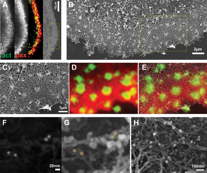
BACKGROUND: Osteoclasts are bone-degrading cells, which play a central role in physiological bone remodeling. Unbalanced osteoclast activity is largely responsible for pathological conditions such as osteoporosis. Osteoclasts develop specialized adhesion structures, the so-called podosomes, which subsequently undergo dramatic reorganization into sealing zones. These ring-like adhesion structures, which delimit the resorption site, effectively seal the cell to the substrate forming a diffusion barrier. The structural integrity of the sealing zone is essential for the cell ability to degrade bone, yet its structural organization is poorly understood. PRINCIPAL FINDINGS: Combining high-resolution scanning electron microscopy with fluorescence microscopy performed on the same sample, we mapped the molecular architecture of the osteoclast resorptive apparatus from individual podosomes to the sealing zone, at an unprecedented resolution. Podosomes are composed of an actin-bundle core, flanked by a ring containing adhesion proteins connected to the core via dome-like radial actin fibers. The sealing zone, hallmark of bone-resorbing osteoclasts, consists of a dense array of podosomes communicating through a network of actin filaments, parallel to the substrate and anchored to the adhesive plaque domain via radial actin fibers. SIGNIFICANCE: The sealing zone of osteoclasts cultured on bone is made of structural units clearly related to individual podosomes. It differs from individual or clustered podosomes in the higher density and degree of inter-connectivity of its building blocks, thus forming a unique continuous functional structure connecting the cell to its extracellular milieu. Through this continuous structure, signals reporting on the substrate condition may be transmitted to the whole cell, modulating the cell response under physiological and pathological conditions.
Overview
The claims and statistics of this paper haven’t been checked yet.
Run the check — free, no account needed. It reads the open-access text and checks the sources it cites.
Integrity signals
Registry
Cites
Statistics
Figures
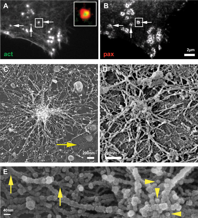
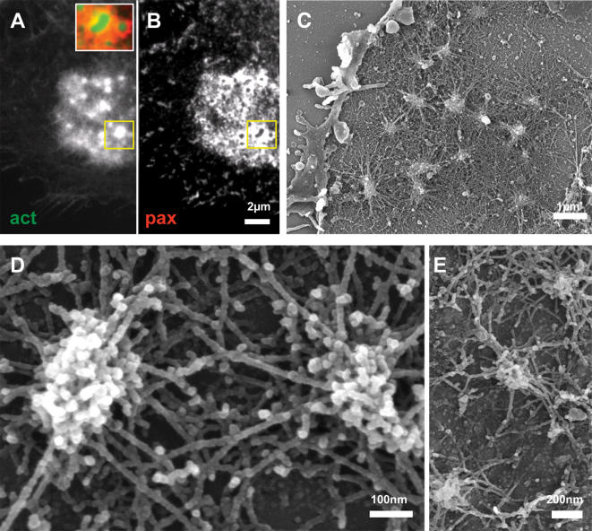
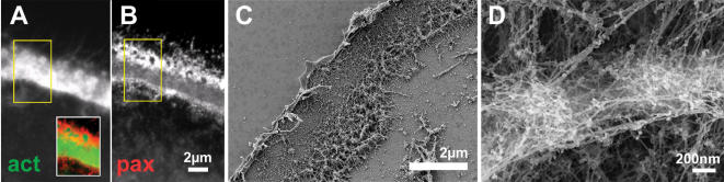
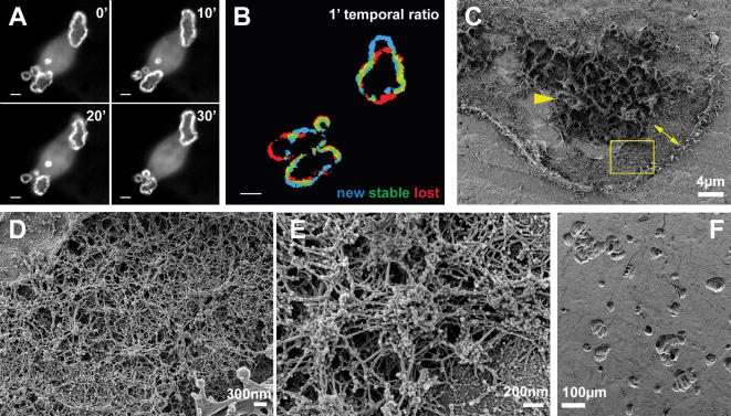
Verification
Claims the paper makes
The claims and statistics of this paper haven’t been checked yet.
Run the check — free, no account needed. It reads the open-access text and checks the sources it cites.
Claims other papers make about this one
Each is checked against this paper’s own text: does it say what the citing paper says it does?
In the citing paper
“It is important to mention that these domains are only formed when osteoclasts are in contact with extracellular mineralized matrix, in a process which α v β 3-integrin, as well as the CD44, mediates the attachment of the osteoclast podosomes to the bone surface [96–99].”
From Biology of Bone Tissue: Structure, Function, and Factors That Influence Bone Cells (2015) · cites this paper as [98]
Self-check
Percentages, sums and reported differences re-computed from the paper’s own numbers.
The self-check needs the paper’s full text.
Citations
Cites
Bone Resorption by Osteoclasts
Teitelbaum, S. L. (2000) · Science · doi:10.1126/science.289.5484.1504 · Cited by 3,962
THE MATERIAL BONE: Structure-Mechanical Function Relations
Weiner, S., & Wagner, H. D. (1998) · Annual Review of Materials Science · doi:10.1146/annurev.matsci.28.1.271 · Cited by 2,717
Therapeutic Approaches to Bone Diseases
Rodan, G. A., & Martin, T. J. (2000) · Science · doi:10.1126/science.289.5484.1508 · Cited by 1,767
Genetic regulation of osteoclast development and function
Teitelbaum, S. L., & Ross, F. P. (2003) · Nature Reviews Genetics · doi:10.1038/nrg1122 · Cited by 1,720
The cell biology of osteoclast function
Väänänen, H. K., Zhao, H., Mulari, M. T. K., & Halleen, J. M. (2000) · Journal of Cell Science · doi:10.1242/jcs.113.3.377 · Cited by 750
Podosomes: adhesion hot-spots of invasive cells
Linder, S., & Aepfelbacher, M. (2003) · Trends in Cell Biology · doi:10.1016/s0962-8924(03)00128-4 · Cited by 615
Destaing, O., Saltel, F., Géminard, J.-C., Jurdic, P., & Bard, F. (2003) · Molecular Biology of the Cell · doi:10.1091/mbc.e02-07-0389 · Cited by 547
Molecular diversity of cell-matrix adhesions
Zamir, E., Katz, B.-Z., Aota, S., Yamada, K. M., Geiger, B., & Kam, Z. (1999) · Journal of Cell Science · doi:10.1242/jcs.112.11.1655 · Cited by 459
Self-citationRemoval of Osteoclast Bone Resorption Products by Transcytosis
Salo, J., Lehenkari, P. P., Mulari, M. T. K., Metsikkö, K., & Väänänen, H. K. (1997) · Science · doi:10.1126/science.276.5310.270 · Cited by 427
Trafficking of Matrix Collagens Through Bone-Resorbing Osteoclasts
Nesbitt, S. A., & Horton, M. A. (1997) · Science · doi:10.1126/science.276.5310.266 · Cited by 378
Podosome and sealing zone: Specificity of the osteoclast model
Jurdic, P., Saltel, F., Chabadel, A., & Destaing, O. (2005) · European Journal of Cell Biology · doi:10.1016/j.ejcb.2005.09.008 · Cited by 356
A Functional Link between Dynamin and the Actin Cytoskeleton at Podosomes
Ochoa, G.-C., Slepnev, V. I., Neff, L., Ringstad, N., Takei, K., DANIELL, L. W., Kim, W., Cao, H., McNiven, M. A., Baron, R., et al. (2000) · The Journal of Cell Biology · doi:10.1083/jcb.150.2.377 · Cited by 354
Marchisio, P. C., Cirillo, D. M., Naldini, L., Primavera, M. V., Teti, A. M., & Zambonin-Zallone, A. (1984) · The Journal of Cell Biology · doi:10.1083/jcb.99.5.1696 · Cited by 328
Apatite-mediated Actin Dynamics in Resorbing Osteoclasts
Saltel, F., Destaing, O., Bard, F., Eichert, D., & Jurdic, P. (2004) · Molecular Biology of the Cell · doi:10.1091/mbc.e04-06-0522 · Cited by 267
Gelsolin Deficiency Blocks Podosome Assembly and Produces Increased Bone Mass and Strength
Chellaiah, M. A., Kizer, N. L., Silva, M. J., Alvarez, U., Kwiatkowski, D. J., & Hruska, K. A. (2000) · The Journal of Cell Biology · doi:10.1083/jcb.148.4.665 · Cited by 262
Kinetics of the osteoclast cytoskeleton during the resorption cycle in vitro
Lakkakorpi, P. T., & Väänänen, K. (1991) · Journal of Bone and Mineral Research · doi:10.1002/jbmr.5650060806 · Cited by 248
The osteoclast clear zone is a specialized cell-extracellular matrix adhesion structure
Väänänen, H. K., & Horton, M. A. (1995) · Journal of Cell Science · doi:10.1242/jcs.108.8.2729 · Cited by 217
Zambonin-Zallone, A., Teti, A. M., Grano, M., Rubinacci, A., Abbadini, M., Gaboli, M. P., & Marchisio, P. C. (1989) · Experimental Cell Research · doi:10.1016/0014-4827(89)90266-8 · Cited by 201
Podosomes in osteoclast-like cells
Pfaff, M., & Jurdic, P. (2001) · Journal of Cell Science · doi:10.1242/jcs.114.15.2775 · Cited by 169
Flores-Gama, C., Rosales, L. M., Ouellet, G., Dou, Y., Thijssen, S., Usvyat, L. A., Zhang, H., Kuntsevich, V. I., Levin, N. W., & Kotanko, P. (2017) · Blood Purification · doi:10.1159/000452731 · Cited by 161
Macrophage podosomes assemble at the leading lamella by growth and fragmentation
Evans, J. G., Correia, I., Krasavina, O., Watson, N., & Matsudaira, P. T. (2003) · The Journal of Cell Biology · doi:10.1083/jcb.200212037 · Cited by 159
The distribution of podosomes in osteoclasts cultured on bone laminae: Effect of retinol
Zambonin-Zallone, A., Teti, A. M., Carano, A., & Marchisio, P. C. (1988) · Journal of Bone and Mineral Research · doi:10.1002/jbmr.5650030507 · Cited by 138
Calle, Y., Jones, G. E., Jagger, C. J., Fuller, K., Blundell, M. P., Chow, J. W. M., Chambers, T. J., & Thrasher, A. J. (2004) · Blood · doi:10.1182/blood-2003-04-1259 · Cited by 117
Luxenburg, C., Parsons, J. T., Addadi, L., & Geiger, B. (2006) · Journal of Cell Science · doi:10.1242/jcs.03271 · Cited by 111
Self-citationAvnur, Z., & Geiger, B. (1981) · Journal of Molecular Biology · doi:10.1016/0022-2836(81)90283-7 · Cited by 109
Self-citationAkisaka, T., Yoshida, H., Inoue, S., & Shimizu, K. (2001) · Journal of Bone and Mineral Research · doi:10.1359/jbmr.2001.16.7.1248 · Cited by 108
Cell/substratum adhesions in RSV-transformed rat fibroblasts
Nermut, M. V., Eason, P., Hirst, E. M. A., & Kellie, S. (1991) · Experimental Cell Research · doi:10.1016/0014-4827(91)90111-7 · Cited by 96
Gavazzi, I., Nermut, M. V., & Marchisio, P. C. (1989) · Journal of Cell Science · doi:10.1242/jcs.94.1.85 · Cited by 89
Lakkakorpi, P. T., Helfrich, M., Horton, M. A., & Väänänen, H. K. (1993) · Journal of Cell Science · doi:10.1242/jcs.104.3.663 · Cited by 86
Tyrosine Phosphatase Epsilon Is a Positive Regulator of Osteoclast Function in Vitro and In Vivo
Chiusaroli, R., Knobler, H., Luxenburg, C., Sanjay, A., Granot-Attas, S., Tiran, Z., Miyazaki, T., Harmelin, A., Baron, R., & Elson, A. (2003) · Molecular Biology of the Cell · doi:10.1091/mbc.e03-04-0207 · Cited by 83
Self-citationThe organization of actin in spreading macrophages
Trotter, J. A. (1981) · Experimental Cell Research · doi:10.1016/0014-4827(81)90099-9 · Cited by 73
The development and function of the skeleton and bone metastases
Rodan, G. A. (2003) · Cancer · doi:10.1002/cncr.11147 · Cited by 73
The molecular dynamics of osteoclast adhesions
Luxenburg, C., Addadi, L., & Geiger, B. (2005) · European Journal of Cell Biology · doi:10.1016/j.ejcb.2005.11.002 · Cited by 69
Self-citationTeti, A. M., Migliaccio, S., Taranta, A., Bernardini, S., Rossi, G., Luciani, M., Iacobini, M., Felice, L. D., Boldrini, R., Bosman, C., et al. (1999) · Journal of Bone and Mineral Research · doi:10.1359/jbmr.1999.14.12.2107 · Cited by 43
Zallone, A. Z., Teti, A. M., Gaboli, M. P., & Marchisio, P. C. (1989) · Connective Tissue Research · doi:10.3109/03008208909023882 · Cited by 39
Akisaka, T. (2006) · Journal of Electron Microscopy · doi:10.1093/jmicro/dfl012 · Cited by 24
Use of bone cell cultures to study skeletal pathology
Jackson, M. E., Sundquist, K. T., & Marks, S. C. (1996) · Microscopy Research and Technique · doi:10.1002/(sici)1097-0029(19960201)33:2 · Cited by 3
Ultrastructure and immunolabelling of podosomes (an adhesion structure in RSV-transformed cells)
Gavazzi, I., Marchisio, P., & Nermut, M. V. (1987) · Cell Biology International Reports · doi:10.1016/0309-1651(87)90072-5 · Cited by 1
Cited by
Sorted by citations
Biology of Bone Tissue: Structure, Function, and Factors That Influence Bone Cells
Florencio-Silva, R., Sasso, G. R. D. S., Sasso-Cerri, E., Simões, M. J., & Cerri, P. S. (2015) · BioMed Research International · doi:10.1155/2015/421746 · Cited by 2,044
The 'ins' and 'outs' of podosomes and invadopodia: characteristics, formation and function
Murphy, D. A., & Courtneidge, S. A. (2011) · Nature Reviews Molecular Cell Biology · doi:10.1038/nrm3141 · Cited by 1,092
Matrix Metalloproteinases Shape the Tumor Microenvironment in Cancer Progression
Niland, S., Riscanevo, A. X., & Eble, J. A. (2021) · International Journal of Molecular Sciences · doi:10.3390/ijms23010146 · Cited by 546
Crockett, J. C., Rogers, M. J., Coxon, F. P., Hocking, L. J., & Helfrich, M. (2011) · Journal of Cell Science · doi:10.1242/jcs.063032 · Cited by 532
Assembly and biological role of podosomes and invadopodia
Gimona, M., Buccione, R., Courtneidge, S. A., & Linder, S. (2008) · Current Opinion in Cell Biology · doi:10.1016/j.ceb.2008.01.005 · Cited by 404
Metadata
- Authors
- Luxenburg, C., Geblinger, D., Klein, E., Anderson, K., Hanein, D., Geiger, B., & Addadi, L.
- Affiliations
- Weizmann Institute of Science (IL)
- Sanford Burnham Prebys Medical Discovery Institute (US)
- Corresponding author
- Addadi, Lia
- Venue
- PLoS ONE
- Publisher
- Public Library of Science (PLoS)
- Published
- 2007-01-31
- Volume
- 2
- Issue
- 1
- Pages
- e179
- ISSN
- 1932-6203 (ISSN-L)
- PMC
- PMC1779809
- OpenAlex
- W2026266643
- Cited by
- 285
- References
- 39
- Citations by year
- 2026: 4 · 2025: 4 · 2024: 4 · 2023: 6 · 2022: 7 · 2021: 11 · 2020: 25 · 2019: 20 · 2018: 17 · 2017: 14 · 2016: 19 · 2015: 19 · 2014: 21 · 2013: 19 · 2012: 17
- FWCI
- 7.43
- Type
- journal article
- Language
- en
- Open access
- gold
- doi.org
- Licence
- Locations
- PLoS ONE (publishedVersion, public-domain)
- PubMed Central (publishedVersion, public-domain)
- PubMed (publishedVersion)
- DOAJ (DOAJ: Directory of Open Access Journals) (submittedVersion, cc-by-sa)
- Figshare (submittedVersion, cc-by)
- Funders
- National Institutes of Health (5u54gm064346-10, GM-64346, U54 GM64346)
- National Institute of General Medical Sciences (U54-GM064346, GM-64346, U54 GM 64346)
- Topics
- Life Sciences › Biochemistry, Genetics and Molecular Biology › Molecular Biology › Bone Metabolism and Diseases
- Life Sciences › Biochemistry, Genetics and Molecular Biology › Cell Biology › Cellular Mechanics and Interactions
- Life Sciences › Biochemistry, Genetics and Molecular Biology › Biochemistry › Bone and Dental Protein Studies
- Keywords
- osteoclasts, podosomes, sealing zone, bone resorption, actin cytoskeleton, cell-substrate adhesion, bone remodeling, osteoporosis
- MeSH terms
- Actins, Animals, Cell Adhesion, Cells, Cultured, Cytoskeleton, Immunohistochemistry, Microscopy, Electron, Scanning, Osteoclasts, Recombinant Fusion Proteins, Cell Surface Extensions, Mice, Paxillin
About these checks
Registry, retraction, Cites and statistics results are computed, not judged, with one exception: a printed reference that no index lists is read once by a model before it counts as a citation error. Claims are extracted by a model and kept only when their quote appears verbatim in the text; each cited source is then checked, with a model’s help, for whether it says what the claim relies on. Those verdicts can be wrong — read the claim’s quote and the cited source before relying on one.
Statistics and arithmetic are recomputed, not judged.
Confidence bands are shown only for fields calibrated on our annotated test set. None is calibrated yet, so no band is shown.
What counts as a claim ›How we measure confidence ›How we check this ›
Are you an author, or do you see a problem with this page? Request a review
