Cited by 69

The DOI matches its Crossref record; 1 of the 1 references it cites were found in a registry or an index.

The C-Terminal Sequence of RhoB Directs Protein Degradation through an Endo-Lysosomal Pathway

Pérez-Sala, Dolores, Boya, Patricia, Ramos, Irene, Herrera, Mónica, Stamatakis, Konstantinos·2009·PLoS ONE

Overview

The claims and statistics of this paper haven’t been checked yet.

Run the check — free, no account needed. It reads the open-access text and checks the sources it cites.

A few free checks a day · papers already verified open instantly · results are public

Integrity signals

Registry

Crossref · 10.1371/journal.pone.0008117

Cites

1 of 1 references verified0 citation errors · 0 not checkable

Statistics

Figures

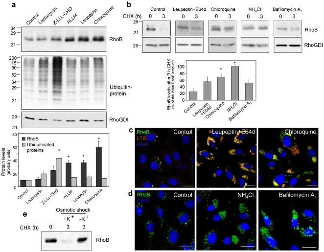
Figure 1 RhoB is degraded through a lysosomal pathway. (a) Confluent serum-deprived bovine aortic endothelial cells (BAEC) were treated with 10 µM lactacystin, 50 nM Z-LLL-CHO, 10 µM ALLM, 50 µg/ml leupeptin or 10 µM chloroquine for 24 h. Cell lysates (20 µg of protein) were analyzed by Western blot. RhoB and ubiquitinated protein levels corrected by RhoGDI levels are shown as average values ± SEM of three experiments (*p<0.05 vs control). (b) BAEC were pre-treated with the inhibitors for 30 min. NH4Cl and bafilomycin A1 were added at 30 mM and 5 nM, final concentrations, respectively. RhoB levels were assessed before and after treatment with CHX (20 µg/ml). Levels of RhoGDI were used as control. The percentage of RhoB remaining after 3 h CHX treatment is shown as average values ± SEM of at least three different experiments (*p<0.05 vs control). (c) BAEC were treated with 50 µg/ml leupeptin plus 5 µg/ml E64d or 10 µM chloroquine for 24 h and visualized by immunofluorescence (IF) of endogenous RhoB (green) and staining with 500 nM LTR (red) and DAPI (blue). Images shown are single confocal sections. Bar, 20 µm. (d) BAEC were treated with 30 mM NH4Cl or 5 nM bafilomycin A1 for 24 h and fixed. Nuclei were stained with DAPI (blue) and RhoB (green) was detected by IF and confocal microscopy. Images are shown as maximal projections of confocal planes of 0.5 µm Z-sections. Bar, 20 µm. (e) BAEC subjected to osmotic shock for 5 min were allowed to recover in the absence or presence of 10 mM potassium chloride (K+) for 3 h, during which CHX (20 µg/ml) was present to block new protein synthesis. RhoB levels were assessed by Western blot.
Figure 1 RhoB is degraded through a lysosomal pathway. (a) Confluent serum-deprived bovine aortic endothelial cells (BAEC) were treated with 10 µM lactacystin, 50 nM Z-LLL-CHO, 10 µM ALLM, 50 µg/ml leupeptin or 10 µM chloroquine for 24 h. Cell lysates (20 µg of protein) were analyzed by Western blot. RhoB and ubiquitinated protein levels corrected by RhoGDI levels are shown as average values ± SEM of three experiments (*p<0.05 vs control). (b) BAEC were pre-treated with the inhibitors for 30 min. NH4Cl and bafilomycin A1 were added at 30 mM and 5 nM, final concentrations, respectively. RhoB levels were assessed before and after treatment with CHX (20 µg/ml). Levels of RhoGDI were used as control. The percentage of RhoB remaining after 3 h CHX treatment is shown as average values ± SEM of at least three different experiments (*p<0.05 vs control). (c) BAEC were treated with 50 µg/ml leupeptin plus 5 µg/ml E64d or 10 µM chloroquine for 24 h and visualized by immunofluorescence (IF) of endogenous RhoB (green) and staining with 500 nM LTR (red) and DAPI (blue). Images shown are single confocal sections. Bar, 20 µm. (d) BAEC were treated with 30 mM NH4Cl or 5 nM bafilomycin A1 for 24 h and fixed. Nuclei were stained with DAPI (blue) and RhoB (green) was detected by IF and confocal microscopy. Images are shown as maximal projections of confocal planes of 0.5 µm Z-sections. Bar, 20 µm. (e) BAEC subjected to osmotic shock for 5 min were allowed to recover in the absence or presence of 10 mM potassium chloride (K+) for 3 h, during which CHX (20 µg/ml) was present to block new protein synthesis. RhoB levels were assessed by Western blot.CC BY · PubMed Central
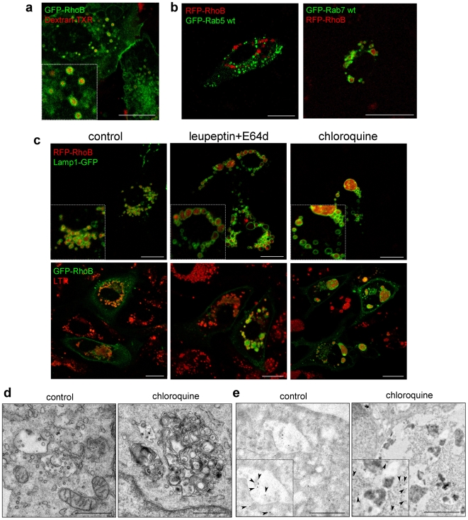
Figure 2 Localization of RhoB along the endo-lysosomal pathway. (a) BAEC transiently transfected with GFP-RhoB were incubated with Texas Red-labeled dextran (Dextran-TXR) for 5 min and visualized by confocal microscopy after 1 h. Bar, 20 µm. (b) BAEC were transiently transfected with the indicated constructs and 24 h later live cells were observed by confocal microscopy. Bars, 15 µm. (c) BAEC were transfected with GFP-RhoB, RFP-RhoB and/or Lamp1-GFP and treated 24 h later with leupeptin plus E64d or chloroquine for further 24 h. When indicated, cells were incubated with 25 nM LTR for 15 min. Live cells were observed by confocal microscopy. Bars, 15 µm. (d) Transmission EM of MVB from control and chloroquine treated BAEC. Bars, 1 µm. (e) IEM analysis of GFP-RhoB in control and choroquine-treated cells by staining with anti-GFP antibody. Bars, 1 µm.
Figure 2 Localization of RhoB along the endo-lysosomal pathway. (a) BAEC transiently transfected with GFP-RhoB were incubated with Texas Red-labeled dextran (Dextran-TXR) for 5 min and visualized by confocal microscopy after 1 h. Bar, 20 µm. (b) BAEC were transiently transfected with the indicated constructs and 24 h later live cells were observed by confocal microscopy. Bars, 15 µm. (c) BAEC were transfected with GFP-RhoB, RFP-RhoB and/or Lamp1-GFP and treated 24 h later with leupeptin plus E64d or chloroquine for further 24 h. When indicated, cells were incubated with 25 nM LTR for 15 min. Live cells were observed by confocal microscopy. Bars, 15 µm. (d) Transmission EM of MVB from control and chloroquine treated BAEC. Bars, 1 µm. (e) IEM analysis of GFP-RhoB in control and choroquine-treated cells by staining with anti-GFP antibody. Bars, 1 µm.CC BY · PubMed Central
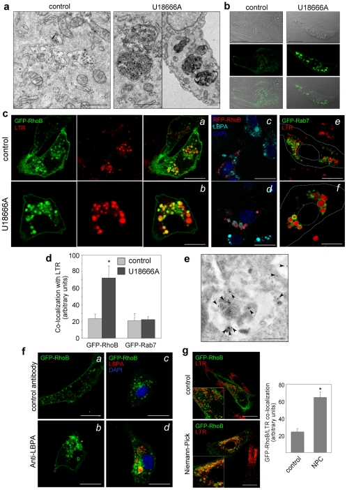
Figure 3 Effect of endosomal lipid dynamics on RhoB distribution. (a) BAEC were treated with vehicle or 10 µM U18666A for 24 h, fixed and analyzed by transmission EM. Bars, 1 µm. (b) Confocal microscopy GFP-RhoB-transfected cells treated as in A. DIC, fluorescent image and their overlay are shown. Bars, 20 µm. (c) BAEC were transfected with the indicated constructs and treated with vehicle or U18666A; (a,b) and (e,f), confocal microscopy of live cells incubated with 25 nM LTR; cell contours are shown by dotted lines in (e,f); (c,d) distribution of LBPA observed by IF. Nuclei were stained with DAPI. Bars, 10 µm. (d) The degree of internalization of GFP-RhoB or GFP-Rab7 into U18666A-induced structures was estimated from co-localization of the corresponding constructs with LTR and it is shown as average values ± SEM from three independent experiments (*p<0.05 vs control). (e) BAEC transfected with GFP-RhoB and treated with U18666A were analyzed by IEM with anti-GFP antibodies. Bar, 0.5 µm. (f) Effect of anti-LBPA on RhoB distribution. BAEC transfected with GFP-RhoB were incubated with a control antibody or with anti-LBPA (hybridoma supernatant diluted 1∶1 in culture medium) for 20 h; (a,b) live cells observed by confocal microscopy; (c,d) accumulation of LBPA observed by IF with anti-mouse Ig antibody. Bars, 20 µm. (g) Control or NPC fibroblasts were transfected with GFP-RhoB. Co-localization with LTR was assessed in live cells. Bars, 20 µm. Co-localization is shown as average values ± SEM of determinations from three experiments (*p<0.05 vs control).
Figure 3 Effect of endosomal lipid dynamics on RhoB distribution. (a) BAEC were treated with vehicle or 10 µM U18666A for 24 h, fixed and analyzed by transmission EM. Bars, 1 µm. (b) Confocal microscopy GFP-RhoB-transfected cells treated as in A. DIC, fluorescent image and their overlay are shown. Bars, 20 µm. (c) BAEC were transfected with the indicated constructs and treated with vehicle or U18666A; (a,b) and (e,f), confocal microscopy of live cells incubated with 25 nM LTR; cell contours are shown by dotted lines in (e,f); (c,d) distribution of LBPA observed by IF. Nuclei were stained with DAPI. Bars, 10 µm. (d) The degree of internalization of GFP-RhoB or GFP-Rab7 into U18666A-induced structures was estimated from co-localization of the corresponding constructs with LTR and it is shown as average values ± SEM from three independent experiments (*p<0.05 vs control). (e) BAEC transfected with GFP-RhoB and treated with U18666A were analyzed by IEM with anti-GFP antibodies. Bar, 0.5 µm. (f) Effect of anti-LBPA on RhoB distribution. BAEC transfected with GFP-RhoB were incubated with a control antibody or with anti-LBPA (hybridoma supernatant diluted 1∶1 in culture medium) for 20 h; (a,b) live cells observed by confocal microscopy; (c,d) accumulation of LBPA observed by IF with anti-mouse Ig antibody. Bars, 20 µm. (g) Control or NPC fibroblasts were transfected with GFP-RhoB. Co-localization with LTR was assessed in live cells. Bars, 20 µm. Co-localization is shown as average values ± SEM of determinations from three experiments (*p<0.05 vs control).CC BY · PubMed Central
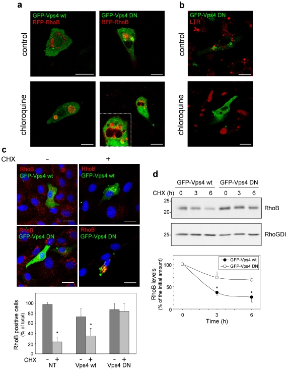
Figure 4 Effect of Vps4 wt and DN on RhoB localization and stability. (a–b) BAEC were transfected with GFP-Vps4 wt or DN, as indicated, and 24 h later, treated in the absence or presence of 10 µM chloroquine for 24 h. In (b), 25 nM LTR was added 15 min before observation of live cells by confocal microscopy. Bars, 15 µm. (c) BAEC were transfected as above and 24 later placed in serum-free medium. After 24 h, control (0 time, -) cells were fixed, and the rest were treated with 20 µg/ml CHX for 8 h (+) to allow degradation of endogenous RhoB, before fixation. Endogenous RhoB (in red) was detected by IF. Nuclei were stained with DAPI. Images shown are overall projections. Bars, 20 µm. The lower panel shows the proportion of RhoB-positive cells among non-transfected (NT) cells or among cells transfected with the indicated constructs before (−) or after CHX (+) treatment. At least 40 cells of each type were monitored from four independent experiments. Results are shown as average values ± SEM (*p<0.05 vs the same condition in the absence of CHX). (d) COS7 cells were transfected with GFP-Vps4 wt or DN, as indicated, and 24 h later split in three dishes for treatment with CHX for the indicated times. RhoB and RhoGDI levels were assessed by Western blot. Results shown are average values ± SEM from three experiments (*p<0.05 vs GFP-Vps4 DN at the same time points).
Figure 4 Effect of Vps4 wt and DN on RhoB localization and stability. (a–b) BAEC were transfected with GFP-Vps4 wt or DN, as indicated, and 24 h later, treated in the absence or presence of 10 µM chloroquine for 24 h. In (b), 25 nM LTR was added 15 min before observation of live cells by confocal microscopy. Bars, 15 µm. (c) BAEC were transfected as above and 24 later placed in serum-free medium. After 24 h, control (0 time, -) cells were fixed, and the rest were treated with 20 µg/ml CHX for 8 h (+) to allow degradation of endogenous RhoB, before fixation. Endogenous RhoB (in red) was detected by IF. Nuclei were stained with DAPI. Images shown are overall projections. Bars, 20 µm. The lower panel shows the proportion of RhoB-positive cells among non-transfected (NT) cells or among cells transfected with the indicated constructs before (−) or after CHX (+) treatment. At least 40 cells of each type were monitored from four independent experiments. Results are shown as average values ± SEM (*p<0.05 vs the same condition in the absence of CHX). (d) COS7 cells were transfected with GFP-Vps4 wt or DN, as indicated, and 24 h later split in three dishes for treatment with CHX for the indicated times. RhoB and RhoGDI levels were assessed by Western blot. Results shown are average values ± SEM from three experiments (*p<0.05 vs GFP-Vps4 DN at the same time points).CC BY · PubMed Central
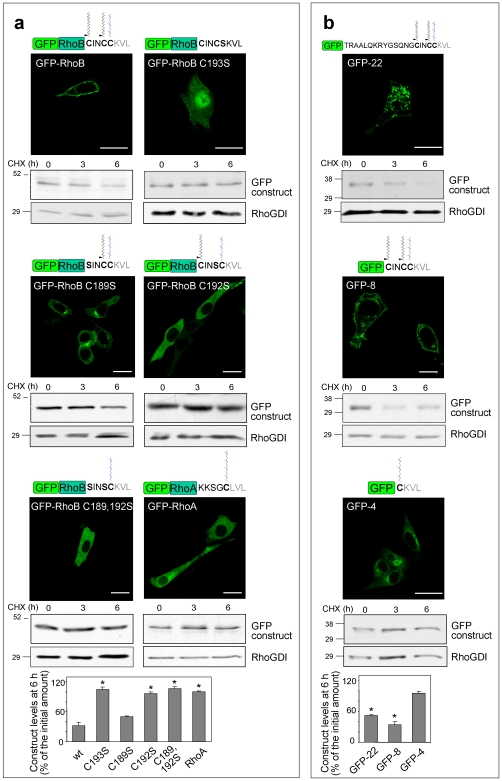
Figure 5 Structural determinants involved in the subcellular localization and degradation of GFP-RhoB. (a,b) NIH-3T3 fibroblasts were transiently transfected with the indicated GFP constructs. 24 h after transfection, the distribution of fluorescent constructs was assessed by confocal microscopy of live cells. Bars, 20 µm. The putative posttranslational modifications of each construct are shown schematically above the panels. Construct stability was assessed by blocking protein synthesis with CHX and estimating the remaining levels by Western blot with anti-GFP antibody. Blots show the levels of the constructs of interest and of RhoGDI, as control. Lower panels show the construct levels remaining after 6 h treatment with CHX. wt, GFP-RhoB; C193S, GFP-RhoB C193S; C189S, GFP-RhoB C189S; C192S, GFP-RhoB C192S; C189,192S, GFP-RhoB C189,192S; RhoA, GFP-RhoA. Results are shown as average values ± SEM of three different experiments (*p<0.05 vs wt levels in (a) and vs GFP-4 levels in (b)).
Figure 5 Structural determinants involved in the subcellular localization and degradation of GFP-RhoB. (a,b) NIH-3T3 fibroblasts were transiently transfected with the indicated GFP constructs. 24 h after transfection, the distribution of fluorescent constructs was assessed by confocal microscopy of live cells. Bars, 20 µm. The putative posttranslational modifications of each construct are shown schematically above the panels. Construct stability was assessed by blocking protein synthesis with CHX and estimating the remaining levels by Western blot with anti-GFP antibody. Blots show the levels of the constructs of interest and of RhoGDI, as control. Lower panels show the construct levels remaining after 6 h treatment with CHX. wt, GFP-RhoB; C193S, GFP-RhoB C193S; C189S, GFP-RhoB C189S; C192S, GFP-RhoB C192S; C189,192S, GFP-RhoB C189,192S; RhoA, GFP-RhoA. Results are shown as average values ± SEM of three different experiments (*p<0.05 vs wt levels in (a) and vs GFP-4 levels in (b)).CC BY · PubMed Central
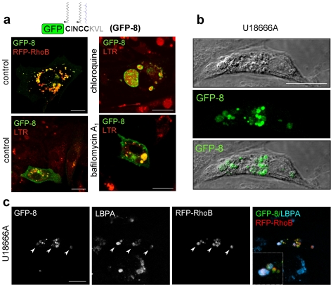
Figure 6 Localization of GFP-CINCCKVL in endo-lysosomal compartments. (a) BAEC transfected with RFP-RhoB and/or GFP-CINCCKVL (GFP-8) were treated with vehicle, 10 µM chloroquine or 5 nM bafilomycin A1, incubated with 25 nM LTR when indicated and visualized live by confocal microscopy. (b) BAEC transfected with GFP-8 were treated with 10 µM U18666A for 24 h. DIC and fluorescence images showing dense lysosomes and GFP-8 accumulations are shown. (c) BAEC transfected with GFP-8 and RFP-RhoB and treated with U18666A as above were fixed and co-localization of GFP-8, RFP-RhoB and LBPA was detected by IF. Bars, 20 µm.
Figure 6 Localization of GFP-CINCCKVL in endo-lysosomal compartments. (a) BAEC transfected with RFP-RhoB and/or GFP-CINCCKVL (GFP-8) were treated with vehicle, 10 µM chloroquine or 5 nM bafilomycin A1, incubated with 25 nM LTR when indicated and visualized live by confocal microscopy. (b) BAEC transfected with GFP-8 were treated with 10 µM U18666A for 24 h. DIC and fluorescence images showing dense lysosomes and GFP-8 accumulations are shown. (c) BAEC transfected with GFP-8 and RFP-RhoB and treated with U18666A as above were fixed and co-localization of GFP-8, RFP-RhoB and LBPA was detected by IF. Bars, 20 µm.CC BY · PubMed Central
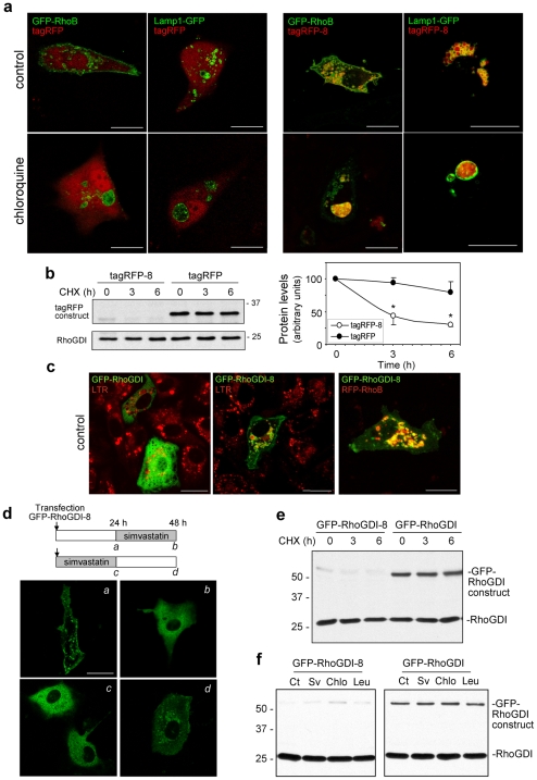
Figure 7 Subcellular localization and stability of chimeric proteins bearing -CINCCKVL C-terminal sequences. (a) BAEC were co-transfected with tagRFP or tagRFP-CINCCKVL (tagRFP-8) and GFP-RhoB or Lamp1-GFP, as indicated, treated for 24 h in the absence or presence of 10 µM chloroquine and directly visualized by confocal microscopy. Images shown are individual confocal sections. Bars, 20 µm. (b) The stability of tagRFP and tagRFP-8 was assessed in BAEC as described in Fig. 5, using an anti-tagRFP antibody. Results are shown as average values ± SD of at least three determinations (*p<0.05 vs tagRFP levels at the same time points). (c) BAEC were transfected with GFP-RhoGDI, GFP-RhoGDI-8 and/or RFP-RhoB, incubated with 25 nM LTR, as indicated, and directly observed by confocal microscopy. Bar, 20 µm. (d) BAEC were transfected with GFP-RhoGDI-8 in the absence (a) or presence (c) of 10 µM simvastatin. 24 h after transfection, confocal images of live cells were obtained (a, c). At that point, simvastatin was withdrawn from (c) and added to (a). After a 24 h additional incubation images of live cells were taken (b, d). Bar, 20 µm. (e,f) BAEC were transiently transfected with GFP-RhoGDI-8 or GFP-RhoGDI. In (e) protein synthesis was blocked by CHX addition. In (f) BAEC were treated with the indicated agents for 24 h. Levels of GFP-RhoGDI-8, GFP-RhoGDI and endogenous RhoGDI, were assessed by Western blot. Sv, simvastatin; Chlo, chloroquine; Leu, leupeptin.
Figure 7 Subcellular localization and stability of chimeric proteins bearing -CINCCKVL C-terminal sequences. (a) BAEC were co-transfected with tagRFP or tagRFP-CINCCKVL (tagRFP-8) and GFP-RhoB or Lamp1-GFP, as indicated, treated for 24 h in the absence or presence of 10 µM chloroquine and directly visualized by confocal microscopy. Images shown are individual confocal sections. Bars, 20 µm. (b) The stability of tagRFP and tagRFP-8 was assessed in BAEC as described in Fig. 5, using an anti-tagRFP antibody. Results are shown as average values ± SD of at least three determinations (*p<0.05 vs tagRFP levels at the same time points). (c) BAEC were transfected with GFP-RhoGDI, GFP-RhoGDI-8 and/or RFP-RhoB, incubated with 25 nM LTR, as indicated, and directly observed by confocal microscopy. Bar, 20 µm. (d) BAEC were transfected with GFP-RhoGDI-8 in the absence (a) or presence (c) of 10 µM simvastatin. 24 h after transfection, confocal images of live cells were obtained (a, c). At that point, simvastatin was withdrawn from (c) and added to (a). After a 24 h additional incubation images of live cells were taken (b, d). Bar, 20 µm. (e,f) BAEC were transiently transfected with GFP-RhoGDI-8 or GFP-RhoGDI. In (e) protein synthesis was blocked by CHX addition. In (f) BAEC were treated with the indicated agents for 24 h. Levels of GFP-RhoGDI-8, GFP-RhoGDI and endogenous RhoGDI, were assessed by Western blot. Sv, simvastatin; Chlo, chloroquine; Leu, leupeptin.CC BY · PubMed Central

Verification

Claims the paper makes

The claims and statistics of this paper haven’t been checked yet.

Run the check — free, no account needed. It reads the open-access text and checks the sources it cites.

A few free checks a day · papers already verified open instantly · results are public

Claims other papers make about this one

Each is checked against this paper’s own text: does it say what the citing paper says it does?

Self-check

Percentages, sums and reported differences re-computed from the paper’s own numbers.

The self-check needs the paper’s full text.

Citations

Cites

Research works printed but not linked: 43

  1. [1]Schwartz AL Ciechanover A 2009 Targeting proteins for destruction by the ubiquitin system: implications for human pathobiology. Annu Rev Pharmacol Toxicol 49 73 96 18834306
  2. [2]Fader CM Colombo MI 2009 Autophagy and multivesicular bodies: two closely related partners. Cell Death Differ 16 70 78 19008921
  3. [3]Mellor H Flynn P Nobes CD Hall A Parker PJ 1998 PRK1 is targeted to endosomes by the small GTPase, RhoB. J Biol Chem 273 4811 4814 9478917
  4. [4]Huang M Duhadaway JB Prendergast GC Laury-Kleintop LD 2007 RhoB regulates PDGFR-beta trafficking and signaling in vascular smooth muscle cells. Arterioscler Thromb Vasc Biol 27 2597 2605 17951322 PMC4384698
  5. [5]Sandilands E Cans C Fincham VJ Brunton VG Mellor H 2004 RhoB and actin polymerization coordinate Src activation with endosome-mediated delivery to the membrane. Dev Cell 7 855 869 15572128
  6. [6]Wherlock M Gampel A Futter C Mellor H 2004 Farnesyltransferase inhibitors disrupt EGF receptor traffic through modulation of the RhoB GTPase. J Cell Sci 117 3221 3231 15226397
  7. [7]Liu AX Rane N Liu JP Prendergast GC 2001 RhoB is dispensable for mouse development, but it modifies susceptibility to tumor formation as well as cell adhesion and growth factor signaling in transformed cells. Mol Cell Biol 21 6906 6912 11564874 PMC99867
  8. [8]Huang M Kamasani U Prendergast GC 2006 RhoB facilitates c-Myc turnover by supporting efficient nuclear accumulation of GSK-3. Oncogene 25 1281 1289 16247449
  9. [9]Engel ME Datta PK Moses HL 1998 RhoB is stabilized by transforming growth factor beta and antagonizes transcriptional activation. J Biol Chem 273 9921 9926 9545335
  10. [10]Stamatakis K Cernuda-Morollón E Hernández-Perera O Pérez-Sala D 2002 Isoprenylation of RhoB is required for its degradation: a novel determinant in the complex regulation of RhoB expression by the mevalonate pathway. J Biol Chem 277 49389 49396 12384510
  11. [11]Fritz G Kaina B 1997 rhoB encoding a UV-inducible Ras-related small GTP-binding protein is regulated by GTPases of the Rho family and independent of JNK, ERK, and p38 MAP kinase. J Biol Chem 272 30637 30644 9388198
  12. [12]Malcolm T Ettehadieh E Sadowski I 2003 Mitogen-responsive expression of RhoB is regulated by RNA stability. Oncogene 22 6142 6150 13679852
  13. [13]Dransart E Olofsson B Cherfils J 2005 RhoGDIs revisited: novel roles in Rho regulation. Traffic 6 957 966 16190977
  14. [14]Robertson D Paterson HF Adamson P Hall A Monaghan P 1995 Ultrastructural localization of ras-related proteins using epitope-tagged plasmids. J Histochem Cytochem 43 471 480 7537292
  15. [15]Michaelson D Silletti J Murphy G D'Eustachio P Rush M 2001 Differential localization of Rho GTPases in live cells: regulation by hypervariable regions and RhoGDI binding. J Cell Biol 152 111 126 11149925 PMC2193662
  16. [16]Wang DA Sebti SM 2005 Palmitoylated cysteine 192 is required for RhoB tumor-suppressive and apoptotic activities. J Biol Chem 280 19243 19249 15713677
  17. [17]Pérez-Sala D Martínez-A C Rebollo A 1999 Novel aspects of Ras proteins biology: regulation and implications. Cell Death Differ 6 722 728 10467345
  18. [18]Wennerberg K Der CJ 2004 Rho-family GTPases: it's not only Rac and Rho (and I like it). J Cell Sci 117 1301 1312 15020670
  19. [19]Bivona TG Quatela SE Bodemann BO Ahearn IM Soskis MJ 2006 PKC regulates a farnesyl-electrostatic switch on K-Ras that promotes its association with Bcl-XL on mitochondria and induces apoptosis. Mol Cell 21 481 493 16483930
  20. [20]Gorfe AA Babakhani A McCammon JA 2007 H-ras protein in a bilayer: interaction and structure perturbation. J Am Chem Soc 129 12280 12286 17880077 PMC2530826
  21. [21]Rock KL Gramm C Rothstein L Clark K Stein R 1994 Inhibitors of the proteasome block degradation of most cell proteins and the generation of peptides presented on MHC class I molecules. Cell 78 761 771 8087844
  22. [22]Hansen SH Sandvig K van Deurs B 1993 Clathrin and HA2 adaptors: effects of potassium depletion, hypertonic medium, and cytosol acidification. J Cell Biol 121 61 72 8458873 PMC2119777
  23. [23]Mizushima N Levine B Cuervo AM Klionsky DJ 2008 Autophagy fights disease through cellular self-digestion. Nature 451 1069 1075 18305538 PMC2670399
  24. [24]Russell MR Nickerson DP Odorizzi G 2006 Molecular mechanisms of late endosome morphology, identity and sorting. Curr Opin Cell Biol 18 422 428 16781134
  25. [25]Falguieres T Luyet PP Gruenberg J 2009 Molecular assemblies and membrane domains in multivesicular endosome dynamics. Exp Cell Res 315 1567 1573 19133258
  26. [26]Kobayashi T Beuchat MH Lindsay M Frias S Palmiter RD 1999 Late endosomal membranes rich in lysobisphosphatidic acid regulate cholesterol transport. Nat Cell Biol 1 113 118 10559883
  27. [27]Sobo K Le Blanc I Luyet PP Fivaz M Ferguson C 2007 Late endosomal cholesterol accumulation leads to impaired intra-endosomal trafficking. PLoS ONE 2 e851 17786222 PMC1952175
  28. [28]Marchetti A Mercanti V Cornillon S Alibaud L Charette SJ 2004 Formation of multivesicular endosomes in Dictyostelium. J Cell Sci 117 6053 6059 15536120
  29. [30]Carstea ED Morris JA Coleman KG Loftus SK Zhang D 1997 Niemann-Pick C1 disease gene: homology to mediators of cholesterol homeostasis. Science 277 228 231 9211849
  30. [31]Sachse M Strous GJ Klumperman J 2004 ATPase-deficient hVPS4 impairs formation of internal endosomal vesicles and stabilizes bilayered clathrin coats on endosomal vacuoles. J Cell Sci 117 1699 1708 15075231
  31. [32]Bishop N Woodman P 2000 ATPase-defective mammalian VPS4 localizes to aberrant endosomes and impairs cholesterol trafficking. Mol Biol Cell 11 227 239 10637304 PMC14770
  32. [33]Gutierrez L Magee AI Marshall CJ Hancock JF 1989 Post-translational processing of p21ras is two-step and involves carboxyl-methylation and carboxy-terminal proteolysis. Embo J 8; 1093-1098 PMC400919 2663468
  33. [34]Fuertes G Villarroya A Knecht E 2003 Role of proteasomes in the degradation of short-lived proteins in human fibroblasts under various growth conditions. Int J Biochem Cell Biol 35 651 664 12672457
  34. [35]Massey AC Zhang C Cuervo AM 2006 Chaperone-mediated autophagy in aging and disease. Curr Top Dev Biol 73 205 235 16782460
  35. [36]Luyet PP Falguieres T Pons V Pattnaik AK Gruenberg J 2008 The ESCRT-I subunit TSG101 controls endosome-to-cytosol release of viral RNA. Traffic 9 2279 2290 18817529
  36. [37]Lafourcade C Sobo K Kieffer-Jaquinod S Garin J van der Goot FG 2008 Regulation of the V-ATPase along the endocytic pathway occurs through reversible subunit association and membrane localization. PLoS ONE 3 e2758 18648502 PMC2447177
  37. [38]Williams RL Urbe S 2007 The emerging shape of the ESCRT machinery. Nat Rev Mol Cell Biol 8 355 368 17450176
  38. [39]Carreno S Gouze ME Schaak S Emorine LJ Maridonneau-Parini I 2000 Lack of palmitoylation redirects p59Hck from the plasma membrane to p61Hck-positive lysosomes. J Biol Chem 275 36223 36229 10967098
  39. [40]Percherancier Y Planchenault T Valenzuela-Fernandez A Virelizier JL Arenzana-Seisdedos F 2001 Palmitoylation-dependent control of degradation, life span, and membrane expression of the CCR5 receptor. J Biol Chem 276 31936 31944 11390405
  40. [41]Braulke T Bonifacino JS 2009 Sorting of lysosomal proteins. Biochim Biophys Acta 1793 605 614 19046998
  41. [42]McCormick PJ Dumaresq-Doiron K Pluviose AS Pichette V Tosato G 2008 Palmitoylation controls recycling in lysosomal sorting and trafficking. Traffic 9 1984 1997 18817523 PMC3500849
  42. [43]Peng Y Tang F Weisman LS 2006 Palmitoylation plays a role in targeting Vac8p to specific membrane subdomains. Traffic 7 1378 1387 16978392
  43. [44]Meiringer CT Auffarth K Hou H Ungermann C 2008 Depalmitoylation of Ykt6 prevents its entry into the multivesicular body pathway. Traffic 9 1510 1521 18541004

Cited by

See all 69

Metadata

Authors
Pérez-Sala, D., Boya, P., Ramos, I., Herrera, M., & Stamatakis, K.
Venue
PLoS ONE
Published
2009-12-02
Volume
4
Issue
12
Pages
e8117
ISSN
1932-6203
Cited by
69
References
1
Language
en

About these checks

Registry, retraction, Cites and statistics results are computed, not judged, with one exception: a printed reference that no index lists is read once by a model before it counts as a citation error. Claims are extracted by a model and kept only when their quote appears verbatim in the text; each cited source is then checked, with a model’s help, for whether it says what the claim relies on. Those verdicts can be wrong — read the claim’s quote and the cited source before relying on one.

Statistics and arithmetic are recomputed, not judged.

Confidence bands are shown only for fields calibrated on our annotated test set. None is calibrated yet, so no band is shown.

What counts as a claim ›How we measure confidence ›How we check this ›

Are you an author, or do you see a problem with this page? Request a review