

The DOI matches its Crossref record; 40 of the 41 references it cites were found in a registry or an index.
The Mitochondrial Fusion-Promoting Factor Mitofusin Is a Substrate of the PINK1/Parkin Pathway
Open accessAbstract
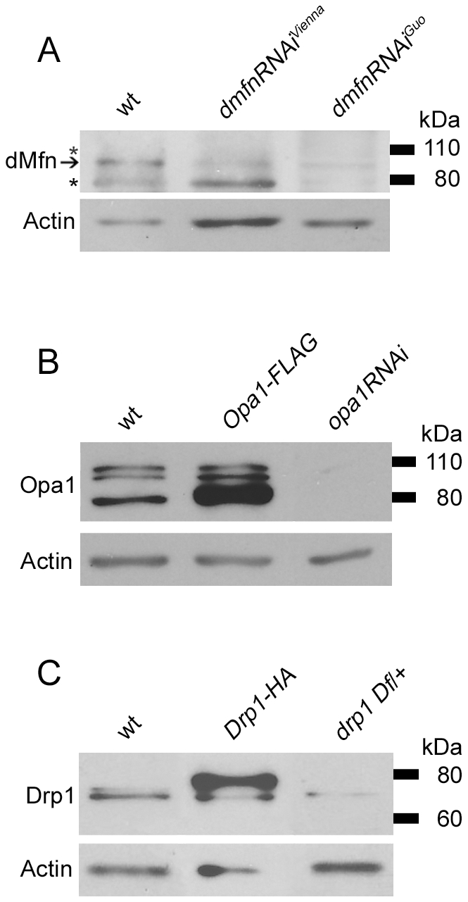
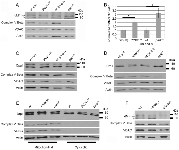
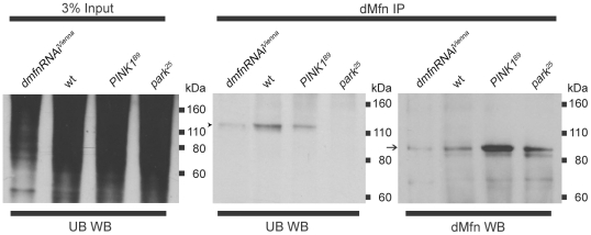
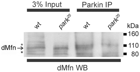
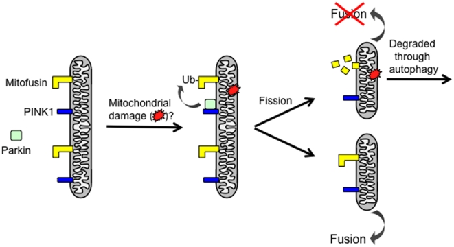
Loss-of-function mutations in the PINK1 or parkin genes result in recessive heritable forms of parkinsonism. Genetic studies of Drosophila orthologs of PINK1 and parkin indicate that PINK1, a mitochondrially targeted serine/threonine kinase, acts upstream of Parkin, a cytosolic ubiquitin-protein ligase, to promote mitochondrial fragmentation, although the molecular mechanisms by which the PINK1/Parkin pathway promotes mitochondrial fragmentation are unknown. We tested the hypothesis that PINK1 and Parkin promote mitochondrial fragmentation by targeting core components of the mitochondrial morphogenesis machinery for ubiquitination. We report that the steady-state abundance of the mitochondrial fusion-promoting factor Mitofusin (dMfn) is inversely correlated with the activity of PINK1 and Parkin in Drosophila. We further report that dMfn is ubiquitinated in a PINK1- and Parkin-dependent fashion and that dMfn co-immunoprecipitates with Parkin. By contrast, perturbations of PINK1 or Parkin did not influence the steady-state abundance of the mitochondrial fission-promoting factor Drp1 or the mitochondrial fusion-promoting factor Opa1, or the subcellular distribution of Drp1. Our findings suggest that dMfn is a direct substrate of the PINK1/Parkin pathway and that the mitochondrial morphological alterations and tissue degeneration phenotypes that derive from mutations in PINK1 and parkin result at least in part from reduced ubiquitin-mediated turnover of dMfn.
Overview
The claims and statistics of this paper haven’t been checked yet.
Run the check — free, no account needed. It reads the open-access text and checks the sources it cites.
Integrity signals
Registry
Cites
Statistics
Figures
Verification
Claims the paper makes
The claims and statistics of this paper haven’t been checked yet.
Run the check — free, no account needed. It reads the open-access text and checks the sources it cites.
Claims other papers make about this one
Each is checked against this paper’s own text: does it say what the citing paper says it does?
In the citing paper
“Once activated, Parkin has been reported to facilitate ubiquitination of numerous OMM proteins, including Mfn1 [126,127,128], Mfn2 [129], VDAC [130], Tom20 (translocase of the outer membrane 20) [121], and Miro [131].”
From Mitochondrial Dynamics and Mitophagy in Skeletal Muscle Health and Aging (2021) · cites this paper as [127]
Self-check
Percentages, sums and reported differences re-computed from the paper’s own numbers.
The self-check needs the paper’s full text.
Citations
Cites
Targeted gene expression as a means of altering cell fates and generating dominant phenotypes
Brand, A. H., & Perrimon, N. (1993) · Development · doi:10.1242/dev.118.2.401 · Cited by 9,510
Parkin is recruited selectively to impaired mitochondria and promotes their autophagy
Narendra, D., Tanaka, A., Suen, D.-F., & Youle, R. J. (2008) · The Journal of Cell Biology · doi:10.1083/jcb.200809125 · Cited by 4,026
PINK1 Is Selectively Stabilized on Impaired Mitochondria to Activate Parkin
Narendra, D. P., Jin, S. M., Tanaka, A., Suen, D.-F., Gautier, C. A., Shen, J., Cookson, M. R., & Youle, R. J. (2010) · PLoS Biology · doi:10.1371/journal.pbio.1000298 · Cited by 2,944
PINK1/Parkin-mediated mitophagy is dependent on VDAC1 and p62/SQSTM1
Geisler, S., Holmström, K. M., Skujat, D., Fiesel, F. C., Rothfuss, O. C., Kahle, P. J., & Springer, W. (2010) · Nature Cell Biology · doi:10.1038/ncb2012 · Cited by 2,835
Drosophila pink1 is required for mitochondrial function and interacts genetically with parkin
Clark, I. E., Dodson, M. W., Jiang, C., Cao, J. H., Huh, J. R., Seol, J. H., Yoo, S. J., Hay, B. A., & Guo, M. (2006) · Nature · doi:10.1038/nature04779 · Cited by 1,788
Recognition of the polyubiquitin proteolytic signal
Thrower, J. S., Hoffman, L. M., Rechsteiner, M., & Pickart, C. M. (2000) · The EMBO Journal · doi:10.1093/emboj/19.1.94 · Cited by 1,769
Mitochondrial dysfunction in Drosophila PINK1 mutants is complemented by parkin
Park, J., Lee, S. B., Lee, S., Kim, Y., Song, S., Kim, S., Bae, E., Kim, J., Shong, M., Kim, J.-M., et al. (2006) · Nature · doi:10.1038/nature04788 · Cited by 1,705
PINK1-dependent recruitment of Parkin to mitochondria in mitophagy
Vives-Bauza, C., Zhou, C., Huang, Y., Cui, M., de Vries, R. L. A., Kim, J., May, J., Tocilescu, M. A., Liu, W., Ko, H. S., et al. (2010) · Proceedings of the National Academy of Sciences · doi:10.1073/pnas.0911187107 · Cited by 1,616
Bender, A., Krishnan, K. J., Morris, C. M., Taylor, G. A., Reeve, A. K., Perry, R. H., Jaros, E., Hersheson, J. S., Betts, J., Klopstock, T., et al. (2006) · Nature Genetics · doi:10.1038/ng1769 · Cited by 1,570
Mitochondrial pathology and apoptotic muscle degeneration in Drosophila parkin mutants
Greene, J. C., Whitworth, A. J., Kuo, I., Andrews, L. A., Feany, M. B., & Pallanck, L. J. (2003) · Proceedings of the National Academy of Sciences · doi:10.1073/pnas.0737556100 · Cited by 1,203
Self-citation
Cited by
Sorted by citations
Items 301–400 of 440
Regulation of Mitochondrial Dynamics by Proteolytic Processing and Protein Turnover
Ali, S., & McStay, G. P. (2018) · Antioxidants · doi:10.3390/antiox7010015 · Cited by 29
A novel role for kynurenine 3-monooxygenase in mitochondrial dynamics
Maddison, D. C., Alfonso-Núñez, M., Swaih, A. M., Breda, C., Campesan, S., Allcock, N., Straatman-Iwanowska, A. A., Kyriacou, C. P., & Giorgini, F. (2020) · PLoS Genetics · doi:10.1371/journal.pgen.1009129 · Cited by 30
Parkin: a potential target to promote healthy ageing
Leduc‐Gaudet, J., Hussain, S. N. A., & Gouspillou, G. (2022) · The Journal of Physiology · doi:10.1113/jp282567 · Cited by 27
Szalárdy, L., Zádori, D., Klivènyi, P., Toldi, J., & Vécsei, L. (2015) · Oxidative Medicine and Cellular Longevity · doi:10.1155/2015/498401 · Cited by 30
Gąssowska-Dobrowolska, M., Olech-Kochańczyk, G., Culmsee, C., & Adamczyk, A. (2024) · Journal of Inflammation Research · doi:10.2147/jir.s468609 · Cited by 29
Molecular Mechanisms of PINK1-Related Neurodegeneration
Pogson, J. H., Ivatt, R. M., & Whitworth, A. J. (2011) · Current Neurology and Neuroscience Reports · doi:10.1007/s11910-011-0187-x · Cited by 26
Dung, V. M., & Thảo, Đ. T. P. (2018) · Advances in experimental medicine and biology · doi:10.1007/978-981-13-0529-0_4 · Cited by 26
Role of Prohibitins in Aging and Therapeutic Potential Against Age-Related Diseases
Belser, M., & Walker, D. W. (2021) · Frontiers in Genetics · doi:10.3389/fgene.2021.714228 · Cited by 26
Marchesan, E., Nardin, A., Mauri, S., Bernardo, G., Chander, V., Paola, S. D., Chinellato, M., von Stockum, S., Chakraborty, J., Herkenne, S., et al. (2024) · Cell Death and Differentiation · doi:10.1038/s41418-023-01251-9 · Cited by 29
Fuchs Endothelial Corneal Dystrophy and Mitochondria
Miyai, T. (2018) · Cornea · doi:10.1097/ico.0000000000001746 · Cited by 23
Soengas, M. S. (2012) · Pigment Cell & Melanoma Research · doi:10.1111/pcmr.12021 · Cited by 23
Analysis of mitochondrial structure and function in the Drosophila larval musculature
Wang, Z.-H., Clark, C. P., & Geisbrecht, E. R. (2015) · Mitochondrion · doi:10.1016/j.mito.2015.11.005 · Cited by 23
Zhuang, Z.-Q., Zhang, Z., Zhang, Y., Ge, H.-H., Sun, S., Zhang, P., & Chen, G.-H. (2021) · Frontiers in Cellular Neuroscience · doi:10.3389/fncel.2020.559182 · Cited by 20
PINK1 rendered temperature sensitive by disease-associated and engineered mutations
Narendra, D. P., Wang, C., Youle, R. J., & Walker, J. E. (2013) · Human Molecular Genetics · doi:10.1093/hmg/ddt106 · Cited by 22
Role of Autophagy in Parkinson’s Disease
Grace, G. Y., Zhang, C., & Lim, K. (2013) · InTech eBooks · doi:10.5772/54927 · Cited by 21
D. melanogaster, mitochondria and neurodegeneration: small model organism, big discoveries
Debattisti, V., & Scorrano, L. (2012) · Molecular and Cellular Neuroscience · doi:10.1016/j.mcn.2012.08.007 · Cited by 21
Protein Quality Control at the Mitochondrial Surface
den Brave, F., Gupta, A., & Becker, T. (2021) · Frontiers in Cell and Developmental Biology · doi:10.3389/fcell.2021.795685 · Cited by 21
Mitochondrial damage triggers the concerted degradation of negative regulators of neuronal autophagy
Basak, B., & Holzbaur, E. L. F. (2025) · Nature Communications · doi:10.1038/s41467-025-62379-5 · Cited by 22
Ligeon, L., Temime‐Smaali, N., & Lafont, F. (2011) · Cellular Microbiology · doi:10.1111/j.1462-5822.2011.01628.x · Cited by 24
Gai, C., Feng, W.-D., Qiang, T., Ma, H., Chai, Y., Zhang, S., Guo, Z., Hu, J., & Sun, H. (2019) · Frontiers in Pharmacology · doi:10.3389/fphar.2019.00372 · Cited by 20
Lim, G. G. Y., Chua, D. S. K., Basil, A. H., Chan, H.-Y., Chai, C., Arumugam, T. V., & Lim, K. (2015) · Journal of Biological Chemistry · doi:10.1074/jbc.m114.622399 · Cited by 19
Culling sick mitochondria from the herd
Pallanck, L. J. (2010) · The Journal of Cell Biology · doi:10.1083/jcb.201011068 · Cited by 20
Self-citationHauser, D. N., Primiani, C. T., & Cookson, M. (2016) · Current Protein and Peptide Science · doi:10.2174/1389203717666160311121954 · Cited by 21
Ubiquitin phosphorylated at Ser57 hyper-activates parkin
George, S., Wang, S. M., Bi, Y., Treidlinger, M., Barber, K. R., Shaw, G. S., & O’Donoghue, P. (2017) · Biochimica et Biophysica Acta (BBA) - General Subjects · doi:10.1016/j.bbagen.2017.06.023 · Cited by 19
PINK1 activation–turning on a promiscuous kinase
Aerts, L., Strooper, B. D., & Morais, V. A. (2015) · Biochemical Society Transactions · doi:10.1042/bst20150002 · Cited by 21
Phosphorylation of MCAD selectively rescuesPINK1deficiencies in behavior and metabolism
Course, M. M., Scott, A. I., Schoor, C., Hsieh, C.-H., Papakyrikos, A. M., Winter, D., Cowan, T. M., & Wang, X. (2018) · Molecular Biology of the Cell · doi:10.1091/mbc.e18-03-0155 · Cited by 19
Pan, X., Hao, E., Zhang, F., Wei, W., Du, Z., Yan, G., Wang, X., Deng, J., & Hou, X. (2024) · Frontiers in Pharmacology · doi:10.3389/fphar.2024.1401961 · Cited by 19
Recent Advances in the Treatment of Genetic Forms of Parkinson’s Disease: Hype or Hope?
Cavallieri, F., Cury, R. G., Guimarães, T. G., Fioravanti, V., Grisanti, S., Rossi, J., Monfrini, E., Zedde, M., Fonzo, A. D., Valzania, F., et al. (2023) · Cells · doi:10.3390/cells12050764 · Cited by 21
Managing risky assets – mitophagy in vivo
Narendra, D. P. (2021) · Journal of Cell Science · doi:10.1242/jcs.240465 · Cited by 16
Nivison, M. P., Ericson, N. G., Green, V. M., Bielas, J. H., Campbell, J. S., & Horner, P. J. (2017) · Experimental Neurology · doi:10.1016/j.expneurol.2017.07.001 · Cited by 19
Buhlman, L. M. (2016) · Mechanisms of Ageing and Development · doi:10.1016/j.mad.2016.06.008 · Cited by 17
Mitochondrial Regulation by PINK1-Parkin Signaling
Imai, Y. (2012) · ISRN Cell Biology · doi:10.5402/2012/926160 · Cited by 15
PINK1/Parkin in Neurodegenerative Disorders
Pandey, M., Saleem, S., Nautiyal, H., Pottoo, F. H., & Javed, M. N. (2020) · Advances in medical diagnosis, treatment, and care (AMDTC) book series · doi:10.4018/978-1-7998-1317-0.ch011 · Cited by 15
Modeling mitochondrial encephalomyopathy in Drosophila
Palladino, M. J. (2010) · Neurobiology of Disease · doi:10.1016/j.nbd.2010.05.009 · Cited by 15
Mechanisms of Cell Death and Relevance to Drug Toxicity
Dara, L., Han, D., & Kaplowitz, N. (2013) · Elsevier eBooks · doi:10.1016/b978-0-12-387817-5.00006-6 · Cited by 15
Proteolytic regulation of mitochondrial dynamics
Dietz, J. V., Bohovych, I., Viana, M. P., & Khalimonchuk, O. (2019) · Mitochondrion · doi:10.1016/j.mito.2019.04.008 · Cited by 15
Patergnani, S., Bonora, M., Bouhamida, E., Danese, A., Marchi, S., Morciano, G., Previati, M., Pedriali, G., Rimessi, A., Anania, G., et al. (2021) · Methods in molecular biology · doi:10.1007/978-1-0716-1433-4_9 · Cited by 15
Bypassing mitochondrial defects rescues Huntington's phenotypes in Drosophila
Campesan, S., Popolo, I. D., Marcou, K., Straatman-Iwanowska, A. A., Repici, M., Boytcheva, K. V., Cotton, V. E., Allcock, N., Rosato, E., Kyriacou, C. P., et al. (2023) · Neurobiology of Disease · doi:10.1016/j.nbd.2023.106236 · Cited by 14
Kurashima, K., Chae, M. S., Inoue, H., Hatakeyama, S., & Tanaka, S. (2012) · Eukaryotic Cell · doi:10.1128/ec.00226-12 · Cited by 14
PARK2 Induces Osteoclastogenesis through Activation of the NF-κB Pathway
Hong, S. J., Jung, S., Jang, J. S., Mo, S., Kwon, J., Kim, M. K., & Kim, H. (2022) · Molecules and Cells · doi:10.14348/molcells.2022.0058 · Cited by 13
Fruitful research: drug target discovery for neurodegenerative diseases in Drosophila
Konsolaki, M. (2013) · Expert Opinion on Drug Discovery · doi:10.1517/17460441.2013.849691 · Cited by 13
Monitoring Mitochondrial Function in Aedes albopictus C6/36 Cell Line during Dengue Virus Infection
Santana-Román, M. E., Maycotte, P., Uribe‐Carvajal, S., Uribe‐Alvarez, C., Alvarado-Medina, N., Khan, M. A., Siddiqui, A., & Pando‐Robles, V. (2021) · Insects · doi:10.3390/insects12100934 · Cited by 15
Mitophagy Regulated by the PINK1-Parkin Pathway
Arano, T., & Imai, Y. (2015) · InTech eBooks · doi:10.5772/61284 · Cited by 14
Diverse Functions of Parkin in Midbrain Dopaminergic Neurons
Song, P., & Krainc, D. (2024) · Movement Disorders · doi:10.1002/mds.29890 · Cited by 13
Taslim, T. H., Hussein, A. M., Keshri, R., Ishibashi, J. R., Chan, T. C., Nguyen, B. N., Liu, S., Brewer, D. K., Harper, S., Lyons, S., et al. (2022) · Stem Cell Reports · doi:10.1016/j.stemcr.2022.11.004 · Cited by 14
Post-translational modification and mitochondrial function in Parkinson’s disease
Luo, S., Wang, D., & Zhang, Z. (2024) · Frontiers in Molecular Neuroscience · doi:10.3389/fnmol.2023.1329554 · Cited by 14
Therapeutic potential of Parkin and its regulation in Parkinson’s disease
Safreena, N., Nair, I. C., & Chandra, G. (2024) · Biochemical Pharmacology · doi:10.1016/j.bcp.2024.116600 · Cited by 11
Reduced expression of PARK2 in manganese-exposed smelting workers
Fan, X., Luo, Y., Fan, Q., & Zheng, W. (2017) · NeuroToxicology · doi:10.1016/j.neuro.2017.08.006 · Cited by 11
Leermakers, P. A., Remels, A. H. V., LANGEN, R. C. J., Schols, A. M. W. J., & Gosker, H. R. (2020) · BMC Pulmonary Medicine · doi:10.1186/s12890-020-1047-8 · Cited by 10
Patterson, J. R., Kim, E. J., Goudreau, J. L., & Lookingland, K. J. (2016) · Brain Research · doi:10.1016/j.brainres.2016.08.030 · Cited by 12
León, M., Prieto, J., Molina‐Navarro, M. M., Garcia‐García, F., Barneo‐Muñoz, M., Ponsoda, X., Sáez, R., Palau, F., Dopazo, J., Belmonte, J. C. I., et al. (2023) · Cell Death Discovery · doi:10.1038/s41420-023-01531-w · Cited by 10
Mitochondrial Defects in Fibroblasts of Pathogenic MAPT Patients
Bharat, V., Hsieh, C.-H., & Wang, X. (2021) · Frontiers in Cell and Developmental Biology · doi:10.3389/fcell.2021.765408 · Cited by 9
Mitophagy and its regulatory mechanisms in the biological effects of nanomaterials
Zhang, R., Yang, H., Guo, M., Niu, S., & Xue, Y. (2024) · Journal of Applied Toxicology · doi:10.1002/jat.4609 · Cited by 9
Mitochondrial Dynamics and Apoptosis
Harwig, M. C., & Youle, R. J. (2011) · Springer eBooks · doi:10.1007/978-94-007-1291-1_4 · Cited by 9
Pharmacological Activities of Bioactive Compounds from Crescentia cujete L. Plant – A Review
(2022) · Biointerface Research in Applied Chemistry · doi:10.33263/briac132.197 · Cited by 8
Crosstalk between myocardial autophagy and sterile inflammation in the development of heart failure
Tang, J., Tam, E., Song, E., Xu, A., & Sweeney, G. (2024) · Autophagy Reports · doi:10.1080/27694127.2024.2320605 · Cited by 7
Mitochondrial Fission and Fusion
Hewitt, V. L., & Whitworth, A. J. (2017) · Elsevier eBooks · doi:10.1016/b978-0-12-803783-6.00003-1 · Cited by 10
Mitochondria and Parkinson's Disease
Simon, D. K., Chu, C. T., & Swerdlow, R. H. (2011) · Parkinson s Disease · doi:10.4061/2011/261791 · Cited by 8
More than 20 years of the discovery of Park2
Mizuno, Y. (2020) · Neuroscience Research · doi:10.1016/j.neures.2020.02.002 · Cited by 8
Endosomal RFFL ubiquitin ligase regulates mitochondrial morphology by targeting mitofusin 2
Narendradev, N. D., Ravindran, R., Jain, P., Chaudhary, S., Velikkakath, A. K. G., Sudharman, A., Janardhanan, A., Nag, T. C., Yadav, S. C., & Srinivasula, S. M. (2025) · Journal of Cell Science · doi:10.1242/jcs.263830 · Cited by 6
Cheramangalam, R. N., Anand, T., Pandey, P., Balasubramanian, D., Varghese, R., Singhal, N., Nagarkar-Jaiswal, S., & Jaiswal, M. (2023) · PLoS Genetics · doi:10.1371/journal.pgen.1010493 · Cited by 6
Bao, F., Zhou, L., Xiao, J., & Liu, X. (2022) · Biochemical Society Transactions · doi:10.1042/bst20220726 · Cited by 6
The role of mtDNA damage in mitochondrial dysfunction
Furda, A. M. (2011) · D-Scholarship@Pitt (University of Pittsburgh) · Cited by 6
Antioxidant modulation in restoring mitochondrial function in neurodegeneration
Arrozi, A. P., Ngah, W. Z. W., Yusof, Y. A. M., Damanhuri, H. A., & Makpol, S. (2016) · International Journal of Neuroscience · doi:10.1080/00207454.2016.1178261 · Cited by 7
Zhu, D., Han, F., Sun, L., Agnihotri, S. K., Hu, Y., & Büeler, H. (2022) · Frontiers in Oncology · doi:10.3389/fonc.2022.893396 · Cited by 6
Swinter, K., Salah, D., Rathnayake, R., & Gunawardena, S. (2023) · Cells · doi:10.3390/cells12192406 · Cited by 5
Mitochondrial Dynamics and Autophagy
Stiles, L., Ferree, A. W., & Shirihai, O. S. (2011) · Springer eBooks · doi:10.1007/978-94-007-1291-1_3 · Cited by 4
PARKIN/PINK1 Pathway for the Selective Isolation and Degradation of Impaired Mitochondria
Narendra, D. P. (2016) · Springer eBooks · doi:10.1007/978-3-319-42139-1_8 · Cited by 4
Ubiquitin precursor with C-terminal extension promotes proteostasis and longevity
Altin, S., Simões, T., Behrendt, C., Anton, V., Domke, D., Voeltzke, K., Kumar, R., Nolte, H., Hermanns, T., Brocke‐Ahmadinejad, N., et al. (2025) · Molecular Cell · doi:10.1016/j.molcel.2025.08.032 · Cited by 4
Molecular mechanisms of mitochondrial function in neurodegenerative diseases
Wilkins, H., & Patergnani, S. (2025) · Molecular Biology Reports · doi:10.1007/s11033-025-11303-7 · Cited by 3
Nitta, Y., Osaka, J., Maki, R., Hakeda‐Suzuki, S., Suzuki, E., Ueki, S., Suzuki, T., & Sugie, A. (2023) · eLife · doi:10.7554/elife.87880 · Cited by 3
Impact of Type 2 Diabetes on Skeletal Muscle Mass and Quality
Sala, D., & Antonio Zorzano, A. (2016) · Elsevier eBooks · doi:10.1016/b978-0-12-801585-8.00007-5 · Cited by 3
Logan, D. C., & Paszkiewicz, G. (2017) · Wiley eBooks · doi:10.1002/9781118906583.ch4 · Cited by 3
Mastering cardiomyocyte mitophagy: molecular governance, pathological derailment and therapeutics
Liu, P., Wu, H., Ren, H., Wang, J., & Yang, F. (2026) · PeerJ · doi:10.7717/peerj.20700 · Cited by 2
Anitha, P. (2020) · Bioinformation · doi:10.6026/97320630016189 · Cited by 2
The Dynamic Chondriome: Control of Number, Shape, Size and Motility of Mitochondria
Logan, D. C., & Paszkiewicz, G. (2018) · Wiley eBooks · doi:10.1002/9781119312994.apr0546 · Cited by 2
Asiago, J. M., Doyle, T. B., Mishra, V. R., Jacquet, A. D. R., & Rochet, J. (2017) · Royal Society of Chemistry eBooks · doi:10.1039/9781782622888-00325 · Cited by 1
Asthma can Promote Cardiomyocyte Mitophagy in a Rat Model
Amini, H., Saghati, S., Hassanpour, M., Amini, M., Ahmadi, M., Keyhanmanesh, ., Panahi, Y., & Rahbarghazi, R. (2022) · Cardiovascular Toxicology · doi:10.1007/s12012-022-09757-3 · Cited by 1
O’Callaghan, B., Melandri, D., Šoltić, D., Cosker, K., Soutar, M. P. M., Evans, J. R., Clarke, H., Robin, J., Gandhi, S., Birsa, N., et al. (2026) · Autophagy Reports · doi:10.1080/27694127.2026.2685472
Mitochondrial Fission-Fusion and Parkinson’s Disease: A Dynamic Question of Compensatory Networks
Chu, C. T., & Berman, S. (2011) · Springer eBooks · doi:10.1007/978-94-007-1291-1_7 · Cited by 1
Control of Autophagy in Parkinson’s Disease
González‐Polo, R. A., Gómez‐Sánchez, R., Pizarro‐Estrella, E., Yakhine-Diop, S. M. S., Rodríguez‐Arribas, M., & Fuentes, J. M. (2015) · Current topics in neurotoxicity · doi:10.1007/978-3-319-13939-5_6 · Cited by 1
Indigestible mitochondria cause heartburn
Gottlieb, R. A., & Linton, P. (2012) · Cell Research · doi:10.1038/cr.2012.96 · Cited by 1
Kinase Signaling Dysfunction in Parkinson's Disease: A Reverse Genetic Approach in Drosophila
Huang, Y., Shenoy, S. A., Lu, B., Liu, W., & Li, C. (2012) · Journal of Neurogenetics · doi:10.3109/01677063.2012.672499 · Cited by 1
PARK2 Induces Autophagy Removal of Impaired Mitochondria via Ubiquitination
Lim, K., Chua, D. S. K., Gallart‐Palau, X., & Yao, T. (2014) · Elsevier eBooks · doi:10.1016/b978-0-12-405528-5.00011-0 · Cited by 1
Sebastián, D., & Zorzano, A. (2018) · Springer eBooks · doi:10.1007/978-3-319-73344-9_3 · Cited by 1
Role of Mitochondria and Oxidative Stress in Parkinson’s Disease
Santos, R. X., Correia, S. C., Cardoso, S., Carvalho, C., Candeias, E., Plácido, A. I., Duarte, A. I., Santos, M. S., & Moreira, P. I. (2014) · CRC Press eBooks · doi:10.1201/b16653-26 · Cited by 1
Parkin and Parkinson’s Disease
Tay, S.-P., Grace, G. Y., Calvin, W. S., & Lim, K. (2011) · InTech eBooks · doi:10.5772/16990 · Cited by 1
Development of Treatments and Therapies to Target Mitochondrial Dysfunction
Helliwell, S. B. (2016) · Springer eBooks · doi:10.1007/978-3-319-28637-2_15 · Cited by 1
Seeking anIn VivoNeuronal Context for the PINK1/Parkin Pathway
Ader, N. R. (2016) · Journal of Neuroscience · doi:10.1523/jneurosci.2525-16.2016 · Cited by 1
Stem cell quiescence requires PRC2/PRC1-mediated mitochondrial checkpointPreprint
Ishibashi, J. R., Taslim, T. H., Hussein, A., Brewer, D., Liu, S., Harper, S., Nguyen, B. N., Dang, J., Chen, A., del Castillo, D., et al. (2021) · bioRxiv (Cold Spring Harbor Laboratory) · doi:10.1101/2021.04.21.440825 · Cited by 1
Interaction Between Mitochondria and Autophagy
Gegg, M. E. (2015) · Current topics in neurotoxicity · doi:10.1007/978-3-319-13939-5_3
Microdevices for Isolating Single Axons for the Study of Axonal Transport in Dopaminergic Neurons
Lu, X. (2013) · Open Scholarship Institutional Repository (Washington University in St. Louis) · doi:10.7936/k7ft8j1n
Neuronal Mitochondrial Transport
Knight, A. L., Chen, Y., Sun, T., & Sheng, Z. (2015) · Wiley eBooks · doi:10.1002/9781119017127.ch7
A Get‐Together to Tear It Apart
McLelland, G., & Fon, E. A. (2015) · Wiley eBooks · doi:10.1002/9781119017127.ch12
Ubiquitination of Mitofusins in PINK1/Parkin-Mediated Mitophagy
Gegg, M. E. (2014) · Elsevier eBooks · doi:10.1016/b978-0-12-405528-5.00012-2
Functions and Kinetics of Mitochondrial Fusion and Fission in the Axon: a Quantitative Study
Yu, Y. (2015) · KiltHub Repository · doi:10.1184/r1/6717419
MECANISMOS DE ADAPTACIÓN DE LA ACTIVIDAD MITOCONDRIAL EN RESPUESTA A ESTRÉS
Gómez, A. T. (2016) · RiuNet (Universitat Politècnica de València) · doi:10.4995/thesis/10251/64873
Birsa, N., Norkett, R., Wauer, T., Mevissen, T. E. T., Wu, H.-C., Foltynie, T., Bhatia, K. P., Hirst, W. D., Komander, D., Plun‐Favreau, H., et al. (2014)
The Emerging Role of Mitochondrial Dynamics in Cardiovascular Disease
Marín-García, J. (2014) · Elsevier eBooks · doi:10.1016/b978-0-12-404599-6.00015-9
Understanding the Role of Rhomboid Proteases and Their Substrates in Parkinson's Disease
Morrison, M. A. (2013) · ERA: Education and Research Archive (University of Alberta) · doi:10.7939/r3kd1qt41
Metadata
- Authors
- Poole, A. C., Thomas, R. E., Yu, S., Vincow, E. S., & Pallanck, L.
- Affiliations
- University of Washington (US)
- Corresponding author
- Pallanck, Leo
- Venue
- PLoS ONE
- Publisher
- Public Library of Science (PLoS)
- Published
- 2010-04-07
- Volume
- 5
- Issue
- 4
- Pages
- e10054
- ISSN
- 1932-6203 (ISSN-L)
- PMC
- PMC2850930
- OpenAlex
- W2054556550
- Cited by
- 440
- References
- 41
- Citations by year
- 2026: 4 · 2025: 4 · 2024: 17 · 2023: 14 · 2022: 13 · 2021: 26 · 2020: 35 · 2019: 16 · 2018: 26 · 2017: 27 · 2016: 30 · 2015: 39 · 2014: 38 · 2013: 44 · 2012: 54
- FWCI
- 28.77
- Type
- journal article
- Language
- en
- Open access
- gold
- doi.org
- Locations
- PLoS ONE (publishedVersion, cc-by)
- PubMed Central (publishedVersion, cc-by)
- PubMed (publishedVersion)
- DOAJ (DOAJ: Directory of Open Access Journals) (submittedVersion)
- Funders
- National Institutes of Health
- National Institute on Aging (T32AG000057)
- National Institute of General Medical Sciences (R01GM086394)
- Topics
- Health Sciences › Medicine › Clinical Neurology › Parkinson's Disease Mechanisms and Treatments
- Life Sciences › Biochemistry, Genetics and Molecular Biology › Molecular Biology › Mitochondrial Function and Pathology
- Life Sciences › Biochemistry, Genetics and Molecular Biology › Cell Biology › Autophagy in Disease and Therapy
- Keywords
- mitofusins, PINK1, Parkin, PINK1/Parkin pathway, mitochondrial fusion, mitochondrial fragmentation, ubiquitination, parkinsonism, Drp1, OPA1, Drosophila, mitochondrial morphogenesis
- MeSH terms
- PTEN-Induced Putative Kinase, Membrane Proteins, Mitochondria, Mutation, Protein Serine-Threonine Kinases, Drosophila Proteins, Ubiquitin-Protein Ligases, Immunoprecipitation, Ubiquitination, Protein Stability
About these checks
Registry, retraction, Cites and statistics results are computed, not judged, with one exception: a printed reference that no index lists is read once by a model before it counts as a citation error. Claims are extracted by a model and kept only when their quote appears verbatim in the text; each cited source is then checked, with a model’s help, for whether it says what the claim relies on. Those verdicts can be wrong — read the claim’s quote and the cited source before relying on one.
Statistics and arithmetic are recomputed, not judged.
Confidence bands are shown only for fields calibrated on our annotated test set. None is calibrated yet, so no band is shown.
What counts as a claim ›How we measure confidence ›How we check this ›
Are you an author, or do you see a problem with this page? Request a review
