

The DOI matches its Crossref record; 40 of the 41 references it cites were found in a registry or an index.
The Mitochondrial Fusion-Promoting Factor Mitofusin Is a Substrate of the PINK1/Parkin Pathway
Open accessAbstract
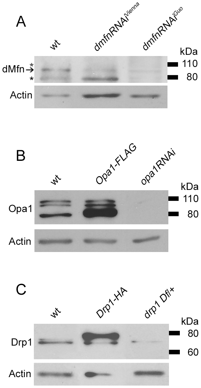
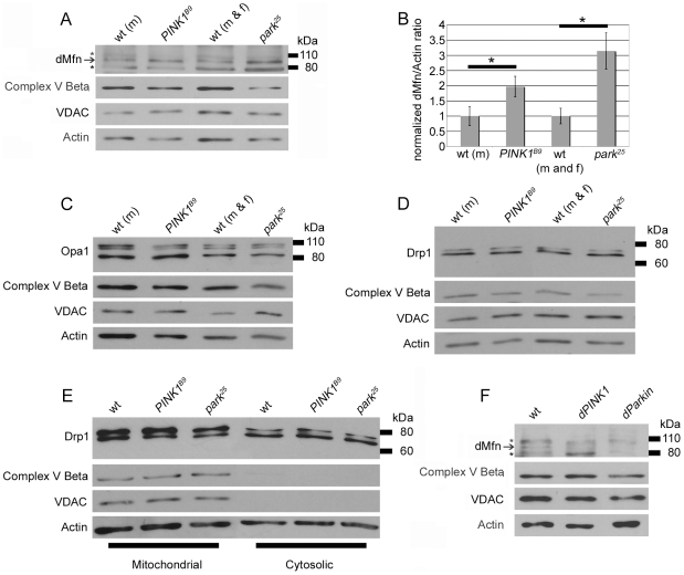
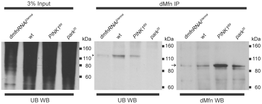
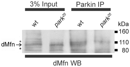
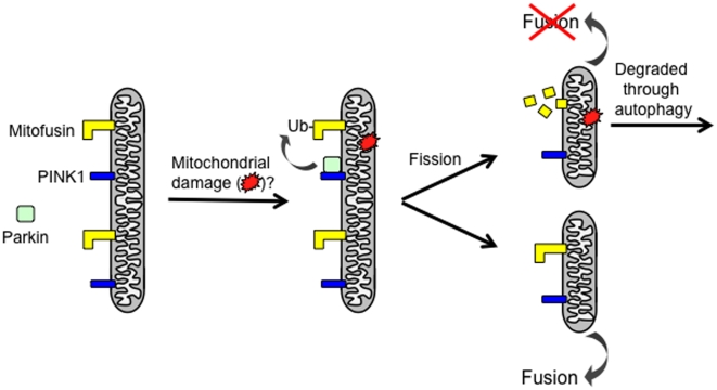
Loss-of-function mutations in the PINK1 or parkin genes result in recessive heritable forms of parkinsonism. Genetic studies of Drosophila orthologs of PINK1 and parkin indicate that PINK1, a mitochondrially targeted serine/threonine kinase, acts upstream of Parkin, a cytosolic ubiquitin-protein ligase, to promote mitochondrial fragmentation, although the molecular mechanisms by which the PINK1/Parkin pathway promotes mitochondrial fragmentation are unknown. We tested the hypothesis that PINK1 and Parkin promote mitochondrial fragmentation by targeting core components of the mitochondrial morphogenesis machinery for ubiquitination. We report that the steady-state abundance of the mitochondrial fusion-promoting factor Mitofusin (dMfn) is inversely correlated with the activity of PINK1 and Parkin in Drosophila. We further report that dMfn is ubiquitinated in a PINK1- and Parkin-dependent fashion and that dMfn co-immunoprecipitates with Parkin. By contrast, perturbations of PINK1 or Parkin did not influence the steady-state abundance of the mitochondrial fission-promoting factor Drp1 or the mitochondrial fusion-promoting factor Opa1, or the subcellular distribution of Drp1. Our findings suggest that dMfn is a direct substrate of the PINK1/Parkin pathway and that the mitochondrial morphological alterations and tissue degeneration phenotypes that derive from mutations in PINK1 and parkin result at least in part from reduced ubiquitin-mediated turnover of dMfn.
Overview
The claims and statistics of this paper haven’t been checked yet.
Run the check — free, no account needed. It reads the open-access text and checks the sources it cites.
Integrity signals
Registry
Cites
Statistics
Figures
Verification
Claims the paper makes
The claims and statistics of this paper haven’t been checked yet.
Run the check — free, no account needed. It reads the open-access text and checks the sources it cites.
Claims other papers make about this one
Each is checked against this paper’s own text: does it say what the citing paper says it does?
In the citing paper
“Once activated, Parkin has been reported to facilitate ubiquitination of numerous OMM proteins, including Mfn1 [126,127,128], Mfn2 [129], VDAC [130], Tom20 (translocase of the outer membrane 20) [121], and Miro [131].”
From Mitochondrial Dynamics and Mitophagy in Skeletal Muscle Health and Aging (2021) · cites this paper as [127]
Self-check
Percentages, sums and reported differences re-computed from the paper’s own numbers.
The self-check needs the paper’s full text.
Citations
Cites
Targeted gene expression as a means of altering cell fates and generating dominant phenotypes
Brand, A. H., & Perrimon, N. (1993) · Development · doi:10.1242/dev.118.2.401 · Cited by 9,510
Parkin is recruited selectively to impaired mitochondria and promotes their autophagy
Narendra, D., Tanaka, A., Suen, D.-F., & Youle, R. J. (2008) · The Journal of Cell Biology · doi:10.1083/jcb.200809125 · Cited by 4,026
PINK1 Is Selectively Stabilized on Impaired Mitochondria to Activate Parkin
Narendra, D. P., Jin, S. M., Tanaka, A., Suen, D.-F., Gautier, C. A., Shen, J., Cookson, M. R., & Youle, R. J. (2010) · PLoS Biology · doi:10.1371/journal.pbio.1000298 · Cited by 2,944
PINK1/Parkin-mediated mitophagy is dependent on VDAC1 and p62/SQSTM1
Geisler, S., Holmström, K. M., Skujat, D., Fiesel, F. C., Rothfuss, O. C., Kahle, P. J., & Springer, W. (2010) · Nature Cell Biology · doi:10.1038/ncb2012 · Cited by 2,835
Drosophila pink1 is required for mitochondrial function and interacts genetically with parkin
Clark, I. E., Dodson, M. W., Jiang, C., Cao, J. H., Huh, J. R., Seol, J. H., Yoo, S. J., Hay, B. A., & Guo, M. (2006) · Nature · doi:10.1038/nature04779 · Cited by 1,788
Recognition of the polyubiquitin proteolytic signal
Thrower, J. S., Hoffman, L. M., Rechsteiner, M., & Pickart, C. M. (2000) · The EMBO Journal · doi:10.1093/emboj/19.1.94 · Cited by 1,769
Mitochondrial dysfunction in Drosophila PINK1 mutants is complemented by parkin
Park, J., Lee, S. B., Lee, S., Kim, Y., Song, S., Kim, S., Bae, E., Kim, J., Shong, M., Kim, J.-M., et al. (2006) · Nature · doi:10.1038/nature04788 · Cited by 1,705
PINK1-dependent recruitment of Parkin to mitochondria in mitophagy
Vives-Bauza, C., Zhou, C., Huang, Y., Cui, M., de Vries, R. L. A., Kim, J., May, J., Tocilescu, M. A., Liu, W., Ko, H. S., et al. (2010) · Proceedings of the National Academy of Sciences · doi:10.1073/pnas.0911187107 · Cited by 1,616
Bender, A., Krishnan, K. J., Morris, C. M., Taylor, G. A., Reeve, A. K., Perry, R. H., Jaros, E., Hersheson, J. S., Betts, J., Klopstock, T., et al. (2006) · Nature Genetics · doi:10.1038/ng1769 · Cited by 1,570
Mitochondrial pathology and apoptotic muscle degeneration in Drosophila parkin mutants
Greene, J. C., Whitworth, A. J., Kuo, I., Andrews, L. A., Feany, M. B., & Pallanck, L. J. (2003) · Proceedings of the National Academy of Sciences · doi:10.1073/pnas.0737556100 · Cited by 1,203
Self-citation
Cited by
Sorted by citations
Mitochondria: In Sickness and in Health
Nunnari, J., & Suomalainen, A. (2012) · Cell · doi:10.1016/j.cell.2012.02.035 · Cited by 3,479
Youle, R. J., & Narendra, D. P. (2011) · Nature Reviews Molecular Cell Biology · doi:10.1038/nrm3028 · Cited by 3,273
The Role of Atg Proteins in Autophagosome Formation
Mizushima, N., Yoshimori, T., & Ohsumi, Y. (2011) · Annual Review of Cell and Developmental Biology · doi:10.1146/annurev-cellbio-092910-154005 · Cited by 2,978
The Roles of PINK1, Parkin, and Mitochondrial Fidelity in Parkinson’s Disease
Pickrell, A. M., & Youle, R. J. (2015) · Neuron · doi:10.1016/j.neuron.2014.12.007 · Cited by 2,310
The pathways of mitophagy for quality control and clearance of mitochondria
Ashrafi, G., & Schwarz, T. L. (2013) · Cell Death & Differentiation · doi:10.1038/cdd.2012.81 · Cited by 1,900
Autophagy, mitochondria and oxidative stress: cross-talk and redox signalling.
Lee, J., Giordano, S., & Zhang, J. (2012) · Biochemical Journal · doi:10.1042/bj20111451 · Cited by 1,438
Molecular mechanisms and physiological functions of mitophagy
Onishi, M., Yamano, K., Sato, M., Matsuda, N., & Okamoto, K. (2021) · The EMBO Journal · doi:10.15252/embj.2020104705 · Cited by 1,469
Proteasome and p97 mediate mitophagy and degradation of mitofusins induced by Parkin
Tanaka, A., Cleland, M. M., Xu, S., Narendra, D. P., Suen, D.-F., Karbowski, M., & Youle, R. J. (2010) · Journal of Cell Biology · doi:10.1083/jcb.201007013 · Cited by 1,369
Fusion and fission: interlinked processes critical for mitochondrial health
Chan, D. C. (2012) · Annual Review of Genetics · doi:10.1146/annurev-genet-110410-132529 · Cited by 1,196
PINK1 and Parkin Target Miro for Phosphorylation and Degradation to Arrest Mitochondrial Motility
Wang, X., Winter, D., Ashrafi, G., Schlehe, J. S., Wong, Y. L., Selkoe, D. J., Rice, S. E., Steen, J. A. J., LaVoie, M. J., & Schwarz, T. L. (2011) · Cell · doi:10.1016/j.cell.2011.10.018 · Cited by 1,198
Landscape of the PARKIN-dependent ubiquitylome in response to mitochondrial depolarization
Sarraf, S. A., Raman, M., Guarani-Pereira, V., Sowa, M. E., Huttlin, E. L., Gygi, S. P., & Harper, J. W. (2013) · Nature · doi:10.1038/nature12043 · Cited by 1,010
Broad activation of the ubiquitin–proteasome system by Parkin is critical for mitophagy
Chan, N. C., Salazar, A. M., Pham, A. H., Sweredoski, M. J., Kolawa, N. J., Graham, R. L. J., Hess, S., & Chan, D. C. (2011) · Human Molecular Genetics · doi:10.1093/hmg/ddr048 · Cited by 993
Mitochondrial dynamics and mitochondrial quality control
Ni, H.-M., Williams, J. A., & Ding, W.-X. (2015) · Redox Biology · doi:10.1016/j.redox.2014.11.006 · Cited by 931
Gegg, M. E., Cooper, J. M., Chau, K.-Y., Rojo, M., Schapira, A. H. V., & Taanman, J.-W. (2010) · Human Molecular Genetics · doi:10.1093/hmg/ddq419 · Cited by 910
Mitochondrial transport in neurons: impact on synaptic homeostasis and neurodegeneration
Sheng, Z., & Cai, Q. (2012) · Nature reviews. Neuroscience · doi:10.1038/nrn3156 · Cited by 940
Wong, Y. C., & Holzbaur, E. L. F. (2014) · Proceedings of the National Academy of Sciences · doi:10.1073/pnas.1405752111 · Cited by 812
Kubli, D. A., & Gustafsson, Å. B. (2012) · Circulation Research · doi:10.1161/circresaha.112.265819 · Cited by 825
The Interplay Between Mitochondrial Dynamics and Mitophagy
Twig, G., & Shirihai, O. S. (2011) · Antioxidants & Redox Signaling · doi:10.1089/ars.2010.3779 · Cited by 778
PINK1- and Parkin-mediated mitophagy at a glance
Jin, S. M., & Youle, R. J. (2012) · Journal of Cell Science · doi:10.1242/jcs.093849 · Cited by 656
Yoshii, S. R., Kishi, C., Ishihara, N., & Mizushima, N. (2011) · Journal of Biological Chemistry · doi:10.1074/jbc.m110.209338 · Cited by 612
Okatsu, K., Oka, T., Iguchi, M., Imamura, K., Kosako, H., Tani, N., Kimura, M., Go, E., Koyano, F., Funayama, M., et al. (2012) · Nature Communications · doi:10.1038/ncomms2016 · Cited by 629
The role of PINK1–Parkin in mitochondrial quality control
Narendra, D. P., & Youle, R. J. (2024) · Nature Cell Biology · doi:10.1038/s41556-024-01513-9 · Cited by 600
Mitochondrial Trafficking in Neurons
Schwarz, T. L. (2013) · Cold Spring Harbor Perspectives in Biology · doi:10.1101/cshperspect.a011304 · Cited by 525
Ge, P., Dawson, V. L., & Dawson, T. M. (2020) · Molecular Neurodegeneration · doi:10.1186/s13024-020-00367-7 · Cited by 496
The PINK1-Parkin pathway promotes both mitophagy and selective respiratory chain turnover in vivo
Vincow, E. S., Merrihew, G., Thomas, R. E., Shulman, N. J., Beyer, R. P., MacCoss, M. J., & Pallanck, L. J. (2013) · Proceedings of the National Academy of Sciences · doi:10.1073/pnas.1221132110 · Cited by 478
Self-citationMitophagy is triggered by mild oxidative stress in a mitochondrial fission dependent manner
Frank, M., Duvezin-Caubet, S., Koob, S., Occhipinti, A., Jagasia, R., Petcherski, A., Ruonala, M. O., Priault, M., Salin, B., & Reichert, A. S. (2012) · Biochimica et Biophysica Acta (BBA) - Molecular Cell Research · doi:10.1016/j.bbamcr.2012.08.007 · Cited by 468
Hsieh, C.-H., Shaltouki, A., Gonzalez, A. E., da Cruz, A. B., Burbulla, L. F., Lawrence, E. S., Schüle, B., Krainc, D., Palmer, T. D., & Wang, X. (2016) · Cell stem cell · doi:10.1016/j.stem.2016.08.002 · Cited by 475
Recent Advances in the Genetics of Parkinson's Disease
Martin, I., Dawson, V. L., & Dawson, T. M. (2011) · Annual Review of Genomics and Human Genetics · doi:10.1146/annurev-genom-082410-101440 · Cited by 451
Mitochondrial quality control: a matter of life and death for neurons
Rugarli, E. I., & Langer, T. (2012) · The EMBO Journal · doi:10.1038/emboj.2012.38 · Cited by 423
The Mitochondrial Basis of Aging and Age-Related Disorders
Srivastava, S. (2017) · Genes · doi:10.3390/genes8120398 · Cited by 446
Mitochondrial trafficking and anchoring in neurons: New insight and implications
Sheng, Z. (2014) · The Journal of Cell Biology · doi:10.1083/jcb.201312123 · Cited by 429
Targeting Mitochondrial Dysfunction: Role for PINK1 and Parkin in Mitochondrial Quality Control
Narendra, D. P., & Youle, R. J. (2011) · Antioxidants & Redox Signaling · doi:10.1089/ars.2010.3799 · Cited by 367
Role of Mitochondria in Nonalcoholic Fatty Liver Disease
Nassir, F., & Ibdah, J. A. (2014) · International Journal of Molecular Sciences · doi:10.3390/ijms15058713 · Cited by 369
Gelmetti, V., Rosa, P. D., Torosantucci, L., Marini, E. S., Romagnoli, A., Rienzo, M. D., Arena, G., Vignone, D., Fimia, G. M., & Valente, E. M. (2017) · Autophagy · doi:10.1080/15548627.2016.1277309 · Cited by 364
SIRT5 regulation of ammonia-induced autophagy and mitophagy
Polletta, L., Vernucci, E., Carnevale, I., Arcangeli, T., Rotili, D., Palmerio, S., Steegborn, C., Nowak, T., Schutkowski, M., Pellegrini, L., et al. (2015) · Autophagy · doi:10.1080/15548627.2015.1009778 · Cited by 350
Soubannier, V., Rippstein, P. U., Kaufman, B. A., Shoubridge, E. A., & McBride, H. M. (2012) · PLoS ONE · doi:10.1371/journal.pone.0052830 · Cited by 346
Mitochondrial Quality Control Mediated by PINK1 and Parkin: Links to ParkinsonismPreprint
Narendra, D. P., Walker, J. E., & Youle, R. J. (2012) · Cold Spring Harbor Perspectives in Biology · doi:10.1101/cshperspect.a011338 · Cited by 360
Preconditioning Involves Selective Mitophagy Mediated by Parkin and p62/SQSTM1
Huang, C., Andres, A. M., Ratliff, E. P., Hernandez, G., Lee, P. P. W., & Gottlieb, R. A. (2011) · PLoS ONE · doi:10.1371/journal.pone.0020975 · Cited by 347
The Parkinson's disease–linked proteins Fbxo7 and Parkin interact to mediate mitophagy
Burchell, V., Nelson, D. E., Sánchez-Martínez, Á., Delgado-Camprubi, M., Ivatt, R. M., Pogson, J. H., Randle, S. J., Wray, S., Lewis, P. A., Houlden, H., et al. (2013) · Nature Neuroscience · doi:10.1038/nn.3489 · Cited by 342
Rana, A., Rera, M., & Walker, D. W. (2013) · Proceedings of the National Academy of Sciences · doi:10.1073/pnas.1216197110 · Cited by 335
Structure of the human Parkin ligase domain in an autoinhibited state
Wauer, T., & Komander, D. (2013) · The EMBO Journal · doi:10.1038/emboj.2013.125 · Cited by 328
McLelland, G., Soubannier, V., Chen, C. X., McBride, H. M., & Fon, E. A. (2014) · The EMBO Journal · doi:10.1002/embj.201385902 · Cited by 743
Leboucher, G. P., Tsai, Y. C., Yang, M., Shaw, K. C., Zhou, M., Veenstra, T. D., Glickman, M. H., & Weissman, A. M. (2012) · Molecular Cell · doi:10.1016/j.molcel.2012.05.041 · Cited by 334
Yun, J., Puri, R., Yang, H., Lizzio, M. A., Wu, C., Sheng, Z.-H., & Guo, M. (2014) · eLife · doi:10.7554/elife.01958 · Cited by 303
RBR E3 ubiquitin ligases: new structures, new insights, new questions
Spratt, D. E., Walden, H., & Shaw, G. S. (2014) · Biochemical Journal · doi:10.1042/bj20140006 · Cited by 335
How mitochondrial dynamism orchestrates mitophagy
Shirihai, O. S., Song, M., & Dorn, G. W. (2015) · Circulation Research · doi:10.1161/circresaha.116.306374 · Cited by 311
Rana, A., Oliveira, M. P., Khamoui, A. V., Aparicio, R., Rera, M., Rossiter, H. B., & Walker, D. W. (2017) · Nature Communications · doi:10.1038/s41467-017-00525-4 · Cited by 294
BNIP3 Protein Suppresses PINK1 Kinase Proteolytic Cleavage to Promote Mitophagy
Zhang, T., Xue, L., Li, L., Tang, C., Wan, Z., Wang, R., Tan, J., Tan, Y., Han, H., Tian, R., et al. (2016) · Journal of Biological Chemistry · doi:10.1074/jbc.m116.733410 · Cited by 293
Itakura, E., Kishi-Itakura, C., Koyama-Honda, I., & Mizushima, N. (2012) · Journal of Cell Science · doi:10.1242/jcs.094110 · Cited by 286
Mitochondrial Dynamics: Coupling Mitochondrial Fitness with Healthy Aging
Sebastián, D., Palacı́n, M., & Antonio Zorzano, A. (2017) · Trends in Molecular Medicine · doi:10.1016/j.molmed.2017.01.003 · Cited by 292
Parkin and mitophagy in cancer
Bernardini, J. P., Lazarou, M., & Dewson, G. (2016) · Oncogene · doi:10.1038/onc.2016.302 · Cited by 291
Role and Mechanisms of Mitophagy in Liver Diseases
Ma, X., McKeen, T., Zhang, J., & Ding, W. (2020) · Cells · doi:10.3390/cells9040837 · Cited by 288
Mitochondria as central hub of the immune system
Breda, C. N. D. S., Davanzo, G. G., Basso, P. J., Câmara, N. O. S., & Moraes‐Vieira, P. M. (2019) · Redox Biology · doi:10.1016/j.redox.2019.101255 · Cited by 289
Self and Nonself: How Autophagy Targets Mitochondria and Bacteria
Randow, F., & Youle, R. J. (2014) · Cell Host & Microbe · doi:10.1016/j.chom.2014.03.012 · Cited by 281
Fabian, L., & Brill, J. A. (2012) · Spermatogenesis · doi:10.4161/spmg.21798 · Cited by 280
Mitochondrial degradation: Mitophagy and beyond
Uoselis, L., Nguyen, T. N., & Lazarou, M. (2023) · Molecular Cell · doi:10.1016/j.molcel.2023.08.021 · Cited by 270
Mitochondrial Dynamics and Mitophagy in Skeletal Muscle Health and Aging
Leduc-Gaudet, J.-P., Hussain, S. N. A., Barreiro, E., & Gouspillou, G. (2021) · International Journal of Molecular Sciences · doi:10.3390/ijms22158179 · Cited by 268
Mitophagy in Alzheimer’s Disease and Other Age-Related Neurodegenerative Diseases
Cai, Q., & Jeong, Y. Y. (2020) · Cells · doi:10.3390/cells9010150 · Cited by 270
Phosphorylated ubiquitin chain is the genuine Parkin receptor
Okatsu, K., Koyano, F., Kimura, M., Kosako, H., Saeki, Y., Tanaka, K., & Matsuda, N. (2015) · The Journal of Cell Biology · doi:10.1083/jcb.201410050 · Cited by 272
Regulating mitochondrial outer membrane proteins by ubiquitination and proteasomal degradation
Karbowski, M., & Youle, R. J. (2011) · Current Opinion in Cell Biology · doi:10.1016/j.ceb.2011.05.007 · Cited by 256
PINK1 drives Parkin self-association and HECT-like E3 activity upstream of mitochondrial binding
Lazarou, M., Narendra, D. P., Jin, S. M., Tekle, E., Banerjee, S., & Youle, R. J. (2013) · Journal of Cell Biology · doi:10.1083/jcb.201210111 · Cited by 236
Sun, Y., Vashisht, A. A., Tchieu, J. H., Wohlschlegel, J. A., & Dreier, L. (2012) · Journal of Biological Chemistry · doi:10.1074/jbc.m112.419721 · Cited by 255
Kim, N. C., Tresse, E., Kolaitis, R., Molliex, A., Thomas, R. E., Alami, N. H., Wang, B., Joshi, A., Smith, R. B., Ritson, G. P., et al. (2013) · Neuron · doi:10.1016/j.neuron.2013.02.029 · Cited by 248
Self-citationThe ubiquitin signal and autophagy: an orchestrated dance leading to mitochondrial degradation
Yamano, K., Matsuda, N., & Tanaka, K. (2016) · EMBO Reports · doi:10.15252/embr.201541486 · Cited by 253
Mutations in PINK1 and Parkin Impair Ubiquitination of Mitofusins in Human Fibroblasts
Raković, A., Grünewald, A., Kottwitz, J., Brüggemann, N., Pramstaller, P. P., Lohmann, K., & Klein, C. (2011) · PLoS ONE · doi:10.1371/journal.pone.0016746 · Cited by 237
Parkin promotes the ubiquitination and degradation of the mitochondrial fusion factor mitofusin 1
Glauser, L., Sonnay, S., Stafa, K., & Moore, D. J. (2011) · Journal of Neurochemistry · doi:10.1111/j.1471-4159.2011.07318.x · Cited by 230
Evolving and Expanding the Roles of Mitophagy as a Homeostatic and Pathogenic Process
Gustafsson, Å. B., & Dorn, G. W. (2018) · Physiological Reviews · doi:10.1152/physrev.00005.2018 · Cited by 235
Mitochondrial dynamics as regulators of cancer biology
Trotta, A. P., & Chipuk, J. E. (2017) · Cellular and Molecular Life Sciences · doi:10.1007/s00018-016-2451-3 · Cited by 226
Alterations in Mitochondrial Quality Control in Alzheimer’s Disease
Cai, Q., & Tammineni, P. (2016) · Frontiers in Cellular Neuroscience · doi:10.3389/fncel.2016.00024 · Cited by 226
Neuronal Mitophagy in Neurodegenerative Diseases
Martínez‐Vicente, M. (2017) · Frontiers in Molecular Neuroscience · doi:10.3389/fnmol.2017.00064 · Cited by 222
Hirota, Y., Yamashita, S.-I., Kurihara, Y., Jin, X., Aihara, M., Saigusa, T., Kang, D., & Kanki, T. (2015) · Autophagy · doi:10.1080/15548627.2015.1023047 · Cited by 218
Iorio, R., Celenza, G., & Petricca, S. (2021) · Cells · doi:10.3390/cells11010030 · Cited by 222
Mitophagy: A Complex Mechanism of Mitochondrial Removal
Novak, I. (2011) · Antioxidants and Redox Signaling · doi:10.1089/ars.2011.4407 · Cited by 213
Kotiadis, V. N., Duchen, M. R., & Osellame, L. D. (2013) · Biochimica et Biophysica Acta (BBA) - General Subjects · doi:10.1016/j.bbagen.2013.10.041 · Cited by 216
Regulation of ER-mitochondria contacts by Parkin via Mfn2
Basso, V., Marchesan, E., Peggion, C., Chakraborty, J., von Stockum, S., Giacomello, M., Ottolini, D., Debattisti, V., Caicci, F., Tasca, E., et al. (2018) · Pharmacological Research · doi:10.1016/j.phrs.2018.09.006 · Cited by 217
Truban, D., Hou, X., Caulfield, T. R., Fiesel, F. C., & Springer, W. (2016) · Journal of Parkinson’s Disease · doi:10.3233/jpd-160989 · Cited by 237
Birsa, N., Norkett, R., Wauer, T., Mevissen, T. E. T., Wu, H.-C., Foltynie, T., Bhatia, K. P., Hirst, W. D., Komander, D., Plun‐Favreau, H., et al. (2014) · Journal of Biological Chemistry · doi:10.1074/jbc.m114.563031 · Cited by 203
Phosphorylation of Mitochondrial Polyubiquitin by PINK1 Promotes Parkin Mitochondrial Tethering
Shiba‐Fukushima, K., Arano, T., Matsumoto, G., Inoshita, T., Yoshida, S., Ishihama, Y., Ryu, K., Nukina, N., Hattori, N., & Imai, Y. (2014) · PLoS Genetics · doi:10.1371/journal.pgen.1004861 · Cited by 196
Ubiquitin–Proteasome System and mitochondria — Reciprocity
Livnat‐Levanon, N., & Glickman, M. H. (2010) · Biochimica et Biophysica Acta (BBA) - Gene Regulatory Mechanisms · doi:10.1016/j.bbagrm.2010.07.005 · Cited by 197
The interplay between autophagy and ROS in tumorigenesis
Kongara, S., & Karantza, V. (2012) · Frontiers in Oncology · doi:10.3389/fonc.2012.00171 · Cited by 186
Mitochondrial Fission in Human Diseases
Serasinghe, M. N., & Chipuk, J. E. (2016) · Handbook of experimental pharmacology · doi:10.1007/164_2016_38 · Cited by 186
Genetics and genomics of Parkinson’s disease
Lin, M. K., & Farrer, M. J. (2014) · Genome Medicine · doi:10.1186/gm566 · Cited by 183
Cookson, M. R. (2012) · Cold Spring Harbor Perspectives in Medicine · doi:10.1101/cshperspect.a009415 · Cited by 170
Parkin and PINK1 functions in oxidative stress and neurodegeneration
Barodia, S. K., Creed, R. B., & Goldberg, M. S. (2016) · Brain Research Bulletin · doi:10.1016/j.brainresbull.2016.12.004 · Cited by 172
Autophagy in Protein and Organelle TurnoverPreprint
Mizushima, N. (2011) · Cold Spring Harbor Symposia on Quantitative Biology · doi:10.1101/sqb.2011.76.011023 · Cited by 170
Staying cool in difficult times: Mitochondrial dynamics, quality control and the stress response
Shutt, T. E., & McBride, H. M. (2012) · Biochimica et Biophysica Acta (BBA) - Molecular Cell Research · doi:10.1016/j.bbamcr.2012.05.024 · Cited by 167
The cell biology of Parkinson’s disease
Panicker, N., Ge, P., Dawson, V. L., & Dawson, T. M. (2021) · The Journal of Cell Biology · doi:10.1083/jcb.202012095 · Cited by 179
Alpha-synuclein delays mitophagy and targeting Miro rescues neuron loss in Parkinson’s models
Shaltouki, A., Hsieh, C.-H., Kim, M. J., & Wang, X. (2018) · Acta Neuropathologica · doi:10.1007/s00401-018-1873-4 · Cited by 170
Parkin Modulates Endosomal Organization and Function of the Endo-Lysosomal Pathway
Song, P., Trajković, K., Tsunemi, T., & Krainc, D. (2016) · Journal of Neuroscience · doi:10.1523/jneurosci.2569-15.2016 · Cited by 161
Mitophagy: the latest problem for Parkinson's disease
Vives-Bauzá, C., & Przedborski, S. (2010) · Trends in Molecular Medicine · doi:10.1016/j.molmed.2010.11.002 · Cited by 161
Mitochondrial dynamics and cell death in heart failure
Marín-García, J., & Akhmedov, A. T. (2016) · Heart Failure Reviews · doi:10.1007/s10741-016-9530-2 · Cited by 160
Filichia, E., Hoffer, B. J., Qi, X., & Luo, Y. (2016) · Scientific Reports · doi:10.1038/srep32656 · Cited by 157
Mitochondria and autophagy: Critical interplay between the two homeostats
Okamoto, K., & Kondo‐Okamoto, N. (2011) · Biochimica et Biophysica Acta (BBA) - General Subjects · doi:10.1016/j.bbagen.2011.08.001 · Cited by 155
Degradation of engulfed mitochondria is rate-limiting in Optineurin-mediated mitophagy in neurons
Evans, C. S., & Holzbaur, E. L. (2020) · eLife · doi:10.7554/elife.50260 · Cited by 137
“…for organelle degradation (Geisler et al., 2010; Kane et al., 2014; Kazlauskaite et al., 2014; Koyano et al., 2014; Matsuda et al., 2010; Narendra et al., 2008; Narendra et al., 2010b; Ordureau et al., 2015; Ordureau et al., 2014; Poole et al., 2010; Sarraf et al., 2013; VivesBauza et al., 2010).”
Mutant Parkin Impairs Mitochondrial Function and Morphology in Human Fibroblasts
Grünewald, A., Voges, L., Raković, A., Kasten, M., Vandebona, H., Hemmelmann, C., Lohmann, K., Orolicki, S., Ramı́rez, A., Schapira, A. H. V., et al. (2010) · PLoS ONE · doi:10.1371/journal.pone.0012962 · Cited by 153
Parkin and mitochondrial quality control: toward assembling the puzzle
Winklhofer, K. F. (2014) · Trends in Cell Biology · doi:10.1016/j.tcb.2014.01.001 · Cited by 153
FoxO1 Promotes Mitophagy in the Podocytes of Diabetic Male Mice via the PINK1/Parkin Pathway
Li, W., Du, M., Wang, Q., Ma, X., Wu, L., Guo, F., Ji, H., Huang, F., & Qin, G. (2017) · Endocrinology · doi:10.1210/en.2016-1970 · Cited by 146
Shaping the dynamic mitochondrial network
Lackner, L. L. (2014) · BMC Biology · doi:10.1186/1741-7007-12-35 · Cited by 156
Mitochondrial dynamism and heart disease: changing shape and shaping change
Dorn, G. W. (2015) · EMBO Molecular Medicine · doi:10.15252/emmm.201404575 · Cited by 157
Regulation of mitochondrial dynamics in astrocytes: Mechanisms, consequences, and unknowns
Jackson, J. G., & Robinson, M. B. (2017) · Glia · doi:10.1002/glia.23252 · Cited by 153
Metadata
- Authors
- Poole, A. C., Thomas, R. E., Yu, S., Vincow, E. S., & Pallanck, L.
- Affiliations
- University of Washington (US)
- Corresponding author
- Pallanck, Leo
- Venue
- PLoS ONE
- Publisher
- Public Library of Science (PLoS)
- Published
- 2010-04-07
- Volume
- 5
- Issue
- 4
- Pages
- e10054
- ISSN
- 1932-6203 (ISSN-L)
- PMC
- PMC2850930
- OpenAlex
- W2054556550
- Cited by
- 440
- References
- 41
- Citations by year
- 2026: 4 · 2025: 4 · 2024: 17 · 2023: 14 · 2022: 13 · 2021: 26 · 2020: 35 · 2019: 16 · 2018: 26 · 2017: 27 · 2016: 30 · 2015: 39 · 2014: 38 · 2013: 44 · 2012: 54
- FWCI
- 28.77
- Type
- journal article
- Language
- en
- Open access
- gold
- doi.org
- Locations
- PLoS ONE (publishedVersion, cc-by)
- PubMed Central (publishedVersion, cc-by)
- PubMed (publishedVersion)
- DOAJ (DOAJ: Directory of Open Access Journals) (submittedVersion)
- Funders
- National Institutes of Health
- National Institute on Aging (T32AG000057)
- National Institute of General Medical Sciences (R01GM086394)
- Topics
- Health Sciences › Medicine › Clinical Neurology › Parkinson's Disease Mechanisms and Treatments
- Life Sciences › Biochemistry, Genetics and Molecular Biology › Molecular Biology › Mitochondrial Function and Pathology
- Life Sciences › Biochemistry, Genetics and Molecular Biology › Cell Biology › Autophagy in Disease and Therapy
- Keywords
- mitofusins, PINK1, Parkin, PINK1/Parkin pathway, mitochondrial fusion, mitochondrial fragmentation, ubiquitination, parkinsonism, Drp1, OPA1, Drosophila, mitochondrial morphogenesis
- MeSH terms
- PTEN-Induced Putative Kinase, Membrane Proteins, Mitochondria, Mutation, Protein Serine-Threonine Kinases, Drosophila Proteins, Ubiquitin-Protein Ligases, Immunoprecipitation, Ubiquitination, Protein Stability
About these checks
Registry, retraction, Cites and statistics results are computed, not judged, with one exception: a printed reference that no index lists is read once by a model before it counts as a citation error. Claims are extracted by a model and kept only when their quote appears verbatim in the text; each cited source is then checked, with a model’s help, for whether it says what the claim relies on. Those verdicts can be wrong — read the claim’s quote and the cited source before relying on one.
Statistics and arithmetic are recomputed, not judged.
Confidence bands are shown only for fields calibrated on our annotated test set. None is calibrated yet, so no band is shown.
What counts as a claim ›How we measure confidence ›How we check this ›
Are you an author, or do you see a problem with this page? Request a review
