Cited by 176Antimicrobial Peptides and ActivitiesAntifungal resistance and susceptibilityAntimicrobial agents and applications

The DOI matches its Crossref record; 72 of the 75 references it cites were found in a registry or an index.

Human Antimicrobial Peptide LL-37 Inhibits Adhesion of Candida albicans by Interacting with Yeast Cell-Wall Carbohydrates

Open access

Abstract

Candida albicans is the major fungal pathogen of humans. Fungal adhesion to host cells is the first step of mucosal infiltration. Antimicrobial peptides play important roles in the initial mucosal defense against C. albicans infection. LL-37 is the only member of the human cathelicidin family of antimicrobial peptides and is commonly expressed in various tissues and cells, including epithelial cells of both the oral cavity and urogenital tract. We found that, at sufficiently low concentrations that do not kill the fungus, LL-37 was still able to reduce C. albicans infectivity by inhibiting C. albicans adhesion to plastic surfaces, oral epidermoid OECM-1 cells, and urinary bladders of female BALB/c mice. Moreover, LL-37-treated C. albicans floating cells that did not adhere to the underlying substratum aggregated as a consequence of LL-37 bound to the cell surfaces. According to the results of a competition assay, the inhibitory effects of LL-37 on cell adhesion and aggregation were mediated by its preferential binding to mannan, the main component of the C. albicans cell wall, and partially by its ability to bind chitin or glucan, which underlie the mannan layer. Therefore, targeting of cell-wall carbohydrates by LL-37 provides a new strategy to prevent C. albicans infection, and LL-37 is a useful, new tool to screen for other C. albicans components involved in adhesion.

Overview

The claims and statistics of this paper haven’t been checked yet.

Run the check — free, no account needed. It reads the open-access text and checks the sources it cites.

A few free checks a day · papers already verified open instantly · results are public

Integrity signals

Registry

Crossref · 10.1371/journal.pone.0017755

Cites

72 of 72 references verified0 citation errors · 0 not checkable

Statistics

Figures

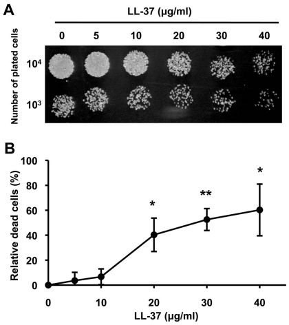
Figure 1 Candidacidal activity of LL-37. (A) Candidacidal activity of LL-37 was determined using a spot assay. The C. albicans SC5314 strain was treated with different concentrations of LL-37, and then cells that had been ten-fold serially diluted were spotted onto YPD agar. All experiments were performed in triplicate. (B) The candidacidal activity of LL-37 was monitored by FUN-1 staining. LL-37-treated cells were stained with FUN-1 for 30 min at 30°C. The number of cells killed by LL-37 was normalized with respect to the number of control cells (no LL-37 treatment) and reported as a percentage. Data are presented as the average of three independent experiments, and the statistical significance of the experimental data in comparison to the control data was determined using Student's t-test (*, p<0.05; **, p<0.01).
Figure 1 Candidacidal activity of LL-37. (A) Candidacidal activity of LL-37 was determined using a spot assay. The C. albicans SC5314 strain was treated with different concentrations of LL-37, and then cells that had been ten-fold serially diluted were spotted onto YPD agar. All experiments were performed in triplicate. (B) The candidacidal activity of LL-37 was monitored by FUN-1 staining. LL-37-treated cells were stained with FUN-1 for 30 min at 30°C. The number of cells killed by LL-37 was normalized with respect to the number of control cells (no LL-37 treatment) and reported as a percentage. Data are presented as the average of three independent experiments, and the statistical significance of the experimental data in comparison to the control data was determined using Student's t-test (*, p<0.05; **, p<0.01).CC BY · PubMed Central
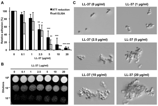
Figure 2 Inhibition of C. albicans adhesion by LL-37. (A) Cell adhesion to an abiotic surface (polystyrene). C. albicans cells were incubated in RPMI-1640 in polystyrene wells for 30 min and then washed three times with PBS to remove unattached cells. The attached cells were detected by measuring the reduction of XTT and by whole-cell ELISA using an antibody against C. albicans. All experiments were done in triplicate, and each was repeated three (XTT reduction assay) or four times (cell ELISA). The statistical significance for the number of treated vs. control cells was determined using Student's t-test (*, p<0.05; **, p<0.01; ***, p<0.001). (B) Spot assay to determine the number of viable, floating cells after LL-37 treatment. Different concentrations of LL-37 were incubated with cells in RPMI-1640 for 30 min; then the floating cells were collected, diluted onto YPD agar, and incubated at 30°C overnight. All experiments were done in triplicate, and each was repeated twice. (C) The morphology of the floating cells after LL-37 treatment was visualized by microscopy (400× magnification).
Figure 2 Inhibition of C. albicans adhesion by LL-37. (A) Cell adhesion to an abiotic surface (polystyrene). C. albicans cells were incubated in RPMI-1640 in polystyrene wells for 30 min and then washed three times with PBS to remove unattached cells. The attached cells were detected by measuring the reduction of XTT and by whole-cell ELISA using an antibody against C. albicans. All experiments were done in triplicate, and each was repeated three (XTT reduction assay) or four times (cell ELISA). The statistical significance for the number of treated vs. control cells was determined using Student's t-test (*, p<0.05; **, p<0.01; ***, p<0.001). (B) Spot assay to determine the number of viable, floating cells after LL-37 treatment. Different concentrations of LL-37 were incubated with cells in RPMI-1640 for 30 min; then the floating cells were collected, diluted onto YPD agar, and incubated at 30°C overnight. All experiments were done in triplicate, and each was repeated twice. (C) The morphology of the floating cells after LL-37 treatment was visualized by microscopy (400× magnification).CC BY · PubMed Central
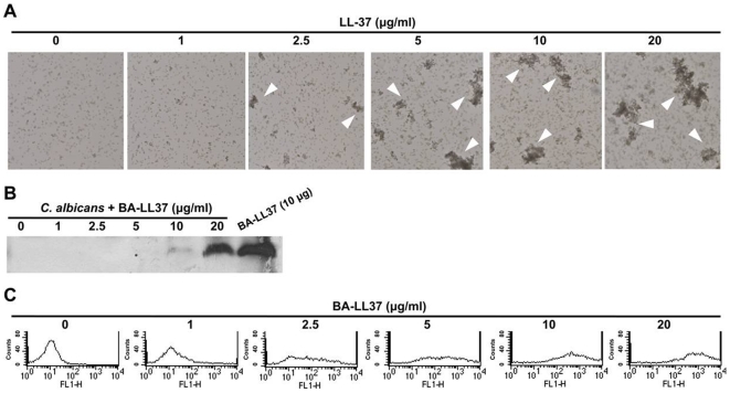
Figure 3 LL-37-induced C. albicans cell aggregation and LL-37 binding to C. albicans. (A) Visualization by microscopy of C. albicans cell aggregation after LL-37 treatment. C. albicans cells were incubated with LL-37 in RPMI-1640, and the floating cells were then injected into μ-Slides and visualized by microscopy (250× magnification). Cell aggregates are indicated by the arrowheads. (B) Western blot showing that LL-37 bound to floating C. albicans cells. C. albicans were incubated with BA-LL37 in RPMI-1640, and the contents of the floating cells were subjected to Tricine SDS-PAGE, blotted, and detected with SA-HRP. BA-LL37 (10 µg) served as the positive control. (C) Flow cytometry showing that LL-37 bound to C. albicans cells. Various concentrations of BA-LL37 were incubated with C. albicans in PBS at 4°C overnight, and then the levels of cell-bound BA-LL37 were assessed by flow cytometry that used SA-DTAF for detection. The data shown are representative of two additional independent experiments. FL1-H means the extent of fluorescence intensity.
Figure 3 LL-37-induced C. albicans cell aggregation and LL-37 binding to C. albicans. (A) Visualization by microscopy of C. albicans cell aggregation after LL-37 treatment. C. albicans cells were incubated with LL-37 in RPMI-1640, and the floating cells were then injected into μ-Slides and visualized by microscopy (250× magnification). Cell aggregates are indicated by the arrowheads. (B) Western blot showing that LL-37 bound to floating C. albicans cells. C. albicans were incubated with BA-LL37 in RPMI-1640, and the contents of the floating cells were subjected to Tricine SDS-PAGE, blotted, and detected with SA-HRP. BA-LL37 (10 µg) served as the positive control. (C) Flow cytometry showing that LL-37 bound to C. albicans cells. Various concentrations of BA-LL37 were incubated with C. albicans in PBS at 4°C overnight, and then the levels of cell-bound BA-LL37 were assessed by flow cytometry that used SA-DTAF for detection. The data shown are representative of two additional independent experiments. FL1-H means the extent of fluorescence intensity.CC BY · PubMed Central
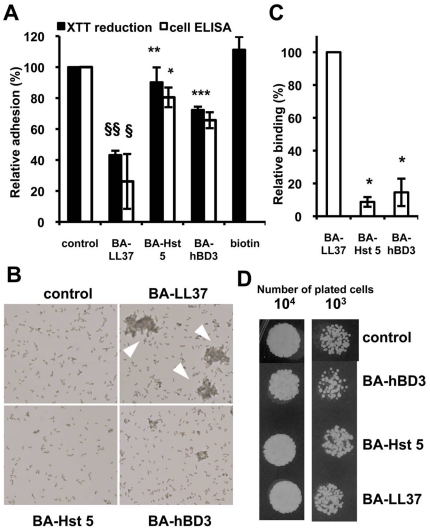
Figure 4 The inhibition of cell adhesion caused by LL-37 is the result of peptide-cell binding and cell-cell aggregation. (A) Effects of BA-LL37, BA-Hst 5, and BA-hBD3 on C. albicans adhesion to polystyrene. The molarity of each peptide was 2.12 µM (∼10 µg/ml of BA-LL37). Cells treated with only biotin served as the negative control. This control was performed for only the XTT assay. Each experiment was performed three times in triplicate. The Student's t-test was used to determine the statistical significance of the results (§, p<0.05 and §§, p<0.01 for BA-LL37-treated vs. control cells; *, p<0.05; **, p<0.01; and ***, p<0.001 for BA-LL37-treated cells vs. BA-Hst 5- or BA-hBD3-treated cells as indicated in the figure). (B) Microscopy of C. albicans cell aggregation after LL-37 treatment. C. albicans cells were incubated in RPMI-1640 with biotinylated AMPs, and the floating cells were collected. Arrowheads indicate BA-LL37-treated cell aggregates. (250× magnification.) (C) Flow cytometry to determine the extent of binding of different AMPs to C. albicans. BA-LL37, BA-Hst 5, or BA-hBD3 (10 µg each) were independently added to C. albicans samples. The peptide/cell complex was reacted in PBS at 4°C overnight. The Student's t-test was used to determine the statistical significance of the results (*, p<0.05 BA-LL37-treated cells vs. BA-Hst 5- or BA-hBD3-treated cells). Each result is the mean ± SD of two independent assays. (D) Spot assay to determine the candidacidal activities of the AMPs. BA-LL37, BA-Hst 5, and BA-hBD3 (2.12 µM) were each incubated with C. albicans for 30 min in RPMI-1640; then mixtures were diluted, spotted onto YPD agar, and incubated overnight. Each experiment was performed three times.
Figure 4 The inhibition of cell adhesion caused by LL-37 is the result of peptide-cell binding and cell-cell aggregation. (A) Effects of BA-LL37, BA-Hst 5, and BA-hBD3 on C. albicans adhesion to polystyrene. The molarity of each peptide was 2.12 µM (∼10 µg/ml of BA-LL37). Cells treated with only biotin served as the negative control. This control was performed for only the XTT assay. Each experiment was performed three times in triplicate. The Student's t-test was used to determine the statistical significance of the results (§, p<0.05 and §§, p<0.01 for BA-LL37-treated vs. control cells; *, p<0.05; **, p<0.01; and ***, p<0.001 for BA-LL37-treated cells vs. BA-Hst 5- or BA-hBD3-treated cells as indicated in the figure). (B) Microscopy of C. albicans cell aggregation after LL-37 treatment. C. albicans cells were incubated in RPMI-1640 with biotinylated AMPs, and the floating cells were collected. Arrowheads indicate BA-LL37-treated cell aggregates. (250× magnification.) (C) Flow cytometry to determine the extent of binding of different AMPs to C. albicans. BA-LL37, BA-Hst 5, or BA-hBD3 (10 µg each) were independently added to C. albicans samples. The peptide/cell complex was reacted in PBS at 4°C overnight. The Student's t-test was used to determine the statistical significance of the results (*, p<0.05 BA-LL37-treated cells vs. BA-Hst 5- or BA-hBD3-treated cells). Each result is the mean ± SD of two independent assays. (D) Spot assay to determine the candidacidal activities of the AMPs. BA-LL37, BA-Hst 5, and BA-hBD3 (2.12 µM) were each incubated with C. albicans for 30 min in RPMI-1640; then mixtures were diluted, spotted onto YPD agar, and incubated overnight. Each experiment was performed three times.CC BY · PubMed Central
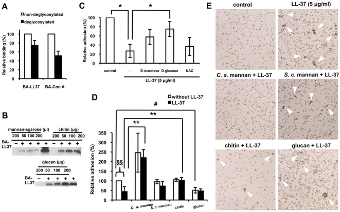
Figure 5 Binding of LL-37 to Candida cell-wall polysaccharides reduces Candida adhesion to polystyrene. (A) Flow cytometry to determine the relative amount of LL-37 bound to deglycosylated C. albicans cells. Cells were mixed with BA-LL37 or BA-Con A, and the binding was measured by SA-DTAF detection. Each result is the mean ± SD of two independent assays. (B) BA-LL37 (10 µg) was incubated with mannan-agarose beads, chitin, or glucan. After washing, the samples were subjected to Tricine SDS-PAGE and detected by western blotting with SA-HRP. BA-LL37 served as the positive control. (C) Monosaccharides reverse the LL-37-mediated inhibition of C. albicans cell adhesion. LL-37 (5 µg/ml) was added to C. albicans in the absence or presence of 400 µM mannose, d-glucose, and N-acetyl-d-glucosamine (NAC), and XTT reduction assays were performed. Each experiment was preformed three times in triplicate. The Student's t-test was used to determine the statistical significance of the results (*, p<0.05 for LL-37-treated cells in the absence or presence of d-glucose and for LL-37-treated cells vs. control cells). (D) C. albicans cell-wall polysaccharides reverse LL-37-mediated inhibition of adhesion. LL-37 (5 µg/ml) was added to C. albicans in the absence or presence of 1 mg/ml of a polysaccharide, and then XTT reduction assays were performed. Each experiment was performed three times in triplicate. The Student's t-test was used to determine the statistical significance of the results (§§, p<0.01 for LL-37-treated cells vs. control cells; #, p<0.05 for cells incubated with S. c. (S. cerevisiae) glucan vs. control cells; **, p<0.01 for LL-37-treated cells in the presence of C. a. (C. albicans) mannan or chitin vs. cells treated only with LL-37). (E) The effects of polysaccharides on the release of LL-37-induced C. albicans cell aggregation. Cells were treated as described above. The floating cells were collected and examined under a microscope (250× magnification). Arrowheads indicate aggregates.
Figure 5 Binding of LL-37 to Candida cell-wall polysaccharides reduces Candida adhesion to polystyrene. (A) Flow cytometry to determine the relative amount of LL-37 bound to deglycosylated C. albicans cells. Cells were mixed with BA-LL37 or BA-Con A, and the binding was measured by SA-DTAF detection. Each result is the mean ± SD of two independent assays. (B) BA-LL37 (10 µg) was incubated with mannan-agarose beads, chitin, or glucan. After washing, the samples were subjected to Tricine SDS-PAGE and detected by western blotting with SA-HRP. BA-LL37 served as the positive control. (C) Monosaccharides reverse the LL-37-mediated inhibition of C. albicans cell adhesion. LL-37 (5 µg/ml) was added to C. albicans in the absence or presence of 400 µM mannose, d-glucose, and N-acetyl-d-glucosamine (NAC), and XTT reduction assays were performed. Each experiment was preformed three times in triplicate. The Student's t-test was used to determine the statistical significance of the results (*, p<0.05 for LL-37-treated cells in the absence or presence of d-glucose and for LL-37-treated cells vs. control cells). (D) C. albicans cell-wall polysaccharides reverse LL-37-mediated inhibition of adhesion. LL-37 (5 µg/ml) was added to C. albicans in the absence or presence of 1 mg/ml of a polysaccharide, and then XTT reduction assays were performed. Each experiment was performed three times in triplicate. The Student's t-test was used to determine the statistical significance of the results (§§, p<0.01 for LL-37-treated cells vs. control cells; #, p<0.05 for cells incubated with S. c. (S. cerevisiae) glucan vs. control cells; **, p<0.01 for LL-37-treated cells in the presence of C. a. (C. albicans) mannan or chitin vs. cells treated only with LL-37). (E) The effects of polysaccharides on the release of LL-37-induced C. albicans cell aggregation. Cells were treated as described above. The floating cells were collected and examined under a microscope (250× magnification). Arrowheads indicate aggregates.CC BY · PubMed Central
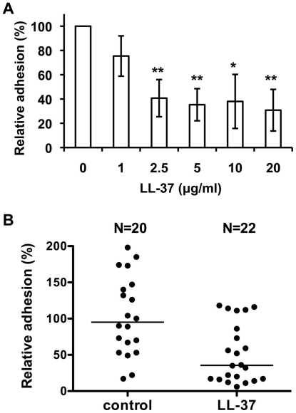
Figure 6 LL-37 inhibits C. albicans attachment to oral epidermal cells and mouse bladder mucosa. (A) C. albicans cells were incubated in RPMI-1640 at 4°C for 30 min with LL-37, after which the cells were incubated with OECM-1 oral epidermal cells for 30 min. The extent of binding by C. albicans was determined by cell ELISA. Each result is expressed as a percentage of that found for untreated cells and is reported as the mean ± SD of four experiments. Each experiment was performed in quadruplicate (*, p<0.05; **, p<0.01 for LL-37-treated cells vs. control cells). (B) Attachment of C. albicans cells to mouse urinary bladders. The number of adhered cells for each bladder was normalized to the average number of cells for all bladders and is reported as a percentage. Three independent experiments were performed with six to eight mice per group. Each horizontal line denotes the median. The difference between 5 µg/ml LL-37-treated cells and controls was significant (p = 0.0011; Student's t-test).
Figure 6 LL-37 inhibits C. albicans attachment to oral epidermal cells and mouse bladder mucosa. (A) C. albicans cells were incubated in RPMI-1640 at 4°C for 30 min with LL-37, after which the cells were incubated with OECM-1 oral epidermal cells for 30 min. The extent of binding by C. albicans was determined by cell ELISA. Each result is expressed as a percentage of that found for untreated cells and is reported as the mean ± SD of four experiments. Each experiment was performed in quadruplicate (*, p<0.05; **, p<0.01 for LL-37-treated cells vs. control cells). (B) Attachment of C. albicans cells to mouse urinary bladders. The number of adhered cells for each bladder was normalized to the average number of cells for all bladders and is reported as a percentage. Three independent experiments were performed with six to eight mice per group. Each horizontal line denotes the median. The difference between 5 µg/ml LL-37-treated cells and controls was significant (p = 0.0011; Student's t-test).CC BY · PubMed Central

Verification

Claims the paper makes

The claims and statistics of this paper haven’t been checked yet.

Run the check — free, no account needed. It reads the open-access text and checks the sources it cites.

A few free checks a day · papers already verified open instantly · results are public

Claims other papers make about this one

Each is checked against this paper’s own text: does it say what the citing paper says it does?

Self-check

Percentages, sums and reported differences re-computed from the paper’s own numbers.

The self-check needs the paper’s full text.

Citations

Cites

Show all 75 references

Cited by

Sorted by citations

See all 176

Metadata

Authors
Tsai, P.-W., Yang, C.-Y., Chang, H.-T., & Lan, C.-Y.
Affiliations
  • National Tsing Hua University (TW)
  • Animal Technology Institute Taiwan (TW)
  • China Medical University (TW)
Corresponding author
  • Chang, Hao-Teng
  • Lan, Chung-Yu
Venue
PLoS ONE
Published
2011-03-14
Volume
6
Issue
3
Pages
e17755
ISSN
1932-6203 (ISSN-L)
OpenAlex
W2167693400
Cited by
175
References
75
Citations by year
2026: 15 · 2025: 9 · 2024: 10 · 2023: 10 · 2022: 13 · 2021: 16 · 2020: 13 · 2019: 10 · 2018: 11 · 2017: 7 · 2016: 12 · 2015: 12 · 2014: 11 · 2013: 12 · 2012: 10
FWCI
10.94
Type
journal article
Language
en
Open access
Locations
  • PLoS ONE (publishedVersion, cc-by)
  • PubMed Central (publishedVersion, cc-by)
  • PubMed (publishedVersion)
  • DOAJ (DOAJ: Directory of Open Access Journals) (submittedVersion)
Funders
  • National Science Council
  • China Medical University
Topics
  • Life Sciences › Immunology and Microbiology › Microbiology › Antimicrobial Peptides and Activities
  • Health Sciences › Medicine › Infectious Diseases › Antifungal resistance and susceptibility
  • Life Sciences › Immunology and Microbiology › Applied Microbiology and Biotechnology › Antimicrobial agents and applications
Keywords
LL-37, cathelicidin, antimicrobial peptides, Candida albicans, fungal adhesion, mannan, cell wall carbohydrates, chitin, glucan, yeast aggregation, oral epithelial cells, urinary bladder, mucosal protection, fungal infection prevention
MeSH terms
Epidermal Cells, Adhesiveness, Animals, Urinary Bladder, Candida albicans, Cell Wall, Epidermis, Humans, Polystyrenes, Protein Binding, Antimicrobial Cationic Peptides, Carbohydrate Metabolism, Microbial Viability, Mice, Cathelicidins

About these checks

Registry, retraction, Cites and statistics results are computed, not judged, with one exception: a printed reference that no index lists is read once by a model before it counts as a citation error. Claims are extracted by a model and kept only when their quote appears verbatim in the text; each cited source is then checked, with a model’s help, for whether it says what the claim relies on. Those verdicts can be wrong — read the claim’s quote and the cited source before relying on one.

Statistics and arithmetic are recomputed, not judged.

Confidence bands are shown only for fields calibrated on our annotated test set. None is calibrated yet, so no band is shown.

What counts as a claim ›How we measure confidence ›How we check this ›

Are you an author, or do you see a problem with this page? Request a review