

The DOI matches its Crossref record; 40 of the 41 references it cites were found in a registry or an index.
Regulation of AKT Phosphorylation at Ser473 and Thr308 by Endoplasmic Reticulum Stress Modulates Substrate Specificity in a Severity Dependent Manner
Open accessAbstract
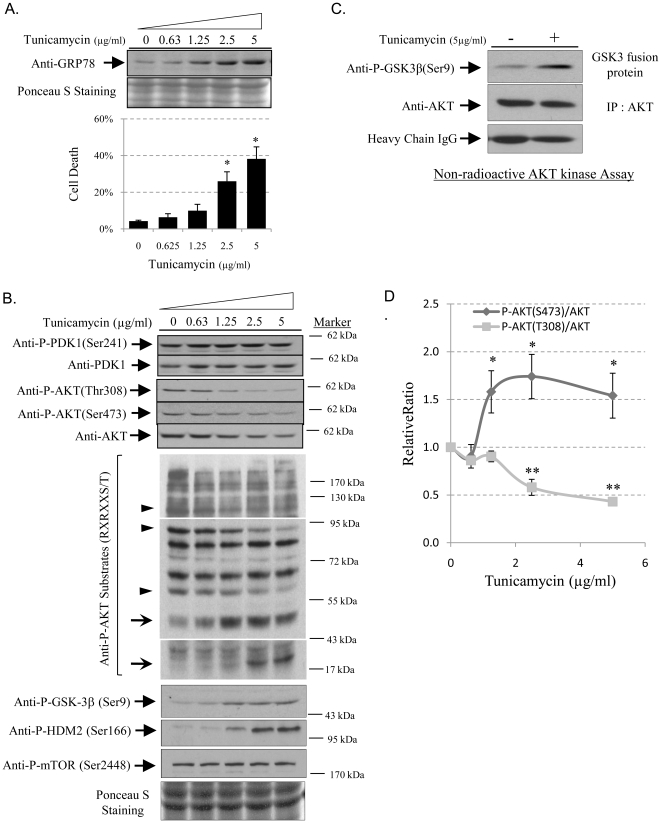
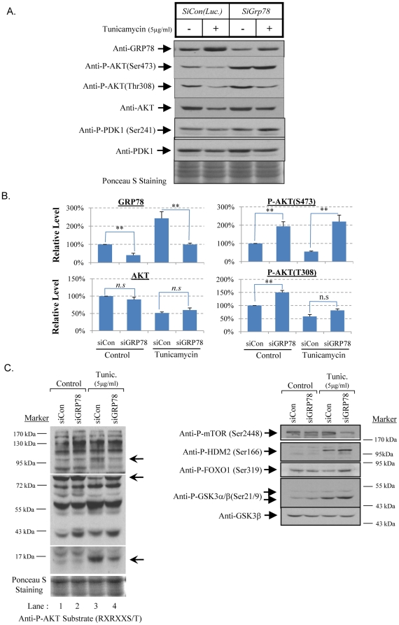
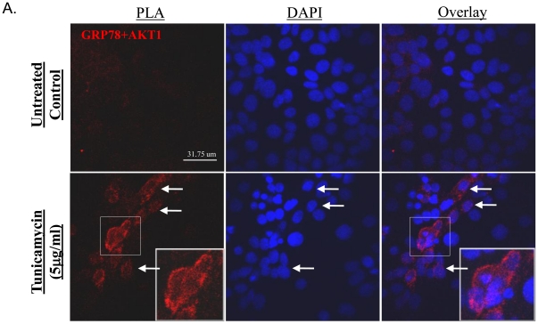
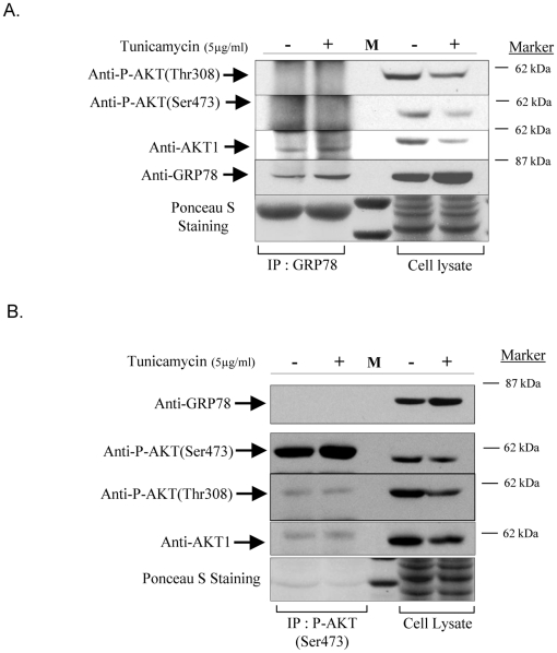
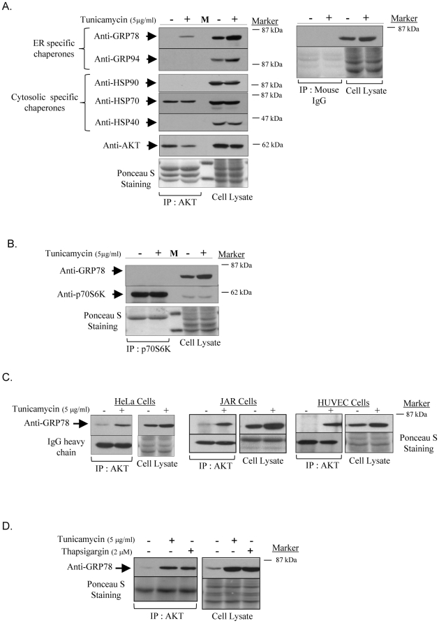
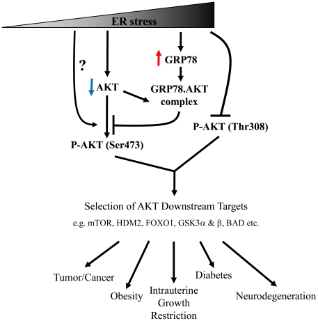
Endoplasmic reticulum (ER) stress is a common factor in the pathophysiology of diverse human diseases that are characterised by contrasting cellular behaviours, from proliferation in cancer to apoptosis in neurodegenerative disorders. Coincidently, dysregulation of AKT/PKB activity, which is the central regulator of cell growth, proliferation and survival, is often associated with the same diseases. Here, we demonstrate that ER stress modulates AKT substrate specificity in a severity-dependent manner, as shown by phospho-specific antibodies against known AKT targets. ER stress also reduces both total and phosphorylated AKT in a severity-dependent manner, without affecting activity of the upstream kinase PDK1. Normalisation to total AKT revealed that under ER stress phosphorylation of Thr308 is suppressed while that of Ser473 is increased. ER stress induces GRP78, and siRNA-mediated knock-down of GRP78 enhances phosphorylation at Ser473 by 3.6 fold, but not at Thr308. Substrate specificity is again altered. An in-situ proximity ligation assay revealed a physical interaction between GRP78 and AKT at the plasma membrane of cells following induction of ER stress. Staining was weak in cells with normal nuclear morphology but stronger in those displaying rounded, condensed nuclei. Co-immunoprecipitation of GRP78 and P-AKT(Ser473) confirmed the immuno-complex consists of non-phosphorylated AKT (Ser473 and Thr308). The interaction is likely specific as AKT did not bind to all molecular chaperones, and GRP78 did not bind to p70 S6 kinase. These findings provide one mechanistic explanation for how ER stress contributes to human pathologies demonstrating contrasting cell fates via modulation of AKT signalling.
Overview
The claims and statistics of this paper haven’t been checked yet.
Run the check — free, no account needed. It reads the open-access text and checks the sources it cites.
Integrity signals
Registry
Cites
Statistics
Figures
Verification
Claims the paper makes
The claims and statistics of this paper haven’t been checked yet.
Run the check — free, no account needed. It reads the open-access text and checks the sources it cites.
Claims other papers make about this one
Each is checked against this paper’s own text: does it say what the citing paper says it does?
In the citing paper
“TSC1/2 also acts on mTORC2, which in turn regulates the activity and substrate specificity of AKT through phosphorylation (597).”
From Placental Origins of Chronic Disease (2016) · cites this paper as [597]
Self-check
Percentages, sums and reported differences re-computed from the paper’s own numbers.
The self-check needs the paper’s full text.
Citations
Cites
Phosphorylation and regulation of Akt/PKB by the rictor-mTOR complex
Sarbassov, D. D., Guertin, D. A., Ali, S. M., & Sabatini, D. M. (2005) · Science · doi:10.1126/science.1106148 · Cited by 6,216
AKT/PKB signaling: navigating downstream
Manning, B. D., & Cantley, L. C. (2007) · Cell · doi:10.1016/j.cell.2007.06.009 · Cited by 6,115
Inhibition of glycogen synthase kinase-3 by insulin mediated by protein kinase B
Cross, D. A. E., Alessi, D. R., Cohen, P., Andjelkovich, M., & Hemmings, B. A. (1995) · Nature · doi:10.1038/378785a0 · Cited by 5,244
Mechanism of activation of protein kinase B by insulin and IGF‐1.
Alessi, D. R., Andjelković, M., Caudwell, B., Cron, P. D., MORRICE, N. A., Cohen, P., & Hemmings, B. A. (1996) · The EMBO Journal · doi:10.1002/j.1460-2075.1996.tb01045.x · Cited by 3,014
Alessi, D. R., James, S. R., Downes, C. P., Holmes, A. B., Gaffney, P. R. J., Reese, C. B., & Cohen, P. (1997) · Current Biology · doi:10.1016/s0960-9822(06)00122-9 · Cited by 2,874
Guertin, D. A., Stevens, D. M., Thoreen, C. C., Burds, A. A., Kalaany, N. Y., Moffat, J., Brown, M., Fitzgerald, K. J., & Sabatini, D. M. (2006) · Developmental Cell · doi:10.1016/j.devcel.2006.10.007 · Cited by 1,467
Direct Regulation of the Akt Proto-Oncogene Product by Phosphatidylinositol-3,4-bisphosphate
Franke, T. F., Kaplan, D. R., Cantley, L. C., & Toker, A. (1997) · Science · doi:10.1126/science.275.5300.665 · Cited by 1,462
Jacinto, E., Facchinetti, V., Liu, D., Soto, N., Wei, S., Jung, S. Y., Huang, Q., Qin, J., & Su, B. (2006) · Cell · doi:10.1016/j.cell.2006.08.033 · Cited by 1,407
A trip to the ER: coping with stress
Rutkowski, D. T., & Kaufman, R. J. (2003) · Trends in Cell Biology · doi:10.1016/j.tcb.2003.11.001 · Cited by 1,375
The nuts and bolts of AGC protein kinases
Pearce, L. R., Komander, D., & Alessi, D. R. (2010) · Nature Reviews Molecular Cell Biology · doi:10.1038/nrm2822 · Cited by 1,294
Cited by
Sorted by citations
ER stress: Autophagy induction, inhibition and selection
Rashid, H.-O., Yadav, R. K., Kim, H., & Chae, H. (2015) · Autophagy · doi:10.1080/15548627.2015.1091141 · Cited by 818
Placental Origins of Chronic Disease
Burton, G. J., Fowden, A. L., & Thornburg, K. L. (2016) · Physiological Reviews · doi:10.1152/physrev.00029.2015 · Cited by 717
Self-citationWhere is mTOR and what is it doing there?
Betz, C., & Hall, M. N. (2013) · The Journal of Cell Biology · doi:10.1083/jcb.201306041 · Cited by 531
An overview of technical considerations for Western blotting applications to physiological research
Bass, J. J., Wilkinson, D. J., Rankin, D., Phillips, B. E., Szewczyk, N. J., Smith, K., & Atherton, P. J. (2016) · Scandinavian Journal of Medicine and Science in Sports · doi:10.1111/sms.12702 · Cited by 432
Stress-induced self-cannibalism: on the regulation of autophagy by endoplasmic reticulum stress
Deegan, S., Saveljeva, S., Gorman, A. M., & Samali, A. (2012) · Cellular and Molecular Life Sciences · doi:10.1007/s00018-012-1173-4 · Cited by 279
Molecular Mechanisms of Glucocorticoid-Induced Insulin Resistance
Beaupere, C., Liboz, A., Fève, B., Blondeau, B., & Guillemain, G. (2021) · International Journal of Molecular Sciences · doi:10.3390/ijms22020623 · Cited by 271
Roberts, R. F., Wade‐Martins, R., & Alegre‐Abarrategui, J. (2015) · Brain · doi:10.1093/brain/awv040 · Cited by 280
Dimasuay, K. G., Aitken, E. H., Rosario, F. J., Njie, M., Glazier, J. D., Rogerson, S. J., Fowkes, F. J. I., Beeson, J. G., Powell, T. L., Jansson, T., et al. (2017) · BMC Medicine · doi:10.1186/s12916-016-0759-3 · Cited by 193
Ghosh, A., Tyson, T., George, S., Hildebrandt, E. N., Steiner, J. A., Madaj, Z., Schulz, E., Machiela, E., McDonald, W. G., Galvis, M. L. E., et al. (2016) · Science Translational Medicine · doi:10.1126/scitranslmed.aag2210 · Cited by 193
Shen, Y., Guerra‐Librero, A., Fernández-Gil, B. I., Florido, J., García‐López, S., Martínez-Ruiz, L., Mendivil‐Perez, M., Soto‐Mercado, V., Acuña-Castroviejo, D., Ortega‐Arellano, H. F., et al. (2017) · Journal of Pineal Research · doi:10.1111/jpi.12461 · Cited by 175
Chaudhari, S. N., & Kipreos, E. T. (2017) · Nature Communications · doi:10.1038/s41467-017-00274-4 · Cited by 147
Szymonowicz, K., Oeck, S., Malewicz, N. M., & Jendrossek, V. (2018) · Cancers · doi:10.3390/cancers10030078 · Cited by 135
Xu, Z., Li, C., Liu, Q.-L., Li, P., & Yang, H. Q. (2018) · International Journal of Molecular Sciences · doi:10.3390/ijms19113658 · Cited by 132
Wiersma, M., Meijering, R. A. M., Qi, X., Zhang, D., Liu, T., Hoogstra‐Berends, F., Sibon, O. C. M., Henning, R. H., Nattel, S., & Brundel, B. J. J. M. (2017) · Journal of the American Heart Association · doi:10.1161/jaha.117.006458 · Cited by 129
Sood, A., Salih, S., Roh, D. S., Lacharme‐Lora, L., Parry, M., Hardiman, B., Keehan, R., Grümmer, R., Winterhager, E., Gokhale, P. J., et al. (2011) · Nature Nanotechnology · doi:10.1038/nnano.2011.188 · Cited by 122
Yung, H. W., Cox, M., van Patot, M. T., & Burton, G. J. (2012) · The FASEB Journal · doi:10.1096/fj.11-190082 · Cited by 112
Self-citationGifford, J. B., Huang, W., Zeleniak, A. E., Hindoyan, A. A., Wu, H., Donahue, T. R., & Hill, R. (2016) · Molecular Cancer Therapeutics · doi:10.1158/1535-7163.mct-15-0774 · Cited by 104
Ren, H., Zhai, W., Lu, X., & Wang, G. (2021) · Frontiers in Aging Neuroscience · doi:10.3389/fnagi.2021.691881 · Cited by 102
Li, N., Cui, J., Duan, X., Chen, H., & Fan, F. (2012) · Investigative Ophthalmology & Visual Science · doi:10.1167/iovs.11-8670 · Cited by 93
Redaelli, S., Ceccon, M., Zappa, M., Sharma, G. G., Mastini, C., Mauri, M., Nigoghossian, M., Massimino, L., Cordani, N., Farina, F., et al. (2018) · Cancer Research · doi:10.1158/0008-5472.can-18-1867 · Cited by 92
Impaired Mitochondrial ATP Production Downregulates Wnt Signaling via ER Stress Induction
Costa, R., Peruzzo, R., Bachmann, M., Montà, G. D., Vicario, M., Santinon, G., Mattarei, A., Moro, E., Quintana–Cabrera, R., Scorrano, L., et al. (2019) · Cell Reports · doi:10.1016/j.celrep.2019.07.050 · Cited by 90
Hurtado, E., Cilleros‐Mañé, V., Nadal, L., Simó, A., Obis, T., García, N., Santafé, M. M., Tomàs, M., Halievski, K., Jordan, C. L., et al. (2017) · Frontiers in Molecular Neuroscience · doi:10.3389/fnmol.2017.00147 · Cited by 90
Koga, T., Suico, M. A., Shimasaki, S., Watanabe, E., Kai, Y., Koyama, K., Omachi, K., Morino‐Koga, S., Sato, T., Shuto, T., et al. (2015) · Journal of Biological Chemistry · doi:10.1074/jbc.m115.664169 · Cited by 86
Wang, Y., Li, J., Nakahata, S., & Iha, H. (2024) · International Journal of Molecular Sciences · doi:10.3390/ijms25137346 · Cited by 83
HSPA5 negatively regulates lysosomal activity through ubiquitination of MUL1 in head and neck cancer
Kim, S., Kim, H. J., Kim, H., Kim, D. H., Han, J. H., Byeon, H. K., Lee, K., & Kim, C. (2017) · Autophagy · doi:10.1080/15548627.2017.1414126 · Cited by 79
Wen, N., Guo, B., Zheng, H., Xu, L., Liang, H., Wang, Q., Wang, D., Chen, X., Zhang, S., Li, Y., et al. (2019) · International Journal of Oncology · doi:10.3892/ijo.2019.4863 · Cited by 79
Apelin alleviates diabetes-associated endoplasmic reticulum stress in the pancreas of Akita mice
Chen, H., Zheng, C., Zhang, X., Li, J., Li, J., Zheng, L., & Huang, K. (2011) · Peptides · doi:10.1016/j.peptides.2011.06.025 · Cited by 79
Luo, W., Hawse, W. F., Conter, L. J., Trivedi, N. H., Weisel, F. J., Wikenheiser, D. J., Cattley, R. T., & Shlomchik, M. J. (2019) · Nature Immunology · doi:10.1038/s41590-019-0376-3 · Cited by 80
Matheson, H., Veerbeek, J. H. W., Charnock‐Jones, D. S., Burton, G. J., & Yung, H. W. (2016) · The Journal of Physiology · doi:10.1113/jp271073 · Cited by 71
Self-citationFan, Y., Liu, K., Wang, Q., Ruan, Y., Ye, W., & Zhang, Y. (2014) · Experimental Eye Research · doi:10.1016/j.exer.2014.05.004 · Cited by 65
Li, M., Wu, C., Muhammad, J. S., Yan, D., Tsuneyama, K., Hatta, H., Cui, Z., & Inadera, H. (2020) · Redox Biology · doi:10.1016/j.redox.2020.101632 · Cited by 65
Activin and TGFβ use diverging mitogenic signaling in advanced colon cancer
Bauer, J. I., Özden, Ö., Akagi, N., Carroll, T. P., Principe, D. R., Staudacher, J. J., Spehlmann, M. E., Eckmann, L., Grippo, P. J., & Jung, B. H. (2015) · Molecular Cancer · doi:10.1186/s12943-015-0456-4 · Cited by 63
Somarajan, B. I., Khanday, M. A., & Mallick, B. N. (2016) · Frontiers in Neurology · doi:10.3389/fneur.2016.00025 · Cited by 63
Phosphorylation-dependent substrate selectivity of protein kinase B (AKT1)
Balasuriya, N., Davey, N. E., Johnson, J. L., Liu, H., Biggar, K. K., Cantley, L. C., Li, S. S. ‐., & O’Donoghue, P. (2020) · Journal of Biological Chemistry · doi:10.1074/jbc.ra119.012425 · Cited by 61
Sharma, P., Yamada, S., Lualdi, M., Dasso, M., & Kuehn, M. R. (2013) · Cell Reports · doi:10.1016/j.celrep.2013.04.016 · Cited by 64
Tscheschner, H., Meinhardt, E., Schlegel, P., Jungmann, A., Lehmann, L., Müller, O. J., Most, P., Katus, H. A., & Raake, P. W. J. (2019) · PLoS ONE · doi:10.1371/journal.pone.0215992 · Cited by 60
Mathur, A., Elmageed, Z. Y. A., Liu, X., Kostochka, M. L., Zhang, H., Abdel‐Mageed, A. B., & Mondal, D. (2014) · PLoS ONE · doi:10.1371/journal.pone.0103109 · Cited by 54
Brefeldin A inhibits colorectal cancer growth by triggering Bip/Akt‐regulated autophagy
Zhou, L., Gao, W. Q., Wang, K., Huang, Z., Zhang, L., Zhang, Z., Zhou, J., Nice, E. C., & Huang, C. (2019) · The FASEB Journal · doi:10.1096/fj.201801983r · Cited by 54
Kim, S., Grenert, J. P., Patterson, C., & Correia, M. A. (2016) · Scientific Reports · doi:10.1038/srep29423 · Cited by 51
Tanti, G. K., & Goswami, S. K. (2014) · Free Radical Biology and Medicine · doi:10.1016/j.freeradbiomed.2014.07.009 · Cited by 50
Haskovic, M., Coelho, A. I., Bierau, J., Vanoevelen, J. M., Steinbusch, L. K. M., Zimmermann, L. J. I., Villamor-Martínez, E., Berry, G. T., & Rubio‐Gozalbo, M. E. (2019) · Journal of Inherited Metabolic Disease · doi:10.1002/jimd.12202 · Cited by 50
Bao, X., Zheng, W., Sugi, N. H., Agarwala, K. L., Xu, Q., Wang, Z., TenDyke, K., Lee, W., Parent, L. A., Li, W., et al. (2015) · Cancer Biology & Therapy · doi:10.1080/15384047.2015.1019184 · Cited by 48
Enos, R. T., Velázquez, K. T., & Murphy, E. A. (2014) · The Journal of Nutritional Biochemistry · doi:10.1016/j.jnutbio.2014.01.011 · Cited by 48
Cojohari, O., Peppenelli, M. A., & Chan, G. C. T. (2016) · Journal of Virology · doi:10.1128/jvi.00214-16 · Cited by 45
ER stress and cancer: The FOXO forkhead transcription factor link
Alasiri, G. A., Fan, L. Y.-N., Zona, S., Goldsbrough, I., Ke, H., Auner, H. W., & Lam, E. W. ‐. (2017) · Molecular and Cellular Endocrinology · doi:10.1016/j.mce.2017.05.027 · Cited by 45
Fan, Y., Liu, K., Wang, Q., Ruan, Y., Zhang, Y., & Ye, W. (2014) · Europe PMC (PubMed Central) · Cited by 43
PI3K inhibitors protect against glucocorticoid-induced skin atrophy
Agarwal, S., Mirzoeva, S., Readhead, B., Dudley, J. T., & Budunova, I. V. (2019) · EBioMedicine · doi:10.1016/j.ebiom.2019.01.055 · Cited by 42
Jiang, D., Li, D., Cao, L., Wang, L., Zhu, S., Xu, T., Wang, C., & Pan, D. (2014) · PLoS ONE · doi:10.1371/journal.pone.0092398 · Cited by 41
Pelizzaro‐Rocha, K. J., de Jesus, M. B., Ruela-de-Sousa, R. R., Nakamura, C. V., Reis, F. S., de Fátima, Â., & Ferreira‐Halder, C. V. (2013) · Biochimica et Biophysica Acta (BBA) - Molecular Cell Research · doi:10.1016/j.bbamcr.2013.07.010 · Cited by 40
Parker, A. L., Turner, N. A., McCarroll, J. A., & Kavallaris, M. (2016) · Carcinogenesis · doi:10.1093/carcin/bgw058 · Cited by 40
Schötz, U., Balzer, V., Brandt, F.-W., Ziemann, F., Subtil, F. S. B., Rieckmann, T., Köcher, S., Engenhart‐Cabillic, R., Dikomey, E., Wittig, A., et al. (2020) · Cancers · doi:10.3390/cancers12020467 · Cited by 41
Billah, M., Ridiandries, A., Allahwala, U. K., Mudaliar, H., Dona, A. C., Hunyor, S. N., Khachigian, L. M., & Bhindi, R. (2020) · International Journal of Molecular Sciences · doi:10.3390/ijms21051692 · Cited by 41
Borsting, E., Patel, S. V., Declèves, A., Lee, S. J., Rahman, Q. M., Akira, S., Satriano, J. A., Sharma, K., Vallon, V., & Cunard, R. A. (2014) · Journal of the American Society of Nephrology · doi:10.1681/asn.2013070811 · Cited by 40
Bioactive Marine Drugs and Marine Biomaterials for Brain Diseases
Grosso, C., Valentão, P., Ferreres, F., & Andrade, P. (2014) · Marine Drugs · doi:10.3390/md12052539 · Cited by 35
Quantum Dot- Conjugated Anti-GRP78 scFv Inhibits Cancer Growth in Mice
Xu, W., Liu, L., Brown, N. J., Christian, S., & HORNBY, D. P. (2012) · Molecules · doi:10.3390/molecules17010796 · Cited by 39
Chirumamilla, C. S., Perez-Novo, C. A., Ostade, X. V., & Berghe, W. V. (2017) · Proceedings of The Nutrition Society · doi:10.1017/s0029665116002937 · Cited by 38
HCMV modulation of cellular PI3K/AKT/mTOR signaling: New opportunities for therapeutic intervention?
Altman, A. M., Mahmud, J., Nikolovska‐Coleska, Z., & Chan, G. C. T. (2019) · Antiviral Research · doi:10.1016/j.antiviral.2019.01.009 · Cited by 37
Siri, M., Dastghaib, S., Zamani, M., Rahmani-Kukia, N., Geraylow, K. R., Fakher, S., Keshvarzi, F., Mehrbod, P., Ahmadi, M., Mokarram, P., et al. (2021) · International Journal of Molecular Sciences · doi:10.3390/ijms22115992 · Cited by 37
The Interplay Between Autophagy and Regulated Necrosis
Zhang, L., Cui, T., & Wang, X. (2022) · Antioxidants and Redox Signaling · doi:10.1089/ars.2022.0110 · Cited by 38
Gao, Y., Wang, C., Jiang, D. M., An, G., Jin, F. J., Zhang, J., Han, G., Cui, C., & Jiang, P. (2022) · Frontiers in Cell and Developmental Biology · doi:10.3389/fcell.2022.994037 · Cited by 37
Jellyman, J. K., Martin‐Gronert, M. S., Cripps, R. L., Giussani, D. A., Ozanne, S. E., Shen, Q. W., Du, ., Fowden, A. L., & Forhead, A. J. (2012) · PLoS ONE · doi:10.1371/journal.pone.0052363 · Cited by 37
Modulation of the Akt Pathway Reveals a Novel Link with PERK/eIF2α, which Is Relevant during Hypoxia
Blaustein, M., Pérez-Munizaga, D., Sánchez, M. A., Urrutia, C., Grande, A., Risso, G., Srebrow, A., Alfaro, J., & Colman‐Lerner, A. (2013) · PLoS ONE · doi:10.1371/journal.pone.0069668 · Cited by 36
Peppenelli, M. A., Arend, K. C., Cojohari, O., Moorman, N. J., & Chan, G. C. T. (2016) · Journal of Virology · doi:10.1128/jvi.02879-15 · Cited by 36
Hu, S., Hu, H., Wang, R., He, H., & Shui, H. (2021) · International Urology and Nephrology · doi:10.1007/s11255-021-02836-4 · Cited by 35
RetractedBalakrishnan, B., Chen, W., Tang, M., Huang, X., Demirbas, D., Siddiqi, A. M., Berry, G. T., & Lai, K. (2016) · Biochemical and Biophysical Research Communications · doi:10.1016/j.bbrc.2016.01.036 · Cited by 35
Lei, Y., Huang, Y. J., Wen, X., Yin, Z., Zhang, Z., & Klionsky, D. J. (2022) · Antioxidants · doi:10.3390/antiox11020304 · Cited by 34
Lin, M., Chen, S., Huang, R.-Y., Lu, Y.-C., Liao, Y.-R., Reddy, M. V. B., Lee, C.-C., & Wu, T.-S. (2014) · APOPTOSIS · doi:10.1007/s10495-014-1031-y · Cited by 33
Lorenzon, A. R., Yung, H. W., Burton, G. J., & Bevilacqua, E. M. A. F. (2019) · Biochimica et Biophysica Acta (BBA) - Molecular Basis of Disease · doi:10.1016/j.bbadis.2019.01.012 · Cited by 32
Self-citationZhang, L., Kim, S. B., Luitel, K., & Shay, J. W. (2018) · Molecular Cancer Therapeutics · doi:10.1158/1535-7163.mct-17-0887 · Cited by 32
Gu, L., Yi, Z., Zhang, Y., Ma, Z., Zhu, Y., & Gao, J. (2017) · Oncotarget · doi:10.18632/oncotarget.16046 · Cited by 31
Reversal of aberrant PI3K/Akt signaling by Salubrinal in a GalT-deficient mouse model
Balakrishnan, B., Nicholas, C., Siddiqi, A. M., Chen, W., Bales, E. S., Feng, M., Johnson, J., & Lai, K. (2017) · Biochimica et Biophysica Acta (BBA) - Molecular Basis of Disease · doi:10.1016/j.bbadis.2017.08.023 · Cited by 31
Chen, Y., Tao, S., Xu, Z., Yin, W., Zhang, Y.-L., Yin, J., Gao, Y., & Zhang, C. (2017) · Oncotarget · doi:10.18632/oncotarget.16075 · Cited by 30
Soundararajan, R., Hernández‐Cuervo, H., Stearns, T. M., Griswold, A. J., Patil, S. S., Fukumoto, J., Narala, V. R., Galam, L., Lockey, R. F., & Kolliputi, N. (2022) · Frontiers in Pharmacology · doi:10.3389/fphar.2022.980723 · Cited by 26
Li, M., Cheng, W., Nie, T., Lai, H., Hu, X., Luo, J., Li, F., & Li, H. (2017) · Biological Trace Element Research · doi:10.1007/s12011-017-1155-3 · Cited by 26
New insights into the role of melatonin in diabetic cardiomyopathy
Huang, K., Luo, X., Zhong, Y., Deng, L., & Feng, J. (2022) · Pharmacology Research & Perspectives · doi:10.1002/prp2.904 · Cited by 24
Pietrafesa, G., Zio, R. D., Scorza, S. I., Armentano, M. F., Pepe, M., Forleo, C., Procino, G., Gerbino, A., Svelto, M., & Carmosino, M. (2023) · Journal of Translational Medicine · doi:10.1186/s12967-023-04170-y · Cited by 23
Thiébaut, P., Delile, E., Coquerel, D., Brunel, J. M., Renet, S., Tamion, F., & Richard, V. J. (2018) · Vascular Pharmacology · doi:10.1016/j.vph.2018.05.011 · Cited by 23
Peppenelli, M. A., Miller, M. J., Altman, A. M., Cojohari, O., & Chan, G. C. T. (2018) · Antiviral Research · doi:10.1016/j.antiviral.2018.07.015 · Cited by 23
Human Cytomegalovirus Glycoprotein-Initiated Signaling Mediates the Aberrant Activation of Akt
Mahmud, J., Miller, M. J., Altman, A. M., & Chan, G. C. T. (2020) · Journal of Virology · doi:10.1128/jvi.00167-20 · Cited by 22
Sáez, P. J., Villalobos-Labra, R., Westermeier, F., Sobrevía, L., & Farías, M. (2014) · Frontiers in Pharmacology · doi:10.3389/fphar.2014.00189 · Cited by 22
Endoplasmic Reticulum Stress and Autophagy
Kabir, M. F., Kim, H., & Chae, H. (2019) · IntechOpen eBooks · doi:10.5772/intechopen.81381 · Cited by 21
Cojohari, O., Mahmud, J., Altman, A. M., Peppenelli, M. A., Miller, M. J., & Chan, G. C. T. (2020) · Viruses · doi:10.3390/v12060652 · Cited by 21
Genotoxic stress-mediated cell cycle activities for the decision of cellular fate
Erol, A. (2011) · Cell Cycle · doi:10.4161/cc.10.19.17460 · Cited by 21
2-Acetyl-5-tetrahydroxybutyl imidazole (THI) protects 661W cells against oxidative stress
Fabiani, C., Zulueta, A., Bonezzi, F., Casas, J., Ghidoni, R., Signorelli, P., & Caretti, A. (2017) · Naunyn-Schmiedeberg s Archives of Pharmacology · doi:10.1007/s00210-017-1374-3 · Cited by 20
Landa-Galván, H. V., Ríos‐Castro, E., Romero‐García, T., Rueda, A., & Olivares-Reyes, J. A. (2020) · PLoS ONE · doi:10.1371/journal.pone.0228115 · Cited by 20
Adult lead exposure increases blood-retinal permeability: A risk factor for retinal vascular disease
Shen, X., Huang, P., Fox, D. A., Yan, L., Zhao, Z., Wang, W., Wang, J.-Y., Liu, X.-Q., Chen, J., & Luo, W. (2016) · NeuroToxicology · doi:10.1016/j.neuro.2016.09.013 · Cited by 19
Pujari, R., Jose, J., Bhavnani, V., Kumar, N., Shastry, P., & Pal, J. K. (2016) · The International Journal of Biochemistry & Cell Biology · doi:10.1016/j.biocel.2016.05.021 · Cited by 19
Endoplasmic reticulum stress-induced cell death mechanism
Madkour, L. H. (2020) · Elsevier eBooks · doi:10.1016/b978-0-12-822481-6.00012-8 · Cited by 18
García, S., Saldana-Caboverde, A., Anwar, M. R., Raval, A. P., Nissanka, N., Pinto, M., Moraes, C. T., & Díaz, F. (2021) · Human Molecular Genetics · doi:10.1093/hmg/ddab282 · Cited by 18
The hypergonadotropic hypogonadism conundrum of classic galactosemia
Derks, B., Rivera‐Cruz, G., Hagen-Lillevik, S., Vos, E. N., Demirbas, D., Lai, K., Treacy, E. P., Levy, H. L., Wilkins-Haug, L. E., Rubio‐Gozalbo, M. E., et al. (2022) · Human Reproduction Update · doi:10.1093/humupd/dmac041 · Cited by 18
Zhao, T., Jiang, T., Li, X., Chang, S., Sun, Q., Kong, F., Kong, X., Wei, F., He, J., Hao, J., et al. (2023) · Clinical Cancer Research · doi:10.1158/1078-0432.ccr-23-1143 · Cited by 17
Wu, L., Guo, Y., Zhang, Q.-H., Xiang, M.-Q., Deng, W., Ye, Y., Pu, Z., Feng, J., & Huang, G. (2014) · International Journal of Molecular Sciences · doi:10.3390/ijms15010525 · Cited by 16
GRP78 Activity Moderation as a Therapeutic Treatment against Obesity
Pan, D., Yang, Y., Nong, A., Tang, Z., & Li, Q. X. (2022) · International Journal of Environmental Research and Public Health · doi:10.3390/ijerph192315965 · Cited by 15
Rumman, M., Buck, S. A., Polin, L., Dzinic, S. H., Boerner, J. L., & Winer, I. S. (2021) · Cancer Medicine · doi:10.1002/cam4.3858 · Cited by 14
Labor inhibits placental mechanistic target of rapamycin complex 1 signaling
Lager, S., Aye, I. L. M. H., Gaccioli, F., Ramirez, V. I., Jansson, T., & Powell, T. L. (2014) · Placenta · doi:10.1016/j.placenta.2014.10.006 · Cited by 15
Gharbiya, M., Bruscolini, A., Sacchetti, M., Rosso, P., Carito, V., Segatto, M., Fico, E., Tirassa, P., & Lambìase, A. (2018) · Journal of Cellular Physiology · doi:10.1002/jcp.26806 · Cited by 14
Effects of CK2β subunit down-regulation on Akt signalling in HK-2 renal cells
Alcaraz, E., Vilardell, J., Borgo, C., Sarró, E., Plana, M., Marin, O., Pinna, L. A., Bayascas, J. R., Meseguer, A., Salvi, M., et al. (2020) · PLoS ONE · doi:10.1371/journal.pone.0227340 · Cited by 14
Heat Shock Proteins: Potential Modulators and Candidate Biomarkers of Peripartum Cardiomyopathy
Chakafana, G., Spracklen, T. F., Kamuli, S., Zininga, T., Shonhai, A., Ntusi, N., & Sliwa, K. S. (2021) · Frontiers in Cardiovascular Medicine · doi:10.3389/fcvm.2021.633013 · Cited by 14
mTORC2 Is Activated under Hypoxia and Could Support Chronic Myeloid Leukemia Stem Cells
Panuzzo, C., Pironi, L., Maglione, A., Rocco, S., Stanga, S., Riganti, C., Kopecka, J., Ali, M. S., Pergolizzi, B., Bracco, E., et al. (2023) · International Journal of Molecular Sciences · doi:10.3390/ijms24021234 · Cited by 14
Yilmaz, T. E., Işıldar, B., Gezer, A., Kankaya, D., Ersöz, C. C., Kayisli, U. A., & Güzel, E. (2020) · Reproductive BioMedicine Online · doi:10.1016/j.rbmo.2020.09.012 · Cited by 13
Metadata
- Authors
- Yung, H. W., Charnock-Jones, D. S., & Burton, G. J.
- Affiliations
- University of Cambridge (GB)
- Corresponding author
- Burton, Graham J.
- Venue
- PLoS ONE
- Publisher
- Public Library of Science (PLoS)
- Published
- 2011-03-21
- Volume
- 6
- Issue
- 3
- Pages
- e17894
- ISSN
- 1932-6203 (ISSN-L)
- PMC
- PMC3061875
- OpenAlex
- W2058236541
- Cited by
- 158
- References
- 41
- Citations by year
- 2026: 6 · 2025: 5 · 2024: 9 · 2023: 7 · 2022: 12 · 2021: 10 · 2020: 15 · 2019: 14 · 2018: 8 · 2017: 16 · 2016: 12 · 2015: 10 · 2014: 15 · 2013: 7 · 2012: 9
- FWCI
- 5.21
- Type
- journal article
- Language
- en
- Open access
- gold
- doi.org
- Locations
- PLoS ONE (publishedVersion, cc-by)
- PubMed Central (publishedVersion, cc-by)
- PubMed (publishedVersion)
- Apollo (University of Cambridge) (submittedVersion, cc-by)
- DOAJ (DOAJ: Directory of Open Access Journals) (submittedVersion, cc-by-sa)
- Figshare (submittedVersion, cc-by)
- Apollo (cc-by)
- Funders
- Wellcome Trust (084804, 084804/2/08/Z, 084804/Z/08/Z)
- Topics
- Life Sciences › Biochemistry, Genetics and Molecular Biology › Cell Biology › Endoplasmic Reticulum Stress and Disease
- Life Sciences › Biochemistry, Genetics and Molecular Biology › Molecular Biology › PI3K/AKT/mTOR signaling in cancer
- Life Sciences › Biochemistry, Genetics and Molecular Biology › Cell Biology › Cellular transport and secretion
- Keywords
- endoplasmic reticulum stress, Akt phosphorylation, GRP78, PDK1, plasma membrane, apoptosis, cell proliferation, p70 S6 kinase, molecular chaperones, proximity ligation assay
- MeSH terms
- Endoplasmic Reticulum Chaperone BiP, Endoplasmic Reticulum, Heat-Shock Proteins, Humans, Phosphorylation, Serine, Substrate Specificity, Threonine, Down-Regulation, Proto-Oncogene Proteins c-akt
About these checks
Registry, retraction, Cites and statistics results are computed, not judged, with one exception: a printed reference that no index lists is read once by a model before it counts as a citation error. Claims are extracted by a model and kept only when their quote appears verbatim in the text; each cited source is then checked, with a model’s help, for whether it says what the claim relies on. Those verdicts can be wrong — read the claim’s quote and the cited source before relying on one.
Statistics and arithmetic are recomputed, not judged.
Confidence bands are shown only for fields calibrated on our annotated test set. None is calibrated yet, so no band is shown.
What counts as a claim ›How we measure confidence ›How we check this ›
Are you an author, or do you see a problem with this page? Request a review
