

The DOI matches its Crossref record; 32 of the 32 references it cites were found in a registry or an index.
Wnt3a Induces Myofibroblast Differentiation by Upregulating TGF-β Signaling Through SMAD2 in a β-Catenin-Dependent Manner
Open accessAbstract
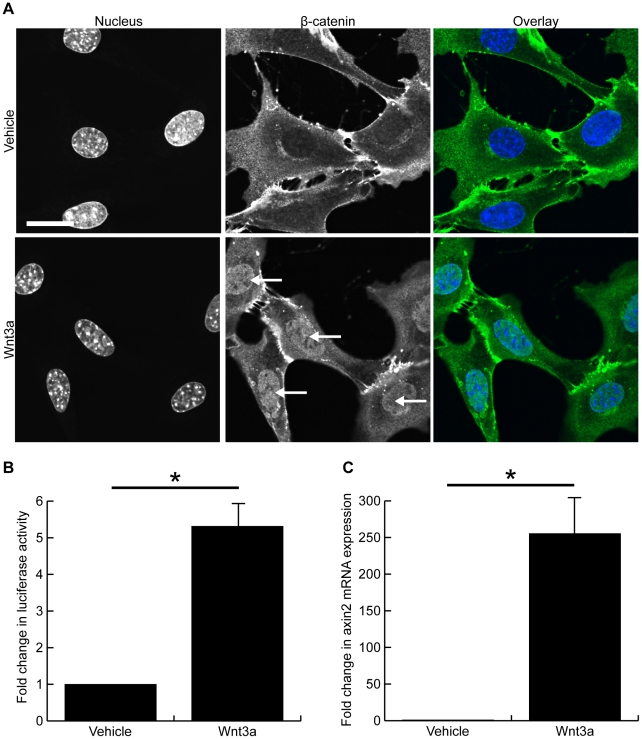
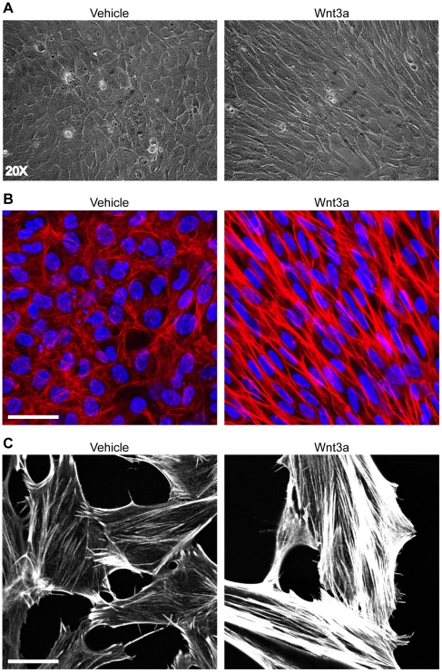
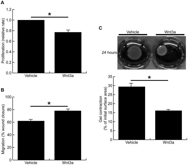
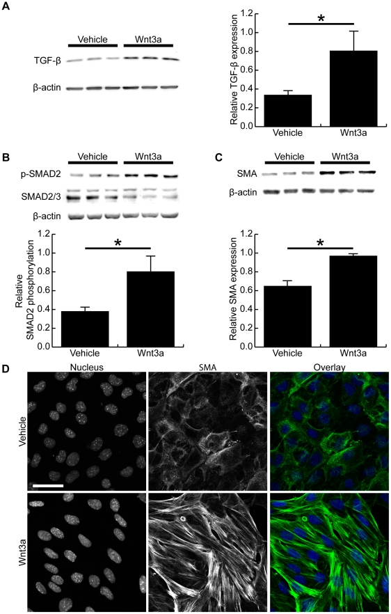
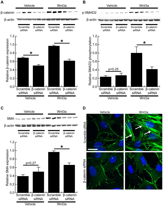
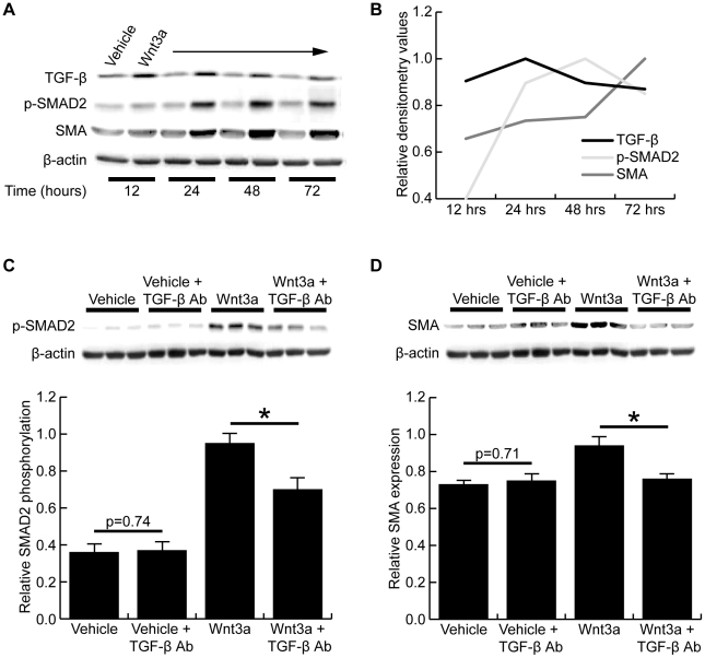
Growing evidence suggests the Wnt family of secreted glycoproteins and their associated signaling pathways, linked to development, are recapitulated during wound repair and regeneration events. However, the role of the Wnt pathway in such settings remains unclear. In the current study, we treated mouse fibroblasts with 250 ng/mL of recombinant Wnt3a for 72 hours and examined its affect on cell morphology and function. Wnt3a induced a spindle-like morphology in fibroblasts characterized by the increased formation of stress fibres. Wnt3a decreased the proliferation of fibroblasts, but significantly increased cell migration as well as fibroblast-mediated contraction of a collagen lattice. Wnt3a significantly increased the expression of TGF-β and its associated signaling through SMAD2. Consistent with this, we observed significantly increased smooth muscle α-actin expression and incorporation of this contractile protein into stress fibres following Wnt3a treatment. Knockdown of β-catenin using siRNA reversed the Wnt3a-induced smooth muscle α-actin expression, suggesting these changes were dependent on canonical Wnt signaling through β-catenin. Neutralization of TGF-β with a blocking antibody significantly inhibited the Wnt3a-induced smooth muscle α-actin expression, indicating these changes were dependent on the increased TGF-β signaling. Collectively, this data strongly suggests Wnt3a promotes the formation of a myofibroblast-like phenotype in cultured fibroblasts, in part, by upregulating TGF-β signaling through SMAD2 in a β-catenin-dependent mechanism. As myofibroblasts are critical regulators of wound healing responses, these findings may have important implications for our understanding of normal and aberrant injury and repair events.
Overview
The claims and statistics of this paper haven’t been checked yet.
Run the check — free, no account needed. It reads the open-access text and checks the sources it cites.
Integrity signals
Registry
Cites
Statistics
Figures
Verification
Claims the paper makes
The claims and statistics of this paper haven’t been checked yet.
Run the check — free, no account needed. It reads the open-access text and checks the sources it cites.
Claims other papers make about this one
Each is checked against this paper’s own text: does it say what the citing paper says it does?
In the citing paper
“The synergistic crosstalk between TGFβ and Wnt signaling in the myofibroblast activation is documented as well as the inhibitory role of SFRP2 in the TGFβ-dependent myofibroblast formation and post-inflammatory fibrosis [138,139].”
From Cellular and Molecular Mechanisms in the Pathogenesis of Classical, Vascular, and Hypermobile Ehlers‒Danlos Syndromes (2019) · cites this paper as [138]
Self-check
Percentages, sums and reported differences re-computed from the paper’s own numbers.
The self-check needs the paper’s full text.
Citations
Cites
Wnt/β-Catenin Signaling in Development and Disease
Clevers, H. (2006) · Cell · doi:10.1016/j.cell.2006.10.018 · Cited by 5,568
The Wnt Signaling Pathway in Development and Disease
Logan, C. Y., & Nusse, R. (2004) · Annual Review of Cell and Developmental Biology · doi:10.1146/annurev.cellbio.20.010403.113126 · Cited by 5,325
Wound Healing--Aiming for Perfect Skin Regeneration
Martin, P. B. (1997) · Science · doi:10.1126/science.276.5309.75 · Cited by 4,842
In vitro scratch assay: a convenient and inexpensive method for analysis of cell migration in vitro
Liang, C.-C., Park, A. Y., & Guan, J.-L. (2007) · Nature Protocols · doi:10.1038/nprot.2007.30 · Cited by 4,696
PERSPECTIVE ARTICLE: Growth factors and cytokines in wound healing
Barrientos, S., Stojadinovic, O., Golinko, M. S., Brem, H., & Tomic‐Canic, M. (2008) · Wound Repair and Regeneration · doi:10.1111/j.1524-475x.2008.00410.x · Cited by 3,583
Convergence of Wnt, ß-Catenin, and Cadherin Pathways
Nelson, W. J., & Nusse, R. (2004) · Science · doi:10.1126/science.1094291 · Cited by 2,537
Desmoulière, A., Geinoz, A., Gabbiani, F., & Gabbiani, G. (1993) · The Journal of Cell Biology · doi:10.1083/jcb.122.1.103 · Cited by 2,269
Hinz, B., Phan, S. H., Thannickal, V. J., Galli, A., Bochaton-Piallat, M.-L., & Gabbiani, G. (2007) · The American Journal of Pathology · doi:10.2353/ajpath.2007.070112 · Cited by 2,068
Caught up in a Wnt storm: Wnt signaling in cancer
Giles, R. H., van Es, J. H., & Clevers, H. (2003) · Biochimica et Biophysica Acta (BBA) - Reviews on Cancer · doi:10.1016/s0304-419x(03)00005-2 · Cited by 1,482
Role of tissue stroma in cancer cell invasion
De Wever, O., & Mareel, M. (2003) · The Journal of Pathology · doi:10.1002/path.1398 · Cited by 1,042
Cited by
Sorted by citations
Frangogiannis, N. G. (2021) · Cardiovascular Research · doi:10.1093/cvr/cvaa324 · Cited by 1,229
Cardiac fibrosis: Cell biological mechanisms, molecular pathways and therapeutic opportunities
Frangogiannis, N. G. (2018) · Molecular Aspects of Medicine · doi:10.1016/j.mam.2018.07.001 · Cited by 888
Signaling pathways and targeted therapy for myocardial infarction
Zhang, Q., Wang, L., Wang, S., Cheng, H., Xu, L., Pei, G., Wang, Y., Fu, C., Jiang, Y., He, C., et al. (2022) · Signal Transduction and Targeted Therapy · doi:10.1038/s41392-022-00925-z · Cited by 882
The role of α-smooth muscle actin in fibroblast-mediated matrix contraction and remodeling
Shinde, A. V., Humeres, C. D., & Frangogiannis, N. G. (2016) · Biochimica et Biophysica Acta (BBA) - Molecular Basis of Disease · doi:10.1016/j.bbadis.2016.11.006 · Cited by 602
An, R., Wang, X., Yang, L., Zhang, J., Wang, N., Xu, F., Hou, Y., Zhang, H., & Zhang, L. (2020) · Toxicology · doi:10.1016/j.tox.2020.152665 · Cited by 462
Signaling in Fibrosis: TGF-β, WNT, and YAP/TAZ Converge
Piersma, B., Bank, R. A., & Boersema, M. (2015) · Frontiers in Medicine · doi:10.3389/fmed.2015.00059 · Cited by 465
Understanding fibrosis in systemic sclerosis: shifting paradigms, emerging opportunities
Bhattacharyya, S., Wei, J., & Varga, J. (2012) · Nature Reviews Rheumatology · doi:10.1038/nrrheum.2011.149 · Cited by 381
WNT Signaling in Cardiac and Vascular Disease
Foulquier, S., Daskalopoulos, E. P., Lluri, G., Hermans, K. C. M., Deb, A., & Blankesteijn, W. M. (2017) · Pharmacological Reviews · doi:10.1124/pr.117.013896 · Cited by 350
Regenerative Scar-Free Skin Wound Healing
Monavarian, M., Kader, S. I., Moeinzadeh, S., & Jabbari, E. (2019) · Tissue Engineering Part B Reviews · doi:10.1089/ten.teb.2018.0350 · Cited by 297
Wei, J., Fang, F., Lam, A. P., Sargent, J. L., Hamburg, E., Hinchcliff, M. E., Gottardi, C. J., Atit, R., Whitfield, M. L., & Varga, J. (2012) · Arthritis & Rheumatism · doi:10.1002/art.34424 · Cited by 226
Vallée, A., Lecarpentier, Y., Guillevin, R., & Vallée, J.-N. (2017) · Oncotarget · doi:10.18632/oncotarget.21234 · Cited by 211
TGF-β and WNT signaling pathways in cardiac fibrosis: non-coding RNAs come into focus
Yousefi, F., Shabaninejad, Z., Vakili, S., Derakhshan, M., Movahedpour, A., Dabiri, H., Ghasemi, Y., Mahjoubin‐Tehran, M., Nikoozadeh, A., Savardashtaki, A., et al. (2020) · Cell Communication and Signaling · doi:10.1186/s12964-020-00555-4 · Cited by 201
TGF-β in fibrosis by acting as a conductor for contractile properties of myofibroblasts
Vallée, A., & Lecarpentier, Y. (2019) · Cell & Bioscience · doi:10.1186/s13578-019-0362-3 · Cited by 205
β-catenin signaling: a novel mediator of fibrosis and potential therapeutic target
Lam, A. P., & Gottardi, C. J. (2011) · Current Opinion in Rheumatology · doi:10.1097/bor.0b013e32834b3309 · Cited by 191
Pivotal Role of TGF-β/Smad Signaling in Cardiac Fibrosis: Non-coding RNAs as Effectual Players
Saadat, S., Noureddini, M., Mahjoubin‐Tehran, M., Nazemi, S., Shojaie, L., Aschner, M., Maleki, B., Abbasi‐Kolli, M., Moghaddam, H. R., Alani, B., et al. (2021) · Frontiers in Cardiovascular Medicine · doi:10.3389/fcvm.2020.588347 · Cited by 204
Błyszczuk, P., Müller‐Edenborn, B., Valenta, T., Osto, E., Stellato, M., Behnke, S., Glatz, K., Basler, K., Lüscher, T. F., Distler, O., et al. (2016) · European Heart Journal · doi:10.1093/eurheartj/ehw116 · Cited by 186
Altorok, N., Tsou, P., Coit, P., Khanna, D., & Sawalha, A. H. (2014) · Annals of the Rheumatic Diseases · doi:10.1136/annrheumdis-2014-205303 · Cited by 162
Genetic studies provide clues on the pathogenesis of idiopathic pulmonary fibrosis
Kropski, J. A., Lawson, W. E., Young, L. R., & Blackwell, T. S. (2012) · Disease Models & Mechanisms · doi:10.1242/dmm.010736 · Cited by 161
Dawson, K., Aflaki, M., & Nattel, S. (2012) · The Journal of Physiology · doi:10.1113/jphysiol.2012.235382 · Cited by 150
Cancer-associated fibroblasts: how do they contribute to metastasis?
Kwa, M. Q., Herum, K. M., & Brakebusch, C. (2019) · Clinical & Experimental Metastasis · doi:10.1007/s10585-019-09959-0 · Cited by 150
A Wnt-TGFβ2 axis induces a fibrogenic program in muscle stem cells from dystrophic mice
Biressi, S., Miyabara, E. H., Gopinath, S. D., Carlig, P. M. M., & Rando, T. A. (2014) · Science Translational Medicine · doi:10.1126/scitranslmed.3008411 · Cited by 152
Cigna, N., Moshai, E. F., Brayer, S., Marchal-Sommé, J., Wémeau-Stervinou, L., Fabre, A., Mal, H., Lesèche, G., Dehoux, M., Soler, P., et al. (2012) · American Journal Of Pathology · doi:10.1016/j.ajpath.2012.08.019 · Cited by 147
Lai, B. F. L., Lu, R. X. Z., Hu, Y., Davenport Huyer, L., Dou, W., Wang, E. Y., Radulovich, N., Tsao, M. S., Sun, Y., & Radisic, M. (2020) · Advanced Functional Materials · doi:10.1002/adfm.202000545 · Cited by 142
Canonical and Noncanonical Wnt Proteins Program Dendritic Cell Responses for Tolerance
Oderup, C., LaJevic, M. D., & Butcher, E. C. (2013) · The Journal of Immunology · doi:10.4049/jimmunol.1203002 · Cited by 135
Crosstalk between TGF-β and WNT signalling pathways during cardiac fibrogenesis
Działo, E., Tkacz, K., & Błyszczuk, P. (2018) · Acta Biochimica Polonica · doi:10.18388/abp.2018_2635 · Cited by 115
Fu, W., Wang, W. E., & Zeng, C. (2018) · Acta Pharmacologica Sinica · doi:10.1038/s41401-018-0060-4 · Cited by 109
Wang, C., Zhu, H., Sun, Z., Xiang, Z., Ge, Y., Ni, C., Luo, Z., Qian, W., & Han, X. (2014) · American Journal of Physiology-Cell Physiology · doi:10.1152/ajpcell.00366.2013 · Cited by 105
Self-citationGumede, D. B., Abrahamse, H., & Houreld, N. N. (2024) · Cell Communication and Signaling · doi:10.1186/s12964-024-01623-9 · Cited by 106
Shentu, T., Huang, T.-S., Cernelc‐Kohan, M., Chan, J., Wong, S. S., Espinoza, C. R., Tan, C., Gramaglia, I., van der Heyde, H., Chien, S., et al. (2017) · Scientific Reports · doi:10.1038/s41598-017-18288-9 · Cited by 100
Vallée, A., Lecarpentier, Y., & Vallée, J.-N. (2021) · Cancers · doi:10.3390/cancers13215557 · Cited by 101
Wnt/β-catenin signaling links embryonic lung development and asthmatic airway remodeling
Hussain, M., Xu, C., Lu, M., Wu, X., Tang, L., & Wu, X. (2017) · Biochimica et Biophysica Acta (BBA) - Molecular Basis of Disease · doi:10.1016/j.bbadis.2017.08.031 · Cited by 99
Hu, B., & Phan, S. H. (2012) · Current Opinion in Rheumatology · doi:10.1097/bor.0b013e32835b1352 · Cited by 98
Awada, H. K., Hwang, M. P., & Wang, Y. (2015) · Biomaterials · doi:10.1016/j.biomaterials.2015.12.025 · Cited by 102
Fibroblast-mediated pathways in cardiac hypertrophy
Fujiu, K., & Nagai, R. (2014) · Journal of Molecular and Cellular Cardiology · doi:10.1016/j.yjmcc.2014.01.013 · Cited by 97
ROS-induced endothelial stress contributes to pulmonary fibrosis through pericytes and Wnt signaling
Andersson‐Sjöland, A., Karlsson, J., & Rydell‐Törmänen, K. (2015) · Laboratory Investigation · doi:10.1038/labinvest.2015.100 · Cited by 91
Myofibroblasts in the Infarct Area: Concepts and Challenges
Daskalopoulos, E. P., Janssen, B. J., & Blankesteijn, W. M. (2012) · Microscopy and Microanalysis · doi:10.1017/s143192761101227x · Cited by 82
Wnt/β-catenin signaling suppresses expressions of Scx, Mkx, and Tnmd in tendon-derived cells
Kishimoto, Y., Ohkawara, B., Sakai, T., Ito, M., Masuda, A., Ishiguro, N., Shukunami, C., Docheva, D., & Ohno, K. (2017) · PLoS ONE · doi:10.1371/journal.pone.0182051 · Cited by 84
Reversal of myofibroblast differentiation: A review
Yang, X., Chen, B., Liu, T., & Chen, X. (2014) · European Journal of Pharmacology · doi:10.1016/j.ejphar.2014.04.007 · Cited by 82
Chiarelli, N., Ritelli, M., Zoppi, N., & Colombi, M. (2019) · Genes · doi:10.3390/genes10080609 · Cited by 82
Pitx2 impairs calcium handling in a dose-dependent manner by modulating Wnt signalling
Lozano-Velasco, E., Hernández‐Torres, F., Daimi, H., Serra, S. A., Herraiz, A., Hove‐Madsen, L., Aránega, A. E., & Franco, D. (2015) · Cardiovascular Research · doi:10.1093/cvr/cvv207 · Cited by 83
Epigenetic Regulation of the Wnt/β-Catenin Signaling Pathway in Cancer
Sharma, A., Mir, R., & Galande, S. (2021) · Frontiers in Genetics · doi:10.3389/fgene.2021.681053 · Cited by 85
Atherosclerotic Calcification: Wnt Is the Hint
Albanese, I., Khan, K., Barratt, B., Kindi, H. N. A., & Schwertani, A. G. (2018) · Journal of the American Heart Association · doi:10.1161/jaha.117.007356 · Cited by 88
Hu, S., Wang, J., Liu, E., Zhang, X., Xiang, J., Li, W., Wei, P., Zeng, J., Zhang, Y., & Ma, X. (2022) · Pharmacological Research · doi:10.1016/j.phrs.2022.106481 · Cited by 80
Remote Activation of the Wnt/β-Catenin Signalling Pathway Using Functionalised Magnetic Particles
Rotherham, M., & Haj, A. J. E. (2015) · PLoS ONE · doi:10.1371/journal.pone.0121761 · Cited by 78
N-Glycosylation Induces the CTHRC1 Protein and Drives Oral Cancer Cell Migration*
Liu, G.-L., Sengupta, P., Jamal, B. T., Yang, H.-Y., Bouchie, M. P., Lindner, V., Varelas, X., & Kukuruzinska, M. (2013) · Journal of Biological Chemistry · doi:10.1074/jbc.m113.473785 · Cited by 67
Chen, X., Wang, H., Liao, H., Hu, W., Gewin, L. S., Mernaugh, G., Zhang, S., Zhang, Z., Vega‐Montoto, L., Vanacore, R., et al. (2014) · Journal of Clinical Investigation · doi:10.1172/jci71668 · Cited by 76
Lecarpentier, Y., CLAES, V. A., Vallée, A., & Hébert, J. (2017) · Clinical and Translational Medicine · doi:10.1186/s40169-017-0144-7 · Cited by 76
Działo, E., Czepiel, M., Tkacz, K., Siedlar, M., Kania, G., & Błyszczuk, P. (2021) · International Journal of Molecular Sciences · doi:10.3390/ijms221810072 · Cited by 72
Choi, S., Yoon, M., & Choi, K. (2021) · Advances in Wound Care · doi:10.1089/wound.2020.1284 · Cited by 72
Guo, X., Sunil, C., Adeyanju, O. A., Parker, A., Huang, S. K., Ikebe, M., Tucker, T. A., Idell, S., & Qian, G. (2022) · Scientific Reports · doi:10.1038/s41598-022-07044-3 · Cited by 65
Atanasova, V. S., Cardona, C. D. J., Hejret, V., Tiefenbacher, A., Mair, T., Tran, L., Pfneissl, J., Draganić, K., Binder, C., Kabiljo, J., et al. (2023) · Cellular and Molecular Gastroenterology and Hepatology · doi:10.1016/j.jcmgh.2023.02.014 · Cited by 69
Valproic Acid Induces Cutaneous Wound Healing In Vivo and Enhances Keratinocyte Motility
Lee, S.-H., Zahoor, M., Hwang, J. K., Min, D. S., & Choi, K. Y. (2012) · PLoS ONE · doi:10.1371/journal.pone.0048791 · Cited by 59
The Dishevelled-binding protein CXXC5 negatively regulates cutaneous wound healing
Lee, S.-H., Kim, M., Kim, H.-Y., Lee, Y. M., Kim, H., Nam, K. A., Roh, M. R., Min, D. S., Chung, K. Y., & Choi, K. (2015) · The Journal of Experimental Medicine · doi:10.1084/jem.20141601 · Cited by 60
Thermodynamic Aspects and Reprogramming Cellular Energy Metabolism during the Fibrosis Process
Vallée, A., Lecarpentier, Y., & Vallée, J.-N. (2017) · International Journal of Molecular Sciences · doi:10.3390/ijms18122537 · Cited by 61
Drugging a Stem Cell Compartment Using Wnt3a Protein as a Therapeutic
Dhamdhere, G. R., Fang, M. Y., Jiang, J., Katherine, L., Cheng, D., Olveda, R., Liu, B., Mulligan, K. A., Carlson, J. C., Ransom, R. C., et al. (2014) · PLoS ONE · doi:10.1371/journal.pone.0083650 · Cited by 58
Lucchetti, D., Calapà, F., Palmieri, V., Fanali, C., Carbone, F., Papa, A., Maria, R. D., de Spirito, M., & Sgambato, A. (2017) · American Journal of Pathology · doi:10.1016/j.ajpath.2017.03.015 · Cited by 58
The molecular pathogenesis of morphoea: from genetics to future treatment targets
Saracino, A., Denton, C. P., & Orteu, C. H. (2016) · British Journal of Dermatology · doi:10.1111/bjd.15001 · Cited by 58
Exosomes: From Potential Culprits to New Therapeutic Promise in the Setting of Cardiac Fibrosis
Tikhomirov, R., Reilly-O’Donnell, B., Catapano, F., Faggian, G. G., Gorelik, J., Martelli, F., & Emanueli, C. (2020) · Cells · doi:10.3390/cells9030592 · Cited by 57
Akkız, H. (2023) · International Journal of Molecular Sciences · doi:10.3390/ijms24043941 · Cited by 54
Multipoint targeting of TGF-β/Wnt transactivation circuit with microRNA 384-5p for cardiac fibrosis
Seo, H., Lee, S., Lee, C. Y., Lee, J., Shin, S., Song, B., Kim, I., Choi, J., Lim, S., Kim, S. W., et al. (2018) · Cell Death and Differentiation · doi:10.1038/s41418-018-0187-3 · Cited by 52
Cunnington, R. H., Northcott, J. M., Ghavami, S., Bathe, K. L., Jahan, F., Kavosh, M., Davies, J., Wigle, J. T., & Dixon, I. (2013) · Journal of Cell Science · doi:10.1242/jcs.126722 · Cited by 50
β-catenin as a regulator and therapeutic target for asthmatic airway remodeling
Kumawat, K., Koopmans, T., & Gosens, R. (2014) · Expert Opinion on Therapeutic Targets · doi:10.1517/14728222.2014.934813 · Cited by 51
Zoppi, N., Chiarelli, N., Binetti, S., Ritelli, M., & Colombi, M. (2018) · Biochimica et Biophysica Acta (BBA) - Molecular Basis of Disease · doi:10.1016/j.bbadis.2018.01.005 · Cited by 49
Contreras, O., Soliman, H. S., Théret, M., Rossi, F. M. V., & Brandan, E. (2020) · Journal of Cell Science · doi:10.1242/jcs.242297 · Cited by 52
Xu, X., Wang, H., Zhang, Y., Liu, Y., Li, Y., Tao, K. X., Wu, C.-T., Jin, J., & Liu, X.-Y. (2014) · Cell & Bioscience · doi:10.1186/2045-3701-4-24 · Cited by 50
Yeganeh, B., Mukherjee, S., Moir, L. M., Kumawat, K., Kashani, H. H., Bagchi, R. A., Baarsma, H. A., Gosens, R., & Ghavami, S. (2012) · Pulmonary Pharmacology & Therapeutics · doi:10.1016/j.pupt.2012.07.006 · Cited by 49
Lee, Y., Wysocki, A. B., Warburton, D., & Tuan, T. (2012) · Birth Defects Research Part C Embryo Today Reviews · doi:10.1002/bdrc.21017 · Cited by 45
LXRα limits TGFβ-dependent hepatocellular carcinoma associated fibroblast differentiation
Morén, A., Bellomo, C., Tsubakihara, Y., Kardassis, D., Mikulits, W., Heldin, C., & Moustakas, A. (2019) · Oncogenesis · doi:10.1038/s41389-019-0140-4 · Cited by 46
Vallée, A., Vallée, J.-N., & Lecarpentier, Y. (2019) · Journal of Molecular and Cellular Cardiology · doi:10.1016/j.yjmcc.2019.05.024 · Cited by 46
1,25 Dihydroxyvitamin D3 Inhibits TGFβ1-Mediated Primary Human Cardiac Myofibroblast Activation
Meredith, A., Boroomand, S., Carthy, J. M., Luo, Z., & McManus, B. M. (2015) · PLoS ONE · doi:10.1371/journal.pone.0128655 · Cited by 42
Self-citationThe role of β-catenin in cardiac diseases
Ni, B., Sun, M., Zhao, J., Wang, J., & Cao, Z. (2023) · Frontiers in Pharmacology · doi:10.3389/fphar.2023.1157043 · Cited by 46
Targeted inhibition of disheveled PDZ domain via NSC668036 depresses fibrotic process
Wang, C., Dai, J., Sun, Z., Shi, C., Cao, H., Chen, X., Gu, S., Li, Z., Qian, W., & Han, X. (2014) · Experimental Cell Research · doi:10.1016/j.yexcr.2014.10.023 · Cited by 41
Somanader, D. V. N., Zhao, P., Widdop, R. E., & Samuel, C. S. (2024) · Biochemical Pharmacology · doi:10.1016/j.bcp.2024.116130 · Cited by 50
Salinomycin and Other Polyether Ionophores Are a New Class of Antiscarring Agent
Woeller, C. F., O’Loughlin, C. W., Roztocil, E., Feldon, S. E., & Phipps, R. P. (2014) · Journal of Biological Chemistry · doi:10.1074/jbc.m114.601872 · Cited by 38
Kim, M., Lee, H. J., Randy, A., Yun, J. H., Oh, S., & Nho, C. W. (2017) · Scientific Reports · doi:10.1038/srep42490 · Cited by 43
Fushimi, S., Nohno, T., Nagatsuka, H., & Katsuyama, H. (2018) · Genes to Cells · doi:10.1111/gtc.12591 · Cited by 40
Molecular determinants of mesenchymal cell activation in fibroproliferative diseases
Penke, L. R. K., & Peters-Golden, M. L. (2019) · Cellular and Molecular Life Sciences · doi:10.1007/s00018-019-03212-3 · Cited by 39
AP-2β/KCTD1 Control Distal Nephron Differentiation and Protect against Renal Fibrosis
Marneros, A. G. (2020) · Developmental Cell · doi:10.1016/j.devcel.2020.05.026 · Cited by 37
Liu, H., Liu, A., Chunli, S., & Li, B. (2016) · Experimental and Therapeutic Medicine · doi:10.3892/etm.2016.2969 · Cited by 38
Zoppi, N., Chiarelli, N., Ritelli, M., & Colombi, M. (2018) · International Journal of Molecular Sciences · doi:10.3390/ijms19040982 · Cited by 37
Li, X., Liu, Y., Tang, Y., & Xia, Z. (2024) · Frontiers in Immunology · doi:10.3389/fimmu.2024.1474688 · Cited by 46
Versican V1 Overexpression Induces a Myofibroblast-Like Phenotype in Cultured Fibroblasts
Carthy, J. M., Meredith, A., Boroomand, S., Abraham, T., Luo, Z., Knight, D. A., & McManus, B. M. (2015) · PLoS ONE · doi:10.1371/journal.pone.0133056 · Cited by 37
Self-citationStrategies Demonstrating Efficacy in Reducing Wound Contraction In Vivo
Sharpe, J. R., & Hewings-Martin, Y. (2013) · Advances in Wound Care · doi:10.1089/wound.2012.0378 · Cited by 37
Interventions in Wnt signaling as a novel therapeutic approach to improve myocardial infarct healing
Hermans, K. C. M., Daskalopoulos, E. P., & Blankesteijn, W. M. (2012) · Fibrogenesis & Tissue Repair · doi:10.1186/1755-1536-5-16 · Cited by 38
Mesenchymal cells in the Lung: Evolving concepts and their role in fibrosis
Ligresti, G., Raslan, A., Hong, J., Caporarello, N., Confalonieri, M., & Huang, S. K. (2023) · Gene · doi:10.1016/j.gene.2022.147142 · Cited by 40
Epigenetics and systemic sclerosis
Altorok, N., & Kahaleh, B. (2015) · Seminars in Immunopathology · doi:10.1007/s00281-015-0504-6 · Cited by 35
Signaling mechanisms regulating fibroblast activation, phenoconversion and fibrosis in the heart.
Maclean, J. A. A., & Pasumarthi, K. B. S. (2014) · PubMed · Cited by 33
Wang, W., Liu, M., Wang, Y., Yang, T., Li, D., Ding, F. H., Sun, H., Bai, G., & Li, Q. (2018) · Evidence-based Complementary and Alternative Medicine · doi:10.1155/2018/2305683 · Cited by 32
Bachmann, J. C., Baumgart, S. J., Uryga, A., Bosteen, M. H., Borghetti, G., Nyberg, M., & Herum, K. M. (2022) · Cells · doi:10.3390/cells11101657 · Cited by 31
Zhu, X., Wang, W., Zhang, X., Bai, J., Chen, G., Li, L., & Li, M. (2015) · PLoS ONE · doi:10.1371/journal.pone.0143255 · Cited by 32
Establishment of in vitro model of corneal scar pathophysiology
Chawla, S., & Ghosh, S. (2017) · Journal of Cellular Physiology · doi:10.1002/jcp.26071 · Cited by 30
Ferrer‐Mayorga, G., Niell, N., Cantero, R., González‐Sancho, J. M., del Peso, L., Múñoz, A., & Larriba, M. J. (2019) · Scientific Reports · doi:10.1038/s41598-019-44574-9 · Cited by 30
Shati, A. A., & Alfaifi, M. Y. (2020) · Clinical and Experimental Pharmacology and Physiology · doi:10.1111/1440-1681.13355 · Cited by 30
van der Velden, J. L. J., Guala, A. S., Leggett, S. E., Sluimer, J., Badura, E. C. H. L., & Janssen-Heininger, Y. M. W. (2012) · American Journal of Respiratory Cell and Molecular Biology · doi:10.1165/rcmb.2011-0297oc · Cited by 30
Kim, J. J., Shin, J. Y., Choi, Y.-H., Lee, S. Y., Jin, M. H., Kim, C. D., Kang, N. G., & Lee, S. (2021) · International Journal of Molecular Sciences · doi:10.3390/ijms22115571 · Cited by 31
Uitterdijk, A., Hermans, K. C. M., de Wijs-Meijler, D. P. M., Daskalopoulos, E. P., Reiss, I. K. M., Duncker, D. J., Blankesteijn, W. M., & Merkus, D. (2015) · Laboratory Investigation · doi:10.1038/labinvest.2015.139 · Cited by 27
Rasmussen, C. H., Petersen, D. R., Moeller, J. B., Hansson, M. D., & Dufva, M. (2015) · PLoS ONE · doi:10.1371/journal.pone.0145389 · Cited by 28
Park, K., Lee, E., & Son, Y. (2013) · Journal of Biomedical Materials Research Part A · doi:10.1002/jbm.a.34881 · Cited by 25
Carthy, J. M., Luo, Z., & McManus, B. M. (2011) · Laboratory Investigation · doi:10.1038/labinvest.2011.164 · Cited by 23
Self-citationBaarsma, H. A., Bos, S. T., Meurs, H., Visser, K. H., Smit, M., Schols, A. M. W. J., LANGEN, R. C. J., Kerstjens, H. A. M., & Gosens, R. (2013) · Respiratory Research · doi:10.1186/1465-9921-14-113 · Cited by 22
Metadata
- Authors
- Carthy, J. M., Garmaroudi, F. S., Luo, Z., & McManus, B. M.
- Affiliations
- University of British Columbia (CA)
- Corresponding author
- McManus, Bruce M.
- ORCID
- McManus, Bruce M.
- Venue
- PLoS ONE
- Publisher
- Public Library of Science (PLoS)
- Published
- 2011-05-18
- Volume
- 6
- Issue
- 5
- Pages
- e19809
- ISSN
- 1932-6203 (ISSN-L)
- PMC
- PMC3097192
- OpenAlex
- W2039378503
- Cited by
- 172
- References
- 32
- Citations by year
- 2026: 9 · 2025: 6 · 2024: 7 · 2023: 9 · 2022: 9 · 2021: 9 · 2020: 17 · 2019: 9 · 2018: 11 · 2017: 15 · 2016: 8 · 2015: 18 · 2014: 17 · 2013: 9 · 2012: 14
- FWCI
- 5.84
- Type
- journal article
- Language
- en
- Open access
- gold
- doi.org
- Locations
- PLoS ONE (publishedVersion, cc-by)
- Europe PMC (PubMed Central) (publishedVersion, cc-by)
- PubMed Central (publishedVersion, cc-by)
- PubMed (publishedVersion)
- DOAJ (DOAJ: Directory of Open Access Journals) (submittedVersion)
- Funders
- Heart and Stroke Foundation of Canada
- Heart and Stroke Foundation of British Columbia and Yukon
- Topics
- Life Sciences › Biochemistry, Genetics and Molecular Biology › Molecular Biology › Wnt/β-catenin signaling in development and cancer
- Life Sciences › Biochemistry, Genetics and Molecular Biology › Molecular Biology › TGF-β signaling in diseases
- Health Sciences › Medicine › Surgery › Wound Healing and Treatments
- Keywords
- Wnt3a, myofibroblast differentiation, TGF-beta signaling, Smad2, β-catenin, Wnt/β-catenin signaling, fibroblasts, smooth muscle actin, stress fibers, cell migration, collagen lattice contraction, wound healing, tissue regeneration
- MeSH terms
- Actins, Animals, Cell Differentiation, Cell Movement, Collagen, Gels, Muscle, Smooth, Phenotype, Phosphorylation, Signal Transduction, Up-Regulation, Transforming Growth Factor beta, Stress Fibers, Cell Shape, Cell Proliferation, Wnt Proteins, beta Catenin, Mice, Smad2 Protein, Myofibroblasts, Wnt3 Protein, Wnt3A Protein
About these checks
Registry, retraction, Cites and statistics results are computed, not judged, with one exception: a printed reference that no index lists is read once by a model before it counts as a citation error. Claims are extracted by a model and kept only when their quote appears verbatim in the text; each cited source is then checked, with a model’s help, for whether it says what the claim relies on. Those verdicts can be wrong — read the claim’s quote and the cited source before relying on one.
Statistics and arithmetic are recomputed, not judged.
Confidence bands are shown only for fields calibrated on our annotated test set. None is calibrated yet, so no band is shown.
What counts as a claim ›How we measure confidence ›How we check this ›
Are you an author, or do you see a problem with this page? Request a review
