Cited by 116Gut microbiota and healthBacteriophages and microbial interactionsGenomics and Phylogenetic Studies

The DOI matches its Crossref record; 91 of the 94 references it cites were found in a registry or an index.

A Novel Extracellular Metallopeptidase Domain Shared by Animal Host-Associated Mutualistic and Pathogenic Microbes

Open access

Abstract

The mucosal microbiota is recognised as an important factor for our health, with many disease states linked to imbalances in the normal community structure. Hence, there is considerable interest in identifying the molecular basis of human-microbe interactions. In this work we investigated the capacity of microbes to thrive on mucosal surfaces, either as mutualists, commensals or pathogens, using comparative genomics to identify co-occurring molecular traits. We identified a novel domain we named M60-like/PF13402 (new Pfam entry PF13402), which was detected mainly among proteins from animal host mucosa-associated prokaryotic and eukaryotic microbes ranging from mutualists to pathogens. Lateral gene transfers between distantly related microbes explained their shared M60-like/PF13402 domain. The novel domain is characterised by a zinc-metallopeptidase-like motif and is distantly related to known viral enhancin zinc-metallopeptidases. Signal peptides and/or cell surface anchoring features were detected in most microbial M60-like/PF13402 domain-containing proteins, indicating that these proteins target an extracellular substrate. A significant subset of these putative peptidases was further characterised by the presence of associated domains belonging to carbohydrate-binding module family 5/12, 32 and 51 and other glycan-binding domains, suggesting that these novel proteases are targeted to complex glycoproteins such as mucins. An in vitro mucinase assay demonstrated degradation of mammalian mucins by a recombinant form of an M60-like/PF13402-containing protein from the gut mutualist Bacteroides thetaiotaomicron. This study reveals that M60-like domains are peptidases targeting host glycoproteins. These peptidases likely play an important role in successful colonisation of both vertebrate mucosal surfaces and the invertebrate digestive tract by both mutualistic and pathogenic microbes. Moreover, 141 entries across various peptidase families described in the MEROPS database were also identified with carbohydrate-binding modules defining a new functional context for these glycan-binding domains and providing opportunities to engineer proteases targeting specific glycoproteins for both biomedical and industrial applications.

Overview

The claims and statistics of this paper haven’t been checked yet.

Run the check — free, no account needed. It reads the open-access text and checks the sources it cites.

A few free checks a day · papers already verified open instantly · results are public

Integrity signals

Registry

Crossref · 10.1371/journal.pone.0030287

Cites

91 of 91 references verified0 citation errors · 0 not checkable

Statistics

Figures

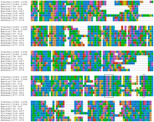
Figure 1 Multiple sequence alignment of proteins with the M60-like/PF13402 domain. Six selected proteins from the PF13402 seed alignment are indicated with their abbreviated species names (first three letters of genus and species name) followed by the position of the PF13402 domain. Three sequences (Vibcho, Bacthe, Enthis) were added and aligned to the PF13402 seed alignment using the MUSCLE profile alignment option in SEAVIEW. Full taxa names and GI and RefSeq accession numbers are: Vibrio cholerae: GI:297579165, ZP_06941093.1; Escherichia coli: GI:91212381, YP_542367.1; Bacillus thuringiensis: GI:228930091, ZP_04093101.1; Photorhabdus asymbiotica: GI:253989814, YP_003041170.1; Bacteroides thetaiotaomicron: GI:29349652, NP_813155.1 (BT4244); Trichomonas vaginalis: GI:123975108, XP_001330197.1; Entamoeba histolytica: GI:67478183, XP_654508.1; Homo sapiens: GI:293651621. NP_001123498.2 and Akkermansia muciniphila: GI:187736004, YP_001878116.1). The colour coding of amino acids indicate residues with similar physicochemical properties as defined in the alignment editor SEAVIEW. The minimal zincin metallopeptidase HEXXH motif and the additional conserved glutamate (E), defining the gluzincins-like motif HEXXHX(8,24)E, are boxed.
Figure 1 Multiple sequence alignment of proteins with the M60-like/PF13402 domain. Six selected proteins from the PF13402 seed alignment are indicated with their abbreviated species names (first three letters of genus and species name) followed by the position of the PF13402 domain. Three sequences (Vibcho, Bacthe, Enthis) were added and aligned to the PF13402 seed alignment using the MUSCLE profile alignment option in SEAVIEW. Full taxa names and GI and RefSeq accession numbers are: Vibrio cholerae: GI:297579165, ZP_06941093.1; Escherichia coli: GI:91212381, YP_542367.1; Bacillus thuringiensis: GI:228930091, ZP_04093101.1; Photorhabdus asymbiotica: GI:253989814, YP_003041170.1; Bacteroides thetaiotaomicron: GI:29349652, NP_813155.1 (BT4244); Trichomonas vaginalis: GI:123975108, XP_001330197.1; Entamoeba histolytica: GI:67478183, XP_654508.1; Homo sapiens: GI:293651621. NP_001123498.2 and Akkermansia muciniphila: GI:187736004, YP_001878116.1). The colour coding of amino acids indicate residues with similar physicochemical properties as defined in the alignment editor SEAVIEW. The minimal zincin metallopeptidase HEXXH motif and the additional conserved glutamate (E), defining the gluzincins-like motif HEXXHX(8,24)E, are boxed.CC BY · PubMed Central
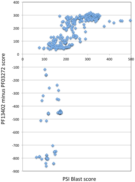
Figure 2 The M60-like/PF13402 and M60-enhancin/PF03272 profiles are derived from distantly related proteins. The bit score for the RefSeq proteins hit by a PSI-Blast search (X-axis) (query: T. vaginalis protein GI:123449825, XP_001313628; e-value ≤1.00E-04) were plotted against the difference of the HMMER bit scores for the PF13402 and PF03272 profiles (PF13402 minus PF03272)(Y-axis). A total of 415 entries were more significant for the PF13402 profile (positive values) and the remaining entries were more significant for the PF03272 profile (negative values). These results indicate that the M60-like/PF13402 and M60-enhancin/PF03272 profiles hit related proteins forming at least two distinct subfamilies defined by these two profiles that are not well discriminated by the PSI-Blast search. As expected the PSI-Blast derived profile is clearly more similar to the PF13402 profile than to the PF03272 profile as the query sequence used for the PSI-Blast search is hit by the PF13402 profile (bit-score 260, e-value 1.40E-74) but is not hit by the PF03272 profile. All values are listed in Table S2.
Figure 2 The M60-like/PF13402 and M60-enhancin/PF03272 profiles are derived from distantly related proteins. The bit score for the RefSeq proteins hit by a PSI-Blast search (X-axis) (query: T. vaginalis protein GI:123449825, XP_001313628; e-value ≤1.00E-04) were plotted against the difference of the HMMER bit scores for the PF13402 and PF03272 profiles (PF13402 minus PF03272)(Y-axis). A total of 415 entries were more significant for the PF13402 profile (positive values) and the remaining entries were more significant for the PF03272 profile (negative values). These results indicate that the M60-like/PF13402 and M60-enhancin/PF03272 profiles hit related proteins forming at least two distinct subfamilies defined by these two profiles that are not well discriminated by the PSI-Blast search. As expected the PSI-Blast derived profile is clearly more similar to the PF13402 profile than to the PF03272 profile as the query sequence used for the PSI-Blast search is hit by the PF13402 profile (bit-score 260, e-value 1.40E-74) but is not hit by the PF03272 profile. All values are listed in Table S2.CC BY · PubMed Central
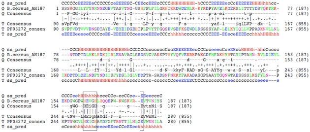
Figure 3 Profile-profile alignment of M60-like/PF13402 and M60-enhancin/PF03272. The alignment was derived from an HHpred search against all databases using the M60-like/PF13402 seed alignment as query (Table S4). The alignment corresponds to the second most significant hit to the enhancin/PF03272 profile (e-value 2.2E-37, score 307.8 and Probability 100%). The shown lines consists of ‘SS_pred’ lines representing sequence secondary structures predicted by PSIPRED, ‘consensus’ lines showing the consensus sequences of the PF13402 domain (shown with the top sequence) and the corresponding PF03272 domain. Amino acid residues are marked in capital letters when occurring with a frequency ≥60% and lower cases when ≥40% in the respective seed alignments. A tilda indicates an un-conserved column. The line in between the two consensus sequences shows the match quality and is defined as follows: ‘ = ’ very bad match, ‘−’ bad, ‘.’ neutral, ‘+’ good match and ‘|’ very good match. The well-conserved zincin HEXXH motif and an additional glutamate (E) residue part of a potential gluzincin are boxed. The upper and lower case letters in ‘SS_pred’ lines for secondary structure predictions show high and low probability respectively where H = helix, E = strand, and C = coil.
Figure 3 Profile-profile alignment of M60-like/PF13402 and M60-enhancin/PF03272. The alignment was derived from an HHpred search against all databases using the M60-like/PF13402 seed alignment as query (Table S4). The alignment corresponds to the second most significant hit to the enhancin/PF03272 profile (e-value 2.2E-37, score 307.8 and Probability 100%). The shown lines consists of ‘SS_pred’ lines representing sequence secondary structures predicted by PSIPRED, ‘consensus’ lines showing the consensus sequences of the PF13402 domain (shown with the top sequence) and the corresponding PF03272 domain. Amino acid residues are marked in capital letters when occurring with a frequency ≥60% and lower cases when ≥40% in the respective seed alignments. A tilda indicates an un-conserved column. The line in between the two consensus sequences shows the match quality and is defined as follows: ‘ = ’ very bad match, ‘−’ bad, ‘.’ neutral, ‘+’ good match and ‘|’ very good match. The well-conserved zincin HEXXH motif and an additional glutamate (E) residue part of a potential gluzincin are boxed. The upper and lower case letters in ‘SS_pred’ lines for secondary structure predictions show high and low probability respectively where H = helix, E = strand, and C = coil.CC BY · PubMed Central
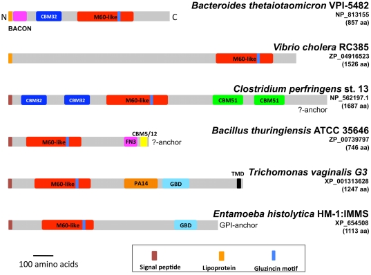
Figure 4 Structural organisation of selected M60-like/PF13402 containing proteins. Selected proteins possessing the PF13402 domain from a diverse taxonomic spread and cellular organisation (Gram-positive and Gram-negative bacteria and eukaryotes) with and without CBM32, CBM5_12 and/or CBM51 are depicted to illustrate their structural diversity. The putative glycan-binding BACON domain found among some Bacteroides spp. proteins and divergent PA14-like and GBD-like domains found among T. vaginalis proteins are also illustrated (see Figure S4). Specie names with accession numbers, protein length and key structural features are shown. The cartoons are drawn to the same scale and all sequences are aligned to their N-terminus. See also Figure 6 for additional structural configurations. For the Clostridium and Bacillus sequences there is no evidence for a cell surface anchoring sequence features indicated by “?-anchor”.
Figure 4 Structural organisation of selected M60-like/PF13402 containing proteins. Selected proteins possessing the PF13402 domain from a diverse taxonomic spread and cellular organisation (Gram-positive and Gram-negative bacteria and eukaryotes) with and without CBM32, CBM5_12 and/or CBM51 are depicted to illustrate their structural diversity. The putative glycan-binding BACON domain found among some Bacteroides spp. proteins and divergent PA14-like and GBD-like domains found among T. vaginalis proteins are also illustrated (see Figure S4). Specie names with accession numbers, protein length and key structural features are shown. The cartoons are drawn to the same scale and all sequences are aligned to their N-terminus. See also Figure 6 for additional structural configurations. For the Clostridium and Bacillus sequences there is no evidence for a cell surface anchoring sequence features indicated by “?-anchor”.CC BY · PubMed Central
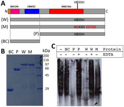
Figure 5 In vitro mucin degradation assay. (A) The structural organisation of the B. thetaiotaomicron BT4244 protein (GI: 29349652; RefSeq: NP_813155.1). The entire wild type (W) protein (minus signal peptide) encoded by the BT4244 gene is contrasted to a mutant (M) (E575D) and two truncated constructs covering either the PF13402 domain (P) or the BACON-CBM32 domains (BC) only. (B) 5 µg of purified recombinant proteins were separated by SDS-PAGE and stained with Coomasie blue for detection. The predicted protein sizes in kDa are: M and W: 95.1, P: 67.3 and BC: 27.8. (C) Bovine submaxillary gland mucins were incubated with 5 µg of purified recombinant proteins W, M, P or BC in the absence (−) or presence (+) of 50 mM EDTA. Following incubation the mucin samples were separated on an agarose gel prior to detection on blots with wheat germ agglutinin lectin.
Figure 5 In vitro mucin degradation assay. (A) The structural organisation of the B. thetaiotaomicron BT4244 protein (GI: 29349652; RefSeq: NP_813155.1). The entire wild type (W) protein (minus signal peptide) encoded by the BT4244 gene is contrasted to a mutant (M) (E575D) and two truncated constructs covering either the PF13402 domain (P) or the BACON-CBM32 domains (BC) only. (B) 5 µg of purified recombinant proteins were separated by SDS-PAGE and stained with Coomasie blue for detection. The predicted protein sizes in kDa are: M and W: 95.1, P: 67.3 and BC: 27.8. (C) Bovine submaxillary gland mucins were incubated with 5 µg of purified recombinant proteins W, M, P or BC in the absence (−) or presence (+) of 50 mM EDTA. Following incubation the mucin samples were separated on an agarose gel prior to detection on blots with wheat germ agglutinin lectin.CC BY · PubMed Central
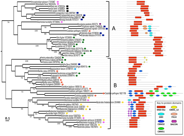
Figure 6 Protein maximum likelihood bootstrap consensus tree for selected M60-like/PF13402 domains. The shown maximum likelihood tree (Log likelihood: −18778.5) was generated as described in the Methods section using an alignment of 57 sequences and 175 residues drawn from an M60-like/PF13402 domain alignment, providing an evolutionary framework for the gene segments encoding these domains. For each sequence the corresponding species name is indicated along with the NCBI GI number and high-ranking taxa - squares are for eukaryotes: black-Metazoa, blue-microbial eukaryotes; circles are for Bacteria: yellow-Bacteroidetes, orange-Firmicutes, green-Proteobacteria (all gamma-proteobacteria), pink-Verrucomicrobia, violet-Tenericutes, cyan-Actinobacteria. Bootstrap support values (≥60%) are indicated below the branches. The scale bar represents the estimated number of changes per site. The domain organisation of the corresponding complete proteins is shown on the right hand side (see also Figure 4 for additional domain configurations). The sequence corresponding to the BT4244 protein used for the mucinase assay (Figure 5) is indicated by a *. The two major clans supported by a bootstrap value of 76% are indicated as clan A and B.
Figure 6 Protein maximum likelihood bootstrap consensus tree for selected M60-like/PF13402 domains. The shown maximum likelihood tree (Log likelihood: −18778.5) was generated as described in the Methods section using an alignment of 57 sequences and 175 residues drawn from an M60-like/PF13402 domain alignment, providing an evolutionary framework for the gene segments encoding these domains. For each sequence the corresponding species name is indicated along with the NCBI GI number and high-ranking taxa - squares are for eukaryotes: black-Metazoa, blue-microbial eukaryotes; circles are for Bacteria: yellow-Bacteroidetes, orange-Firmicutes, green-Proteobacteria (all gamma-proteobacteria), pink-Verrucomicrobia, violet-Tenericutes, cyan-Actinobacteria. Bootstrap support values (≥60%) are indicated below the branches. The scale bar represents the estimated number of changes per site. The domain organisation of the corresponding complete proteins is shown on the right hand side (see also Figure 4 for additional domain configurations). The sequence corresponding to the BT4244 protein used for the mucinase assay (Figure 5) is indicated by a *. The two major clans supported by a bootstrap value of 76% are indicated as clan A and B.CC BY · PubMed Central

Verification

Claims the paper makes

The claims and statistics of this paper haven’t been checked yet.

Run the check — free, no account needed. It reads the open-access text and checks the sources it cites.

A few free checks a day · papers already verified open instantly · results are public

Claims other papers make about this one

Each is checked against this paper’s own text: does it say what the citing paper says it does?

Self-check

Percentages, sums and reported differences re-computed from the paper’s own numbers.

The self-check needs the paper’s full text.

Citations

Cites

Show all 94 references

Cited by

Sorted by citations

See all 116

Metadata

Authors
Nakjang, S., Ndeh, D. A., Wipat, A., Bolam, D. N., & Hirt, R. P.
Affiliations
Newcastle University (GB)
Corresponding author
  • Nakjang, Sirintra
  • Ndeh, Didier A.
  • Wipat, Anil
  • Bolam, David N.
  • Hirt, Robert P.
Venue
PLoS ONE
Published
2012-01-27
Volume
7
Issue
1
Pages
e30287
ISSN
1932-6203 (ISSN-L)
OpenAlex
W2015392436
Cited by
116
References
94
Citations by year
2026: 10 · 2025: 4 · 2024: 12 · 2023: 5 · 2022: 12 · 2021: 7 · 2020: 15 · 2019: 14 · 2018: 5 · 2017: 5 · 2016: 7 · 2015: 3 · 2014: 7 · 2013: 7 · 2012: 3
FWCI
2.57
Type
journal article
Language
en
Open access
Locations
  • PLoS ONE (publishedVersion, cc-by)
  • Europe PMC (PubMed Central) (publishedVersion, cc-by)
  • PubMed Central (publishedVersion, cc-by)
  • PubMed (publishedVersion)
  • Discovery Research Portal (University of Dundee) (publishedVersion, cc-by)
  • DOAJ (DOAJ: Directory of Open Access Journals) (submittedVersion)
  • Figshare (submittedVersion, cc-by)
  • Newcastle University ePrints (Newcastle Univesity) (cc-by)
Funders
  • Wellcome Trust
  • Newcastle University
  • Commonwealth Scholarship Commission
  • Department for International Development
  • Fakultet Medicinskih Nauka, Univerziteta U Kragujevcu
  • Biotechnology and Biological Sciences Research Council (BB/F014163/1)
Topics
  • Life Sciences › Biochemistry, Genetics and Molecular Biology › Molecular Biology › Gut microbiota and health
  • Life Sciences › Immunology and Microbiology › Virology › Bacteriophages and microbial interactions
  • Life Sciences › Biochemistry, Genetics and Molecular Biology › Molecular Biology › Genomics and Phylogenetic Studies
Keywords
extracellular peptidases, mucin degradation, mucin, host-microbe interactions, mucosal microbiota, horizontal gene transfer, carbohydrate-binding module, Bacteroides thetaiotaomicron, host colonization, signal peptide
MeSH terms
Adaptation, Biological, Amino Acid Sequence, Animals, Base Sequence, Digestive System, Humans, Invertebrates, Molecular Sequence Data, Symbiosis, Sequence Homology, Amino Acid, Protein Structure, Tertiary, Metalloproteases, Host-Pathogen Interactions

About these checks

Registry, retraction, Cites and statistics results are computed, not judged, with one exception: a printed reference that no index lists is read once by a model before it counts as a citation error. Claims are extracted by a model and kept only when their quote appears verbatim in the text; each cited source is then checked, with a model’s help, for whether it says what the claim relies on. Those verdicts can be wrong — read the claim’s quote and the cited source before relying on one.

Statistics and arithmetic are recomputed, not judged.

Confidence bands are shown only for fields calibrated on our annotated test set. None is calibrated yet, so no band is shown.

What counts as a claim ›How we measure confidence ›How we check this ›

Are you an author, or do you see a problem with this page? Request a review