

The DOI matches its Crossref record; 41 of the 42 references it cites were found in a registry or an index.
Stress Leads to Contrasting Effects on the Levels of Brain Derived Neurotrophic Factor in the Hippocampus and Amygdala
Open accessAbstract
Recent findings on stress induced structural plasticity in rodents have identified important differences between the hippocampus and amygdala. The same chronic immobilization stress (CIS, 2 h/day) causes growth of dendrites and spines in the basolateral amygdala (BLA), but dendritic atrophy in hippocampal area CA3. CIS induced morphological changes also differ in their temporal longevity--BLA hypertrophy, unlike CA3 atrophy, persists even after 21 days of stress-free recovery. Furthermore, a single session of acute immobilization stress (AIS, 2 h) leads to a significant increase in spine density 10 days, but not 1 day, later in the BLA. However, little is known about the molecular correlates of the differential effects of chronic and acute stress. Because BDNF is known to be a key regulator of dendritic architecture and spines, we investigated if the levels of BDNF expression reflect the divergent effects of stress on the hippocampus and amygdala. CIS reduces BDNF in area CA3, while it increases it in the BLA of male Wistar rats. CIS-induced increase in BDNF expression lasts for at least 21 days after the end of CIS in the BLA. But CIS-induced decrease in area CA3 BDNF levels, reverses to normal levels within the same period. Finally, BDNF is up regulated in the BLA 1 day after AIS and this increase persists even 10 days later. In contrast, AIS fails to elicit any significant change in area CA3 at either time points. Together, these findings demonstrate that both acute and chronic stress trigger opposite effects on BDNF levels in the BLA versus area CA3, and these divergent changes also follow distinct temporal profiles. These results point to a role for BDNF in stress-induced structural plasticity across both hippocampus and amygdala, two brain areas that have also been implicated in the cognitive and affective symptoms of stress-related psychiatric disorders.
Overview
The claims and statistics of this paper haven’t been checked yet.
Run the check — free, no account needed. It reads the open-access text and checks the sources it cites.
Integrity signals
Registry
Cites
Statistics
Figures
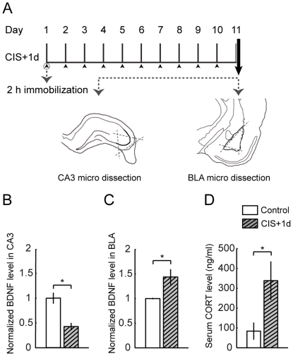
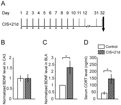
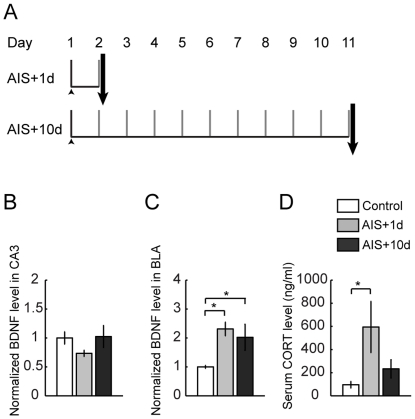
Verification
Claims the paper makes
The claims and statistics of this paper haven’t been checked yet.
Run the check — free, no account needed. It reads the open-access text and checks the sources it cites.
Claims other papers make about this one
Each is checked against this paper’s own text: does it say what the citing paper says it does?
The source backs part of the claim, not all of it — or backs it for a different population, condition or comparison.
In the citing paper
“enhanced BDNF/TrkB signaling in the amygdala leads to maladaptive plasticity resulting in overactivation of this brain region and exacerbated anxiety and fear responses (47, 198–200)”
From Psychedelics and Other Psychoplastogens for Treating Mental Illness. (2021) · cites this paper as [200]
Experimental — accuracy not yet measured
Self-check
Percentages, sums and reported differences re-computed from the paper’s own numbers.
The self-check needs the paper’s full text.
Citations
Cites
A Neurotrophic Model for Stress-Related Mood Disorders
Duman, R. S., & Monteggia, L. M. (2006) · Biological Psychiatry · doi:10.1016/j.biopsych.2006.02.013 · Cited by 3,375
Nestler, E. J., Barrot, M., DiLeone, R. J., Eisch, A. J., Gold, S. J., & Monteggia, L. M. (2002) · Neuron · doi:10.1016/s0896-6273(02)00653-0 · Cited by 3,217
The molecular neurobiology of depression
Krishnan, V., & Nestler, E. J. (2008) · Nature · doi:10.1038/nature07455 · Cited by 2,889
Neurocircuitry of stress: central control of the hypothalamo–pituitary–adrenocortical axis
Herman, J. P., & Cullinan, W. E. (1997) · Trends in Neurosciences · doi:10.1016/s0166-2236(96)10069-2 · Cited by 2,270
Essential role of BDNF in the mesolimbic dopamine pathway in social defeat stress
Berton, O., McClung, C. A., DiLeone, R. J., Krishnan, V., Renthal, W., Russo, S. J., Graham, D., Tsankova, N. M., Bolanos, C. A., Rios, M., et al. (2006) · Science · doi:10.1126/science.1120972 · Cited by 2,129
Nibuya, M., Morinobu, S., & Duman, R. (1995) · The Journal of Neuroscience · doi:10.1523/jneurosci.15-11-07539.1995 · Cited by 2,109
STRESS AND HIPPOCAMPAL PLASTICITY
McEwen, B. S. (1999) · Annual Review of Neuroscience · doi:10.1146/annurev.neuro.22.1.105 · Cited by 2,087
Stress, memory and the amygdala
Roozendaal, B., McEwen, B. S., & Chattarji, S. (2009) · Nature Reviews Neuroscience · doi:10.1038/nrn2651 · Cited by 1,760
Self-citationVyas, A., Mitra, R., Shankaranarayana Rao, B. S., & Chattarji, S. (2002) · The Journal of Neuroscience · doi:10.1523/jneurosci.22-15-06810.2002 · Cited by 1,662
Self-citationBrain-derived neurotrophic factor produces antidepressant effects in behavioral models of depression
Shirayama, Y., Chen, A. C.-H., Nakagawa, S., Russell, D. S., & Duman, R. S. (2002) · The Journal of Neuroscience · doi:10.1523/jneurosci.22-08-03251.2002 · Cited by 1,654
Research works printed but not linked: 1
- BS McEwen, 1999, Stress and hippocampal plasticity., Annual Review of Neuroscience, 22, 105, 10.1146/annurev.neuro.22.1.105 · DOI · Paper page
Cited by
Sorted by citations
Stress Effects on Neuronal Structure: Hippocampus, Amygdala, and Prefrontal Cortex
McEwen, B. S., Nasca, C., & Gray, J. D. (2016) · Neuropsychopharmacology · doi:10.1038/npp.2015.171 · Cited by 1,574
Mechanisms of stress in the brain
McEwen, B. S., Bowles, N. P., Gray, J. D., Hill, M. N., Hunter, R. G., Karatsoreos, I. N., & Nasca, C. (2015) · Nature Neuroscience · doi:10.1038/nn.4086 · Cited by 1,386
Hannibal, K. E., & Bishop, M. D. (2014) · Physical Therapy · doi:10.2522/ptj.20130597 · Cited by 729
The Role of Neural Plasticity in Depression: From Hippocampus to Prefrontal Cortex
Liu, W., Ge, T., Leng, Y., Pan, Z., Fan, J., Yang, W., & Cui, R. (2017) · Neural Plasticity · doi:10.1155/2017/6871089 · Cited by 640
The hippocampal–prefrontal pathway: The weak link in psychiatric disorders?
Godsil, B. P., Kiss, J., Spedding, M., & Jay, T. M. (2013) · European Neuropsychopharmacology · doi:10.1016/j.euroneuro.2012.10.018 · Cited by 459
Metadata
- Authors
- Lakshminarasimhan, H., & Chattarji, S.
- Affiliations
- Tata Institute of Fundamental Research (IN)
- Corresponding author
- Chattarji, Sumantra
- ORCID
- Chattarji, Sumantra
- Venue
- PLoS ONE
- Publisher
- Public Library of Science (PLoS)
- Published
- 2012-01-17
- Volume
- 7
- Issue
- 1
- Pages
- e30481
- ISSN
- 1932-6203 (ISSN-L)
- PMC
- PMC3260293
- OpenAlex
- W2058006510
- Cited by
- 253
- References
- 42
- Citations by year
- 2026: 11 · 2025: 13 · 2024: 12 · 2023: 15 · 2022: 16 · 2021: 29 · 2020: 19 · 2019: 14 · 2018: 18 · 2017: 23 · 2016: 20 · 2015: 15 · 2014: 24 · 2013: 16 · 2012: 8
- FWCI
- 8.44
- Type
- journal article
- Language
- en
- Open access
- gold
- doi.org
- Locations
- PLoS ONE (publishedVersion, cc-by)
- Europe PMC (PubMed Central) (publishedVersion, cc-by)
- PubMed Central (publishedVersion, cc-by)
- PubMed (publishedVersion)
- DOAJ (DOAJ: Directory of Open Access Journals) (submittedVersion, cc-by-sa)
- Funders
- Wellcome Trust (GR 0701339 MA)
- Directorate for Biological Sciences
- National Centre for Biological Sciences
- Topics
- Life Sciences › Neuroscience › Behavioral Neuroscience › Stress Responses and Cortisol
- Life Sciences › Neuroscience › Cellular and Molecular Neuroscience › Nerve injury and regeneration
- Life Sciences › Neuroscience › Cognitive Neuroscience › Memory and Neural Mechanisms
- Keywords
- brain-derived neurotrophic factor, restraint stress, chronic stress, acute stress, hippocampus, amygdala, basolateral amygdala, CA3, dendritic spine density, dendritic atrophy, structural plasticity, stress recovery, stress-related psychiatric disorders
- MeSH terms
- Amygdala, Animals, Body Weight, Hippocampus, Immunoassay, Male, Restraint, Physical, Stress, Physiological, Stress, Psychological, Time Factors, Rats, Wistar, Brain-Derived Neurotrophic Factor, Rats, CA3 Region, Hippocampal
About these checks
Registry, retraction, Cites and statistics results are computed, not judged, with one exception: a printed reference that no index lists is read once by a model before it counts as a citation error. Claims are extracted by a model and kept only when their quote appears verbatim in the text; each cited source is then checked, with a model’s help, for whether it says what the claim relies on. Those verdicts can be wrong — read the claim’s quote and the cited source before relying on one.
Statistics and arithmetic are recomputed, not judged.
Confidence bands are shown only for fields calibrated on our annotated test set. None is calibrated yet, so no band is shown.
What counts as a claim ›How we measure confidence ›How we check this ›
Are you an author, or do you see a problem with this page? Request a review
