

The DOI matches its Crossref record; 79 of the 79 references it cites were found in a registry or an index.
An Antagomir to MicroRNA Let7f Promotes Neuroprotection in an Ischemic Stroke Model
Open accessAbstract
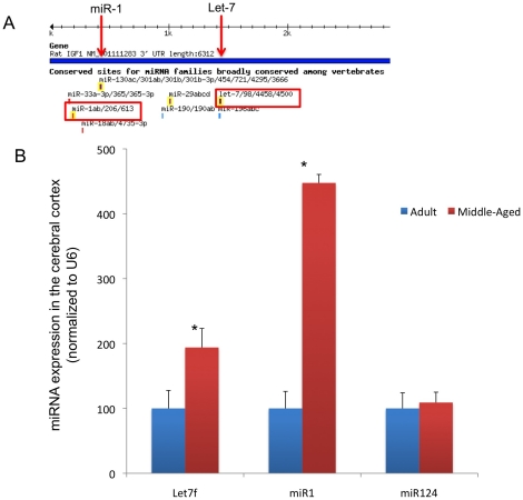
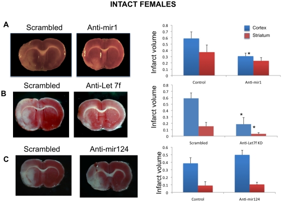
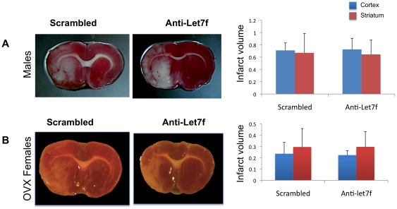
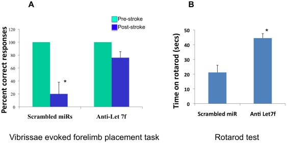
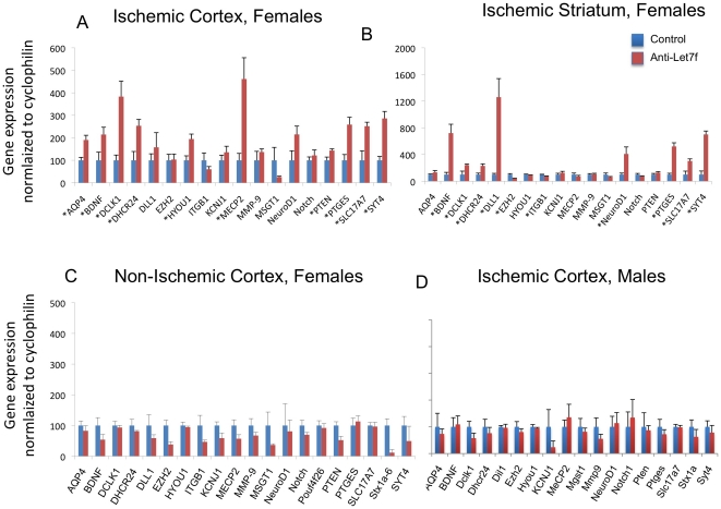
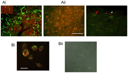
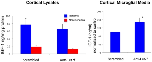
We previously showed that middle-aged female rats sustain a larger infarct following experimental stroke as compared to younger female rats, and paradoxically, estrogen treatment to the older group is neurotoxic. Plasma and brain insulin-like growth factor-1 (IGF-1) levels decrease with age. However, IGF-1 infusion following stroke, prevents estrogen neurotoxicity in middle-aged female rats. IGF1 is neuroprotective and well tolerated, but also has potentially undesirable side effects. We hypothesized that microRNAs (miRNAs) that target the IGF-1 signaling family for translation repression could be alternatively suppressed to promote IGF-1-like neuroprotection. Here, we report that two conserved IGF pathway regulatory microRNAs, Let7f and miR1, can be inhibited to mimic and even extend the neuroprotection afforded by IGF-1. Anti-mir1 treatment, as late as 4 hours following ischemia, significantly reduced cortical infarct volume in adult female rats, while anti-Let7 robustly reduced both cortical and striatal infarcts, and preserved sensorimotor function and interhemispheric neural integration. No neuroprotection was observed in animals treated with a brain specific miRNA unrelated to IGF-1 (anti-miR124). Remarkably, anti-Let7f was only effective in intact females but not males or ovariectomized females indicating that the gonadal steroid environment critically modifies miRNA action. Let7f is preferentially expressed in microglia in the ischemic hemisphere and confirmed in ex vivo cultures of microglia obtained from the cortex. While IGF-1 was undetectable in microglia harvested from the non-ischemic hemisphere, IGF-1 was expressed by microglia obtained from the ischemic cortex and was further elevated by anti-Let7f treatment. Collectively these data support a novel miRNA-based therapeutic strategy for neuroprotection following stroke.
Overview
The claims and statistics of this paper haven’t been checked yet.
Run the check — free, no account needed. It reads the open-access text and checks the sources it cites.
Integrity signals
Registry
Cites
Statistics
Figures
Verification
Claims the paper makes
The claims and statistics of this paper haven’t been checked yet.
Run the check — free, no account needed. It reads the open-access text and checks the sources it cites.
Claims other papers make about this one
Each is checked against this paper’s own text: does it say what the citing paper says it does?
The source backs part of the claim, not all of it — or backs it for a different population, condition or comparison.
In the citing paper
“Recently, miRNAs targeting several genes are involved in regulating microglia behavior and functions in diverse CNS pathologies including ischemic brain injuries (Selvamani et al., 2012; Zhao et al., 2013; Karthikeyan et al., 2018), multiple sclerosis (Ponomarev et al., 2011) and prion disease (Saba et al., 2012; Slota and Booth, 2019).”
From miR-142-3p Regulates BDNF Expression in Activated Rodent Microglia Through Its Target CAMK2A (2020) · cites this paper as Selvamani et al., 2012
Experimental — accuracy not yet measuredIn the citing paper
“Some in vitro and in vivo studies have shown the role of IGF-1 in promoting neuroprotection and neuroregeneration (Li et al., 2010; Selvamani et al., 2012; Sohrabji and Williams, 2013; Bake et al., 2014).”
From The impact of microglial activation on blood-brain barrier in brain diseases (2014) · cites this paper as Selvamani et al., 2012
Self-check
Percentages, sums and reported differences re-computed from the paper’s own numbers.
The self-check needs the paper’s full text.
Citations
Cites
Bartel (2004) · doi:10.1016/s0092-8674(04)00045-5 · Cited by 34,714
Lewis, B. P., Burge, C. B., & Bartel, D. P. (2005) · Cell · doi:10.1016/j.cell.2004.12.035 · Cited by 11,855
The functions of animal microRNAs
Ambros, V. (2004) · Nature · doi:10.1038/nature02871 · Cited by 10,728
Resting microglial cells are highly dynamic surveillants of brain parenchyma in vivo
Nimmerjahn, A., Kirchhoff, F., & Helmchen, F. (2005) · Science · doi:10.1126/science.1110647 · Cited by 5,827
ATP mediates rapid microglial response to local brain injury in vivo
Davalos, D., Grutzendler, J., Yang, G., Kim, J. V., Zuo, Y., Jung, S., Littman, D. R., Dustin, M. L., & Gan, W.-B. (2005) · Nature Neuroscience · doi:10.1038/nn1472 · Cited by 3,913
MicroRNA Targeting Specificity in Mammals: Determinants beyond Seed Pairing
Grimson, A., Farh, K. K.-H., Johnston, W. K., Garrett-Engele, P., Lim, L. P., & Bartel, D. P. (2007) · Molecular Cell · doi:10.1016/j.molcel.2007.06.017 · Cited by 3,766
Inflammatory Blockade Restores Adult Hippocampal Neurogenesis
Monje, M. L., Toda, H., & Palmer, T. D. (2003) · Science · doi:10.1126/science.1088417 · Cited by 2,426
The Diverse Functions of MicroRNAs in Animal Development and Disease
Kloosterman, W. P., & Plasterk, R. H. A. (2006) · Developmental Cell · doi:10.1016/j.devcel.2006.09.009 · Cited by 2,082
Wake, H., Moorhouse, A. J., Jinno, S., Kohsaka, S., & Nabekura, J. (2009) · The Journal of Neuroscience · doi:10.1523/jneurosci.4363-08.2009 · Cited by 1,619
The Endothelial-Specific MicroRNA miR-126 Governs Vascular Integrity and Angiogenesis
Wang, S., Aurora, A. B., Johnson, B. A., Qi, X., McAnally, J., Hill, J. A., Richardson, J. A., Bassel-Duby, R., & Olson, E. N. (2008) · Developmental Cell · doi:10.1016/j.devcel.2008.07.002 · Cited by 1,617
Cited by
Sorted by citations
The biphasic function of microglia in ischemic stroke
Ma, Y., Wang, J., Wang, Y., & Yang, G.-Y. (2017) · Progress in Neurobiology · doi:10.1016/j.pneurobio.2016.01.005 · Cited by 655
The impact of microglial activation on blood-brain barrier in brain diseases
da Fonseca, A. C. C., Matias, D., Garcia, C., Amaral, R., Geraldo, L. H., Freitas, C., & Lima, F. R. S. (2014) · Frontiers in Cellular Neuroscience · doi:10.3389/fncel.2014.00362 · Cited by 572
Roles of microglia in brain development, tissue maintenance and repair
Michell‐Robinson, M. A., Touil, H., Healy, L. M., Owen, D. R., Durafourt, B. A., Bar‐Or, A., Antel, J. P., & Moore, C. S. (2015) · Brain · doi:10.1093/brain/awv066 · Cited by 435
Estrogens, Neuroinflammation, and Neurodegeneration
Villa, A., Vegeto, E., Poletti, A., & Maggi, A. (2016) · Endocrine Reviews · doi:10.1210/er.2016-1007 · Cited by 423
MicroRNA-125b induces tau hyperphosphorylation and cognitive deficits in Alzheimer’s disease
Banzhaf‐Strathmann, J., Benito, E., May, S., Arzberger, T., Tahirovic, S., Kretzschmar, H., Fischer, A., & Edbauer, D. (2014) · The EMBO Journal · doi:10.15252/embj.201387576 · Cited by 347
Metadata
- Authors
- Selvamani, A., Sathyan, P., Miranda, R. C., & Sohrabji, F.
- Affiliations
- Texas A&M Health Science Center (US)
- Corresponding author
- Sohrabji, Farida
- Venue
- PLoS ONE
- Publisher
- Public Library of Science (PLoS)
- Published
- 2012-02-29
- Volume
- 7
- Issue
- 2
- Pages
- e32662
- ISSN
- 1932-6203 (ISSN-L)
- PMC
- PMC3290559
- OpenAlex
- W2071432376
- Cited by
- 217
- References
- 79
- Citations by year
- 2026: 1 · 2025: 4 · 2024: 6 · 2023: 7 · 2022: 11 · 2021: 12 · 2020: 16 · 2019: 19 · 2018: 18 · 2017: 26 · 2016: 20 · 2015: 23 · 2014: 27 · 2013: 22 · 2012: 4
- FWCI
- 8.02
- Type
- journal article
- Language
- en
- Open access
- gold
- doi.org
- Locations
- PLoS ONE (publishedVersion, cc-by)
- Europe PMC (PubMed Central) (publishedVersion, cc-by)
- PubMed Central (publishedVersion, cc-by)
- PubMed (publishedVersion)
- DOAJ (DOAJ: Directory of Open Access Journals) (submittedVersion, cc-by-sa)
- Figshare (submittedVersion, cc-by)
- OakTrust (Texas A&M University Libraries) (submittedVersion)
- Funders
- National Institutes of Health
- National Institute on Aging (R01AG027684, AG027684, AG028303, R01AG028303)
- Topics
- Life Sciences › Biochemistry, Genetics and Molecular Biology › Cancer Research › MicroRNA in disease regulation
- Life Sciences › Neuroscience › Neurology › Neuroinflammation and Neurodegeneration Mechanisms
- Health Sciences › Medicine › Clinical Neurology › Acute Ischemic Stroke Management
- Keywords
- ischemic stroke, neuroprotection, microRNA, let-7f, miR-1, insulin-like growth factor I, IGF-1 signaling, microglia, sex differences, infarct size, sensorimotor function, hemispheric integration, sex steroids
- MeSH terms
- Animals, Behavior, Animal, Disease Models, Animal, Female, Gene Expression Regulation, Insulin-Like Growth Factor I, Ischemia, Male, Sex Factors, Reperfusion Injury, Microglia, Neuroprotective Agents, Stroke, MicroRNAs, Rats
About these checks
Registry, retraction, Cites and statistics results are computed, not judged, with one exception: a printed reference that no index lists is read once by a model before it counts as a citation error. Claims are extracted by a model and kept only when their quote appears verbatim in the text; each cited source is then checked, with a model’s help, for whether it says what the claim relies on. Those verdicts can be wrong — read the claim’s quote and the cited source before relying on one.
Statistics and arithmetic are recomputed, not judged.
Confidence bands are shown only for fields calibrated on our annotated test set. None is calibrated yet, so no band is shown.
What counts as a claim ›How we measure confidence ›How we check this ›
Are you an author, or do you see a problem with this page? Request a review
