Cited by 50

The DOI matches its Crossref record; 2 of the 2 references it cites were found in a registry or an index.

A Late Form of Nucleophagy in Saccharomyces cerevisiae

Mijaljica, Dalibor, Prescott, Mark, Devenish, Rodney J.·2012·PLoS ONE

Overview

The claims and statistics of this paper haven’t been checked yet.

Run the check — free, no account needed. It reads the open-access text and checks the sources it cites.

A few free checks a day · papers already verified open instantly · results are public

Integrity signals

Registry

Crossref · 10.1371/journal.pone.0040013

Cites

2 of 2 references verified0 citation errors · 0 not checkable

Statistics

Figures

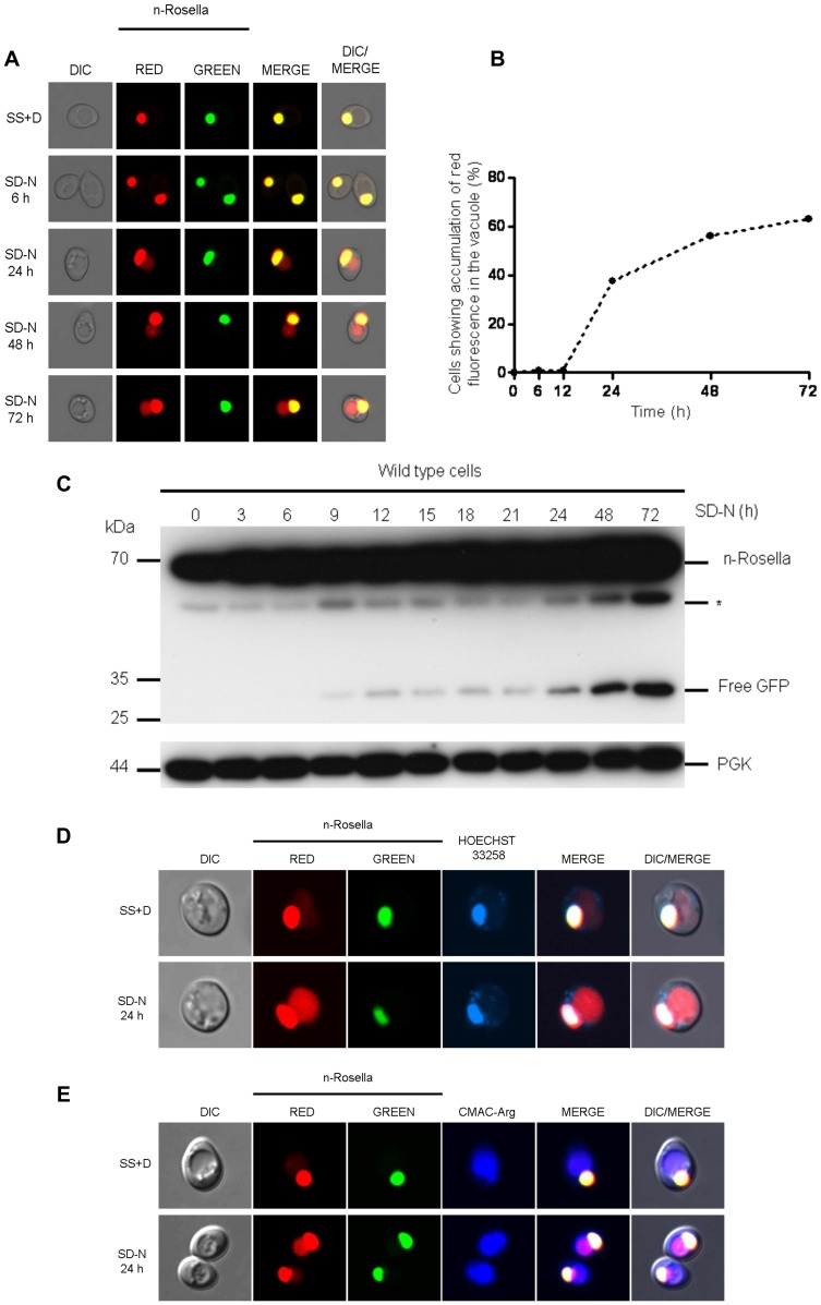
Figure 1 Delivery of n-Rosella to the vacuole of nitrogen-starved wild type cells. (A) Wild type (BY4741) cells expressing n-Rosella were subjected to nitrogen starvation and then sampled at selected time points (0, 6, 24, 48 and 72 hours after commencement of nitrogen starvation). Accumulation of diffuse red fluorescence in the vacuole was taken as evidence of nucleophagy. (B) The percentage of cells showing accumulation of diffuse red fluorescence in the vacuole at various time points is shown. (C) Cells expressing n-Rosella were starved in SD(-N) medium for 0, 3, 6, 9, 12, 15, 18, 21, 24, 48 and 72 hours, and the increase in processed degradation product (free GFP) levels was monitored by immunoblotting as described in Materials and Methods. Cytosolic PGK was detected as a loading control. *indicates the presumptive degradation product of n-Rosella lacking the NAB35 nuclear targeting signal. (D) Staining with Hoechst 33258 was performed to confirm the targeting of n-Rosella (red and green fluorescence) to the nucleus 24 hours after commencement of nitrogen starvation. (E) Staining with the vacuolar lumen dye, CMAC-Arg was performed to confirm the delivery of n-Rosella (diffuse red fluorescence) to the vacuole (24 hours after commencement of nitrogen starvation).
Figure 1 Delivery of n-Rosella to the vacuole of nitrogen-starved wild type cells. (A) Wild type (BY4741) cells expressing n-Rosella were subjected to nitrogen starvation and then sampled at selected time points (0, 6, 24, 48 and 72 hours after commencement of nitrogen starvation). Accumulation of diffuse red fluorescence in the vacuole was taken as evidence of nucleophagy. (B) The percentage of cells showing accumulation of diffuse red fluorescence in the vacuole at various time points is shown. (C) Cells expressing n-Rosella were starved in SD(-N) medium for 0, 3, 6, 9, 12, 15, 18, 21, 24, 48 and 72 hours, and the increase in processed degradation product (free GFP) levels was monitored by immunoblotting as described in Materials and Methods. Cytosolic PGK was detected as a loading control. *indicates the presumptive degradation product of n-Rosella lacking the NAB35 nuclear targeting signal. (D) Staining with Hoechst 33258 was performed to confirm the targeting of n-Rosella (red and green fluorescence) to the nucleus 24 hours after commencement of nitrogen starvation. (E) Staining with the vacuolar lumen dye, CMAC-Arg was performed to confirm the delivery of n-Rosella (diffuse red fluorescence) to the vacuole (24 hours after commencement of nitrogen starvation).CC BY · PubMed Central
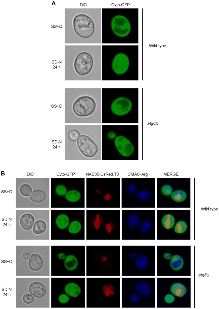
Figure 2 The diffuse red fluorescence (NAB35-DsRed.T3) accumulated in the vacuole is delivered directly from the nucleus. Wild type (BY4741) and atg6Δ cells expressing cyto-GFP alone (A), or co-expressing cyto-GFP and NAB35-DsRed.T3 (B), under growing (SS+D) and nitrogen starvation conditions (SD-N) were sampled after 24 hours. Staining with the vacuolar lumen staining dye, CMAC-Arg was used to confirm delivery of fluorescent reporters to the vacuole.
Figure 2 The diffuse red fluorescence (NAB35-DsRed.T3) accumulated in the vacuole is delivered directly from the nucleus. Wild type (BY4741) and atg6Δ cells expressing cyto-GFP alone (A), or co-expressing cyto-GFP and NAB35-DsRed.T3 (B), under growing (SS+D) and nitrogen starvation conditions (SD-N) were sampled after 24 hours. Staining with the vacuolar lumen staining dye, CMAC-Arg was used to confirm delivery of fluorescent reporters to the vacuole.CC BY · PubMed Central
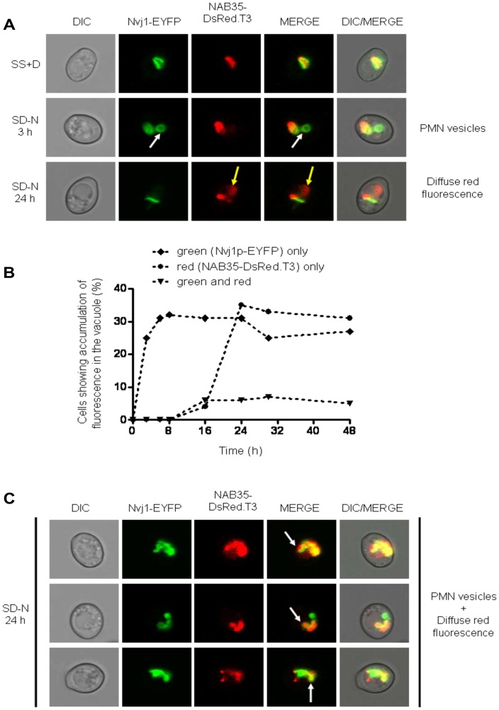
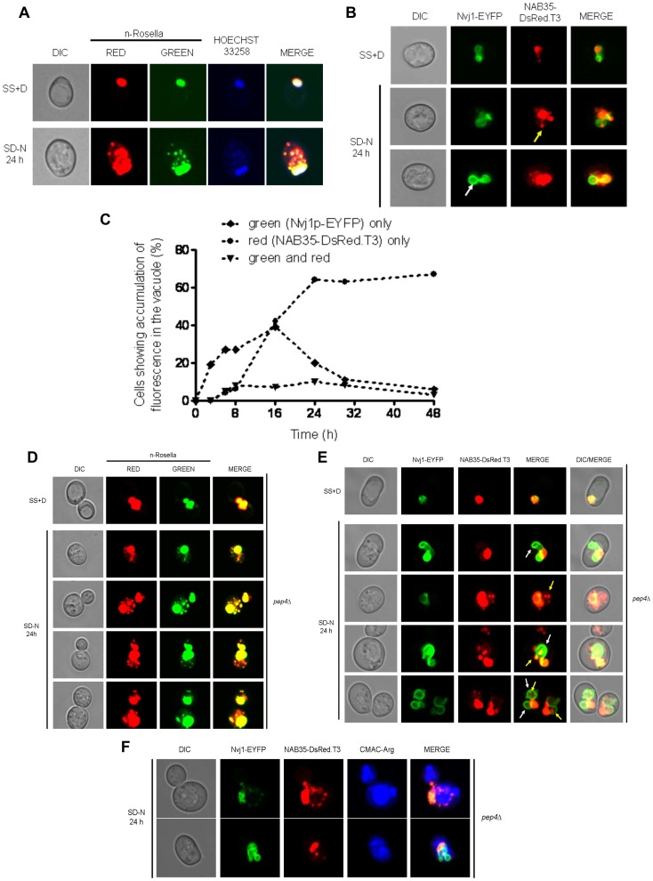
Figure 3 Wild type cells co-expressing Nvj1-EYFP and NAB35-DsRed.T3 nuclear reporters show temporal separation of nucleophagic events. (A) Wild type (BY4741) cells co-expressing both nuclear reporters were imaged under growing (SS+D) and nitrogen starvation (SD-N) conditions (3 and 24 hours after commencement of nitrogen starvation), respectively. The appearance of Nvj1p-EYFP-derived vesicles (PMN vesicles) in the vacuole is highlighted by white arrows, whereas accumulation of NAB35-DsRed.T3-derived fluorescence (diffuse red fluorescence) is indicated by yellow arrows. (B) Percentage of cells showing accumulation of fluorescence in the vacuole over time: ♦ green (Nvj1p-EYFP) only; • red (NAB35-DsRed.T3) only; ▾ green and red. (C) Accumulation of both Nvj1p-EYFP-derived vesicles (PMN vesicles) and accumulation of NAB35-DsRed.T3-derived diffuse red fluorescence in the same cells 24 hours after commencement of nitrogen starvation. The appearance of vacuolar vesicles containing both nuclear reporters is indicated by white arrows.
Figure 3 Wild type cells co-expressing Nvj1-EYFP and NAB35-DsRed.T3 nuclear reporters show temporal separation of nucleophagic events. (A) Wild type (BY4741) cells co-expressing both nuclear reporters were imaged under growing (SS+D) and nitrogen starvation (SD-N) conditions (3 and 24 hours after commencement of nitrogen starvation), respectively. The appearance of Nvj1p-EYFP-derived vesicles (PMN vesicles) in the vacuole is highlighted by white arrows, whereas accumulation of NAB35-DsRed.T3-derived fluorescence (diffuse red fluorescence) is indicated by yellow arrows. (B) Percentage of cells showing accumulation of fluorescence in the vacuole over time: ♦ green (Nvj1p-EYFP) only; • red (NAB35-DsRed.T3) only; ▾ green and red. (C) Accumulation of both Nvj1p-EYFP-derived vesicles (PMN vesicles) and accumulation of NAB35-DsRed.T3-derived diffuse red fluorescence in the same cells 24 hours after commencement of nitrogen starvation. The appearance of vacuolar vesicles containing both nuclear reporters is indicated by white arrows.CC BY · PubMed Central
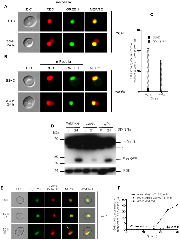
Figure 4 The inactivation of the NVJ1 and VAC8 genes required for PMN does not abrogate the delivery of n-Rosella to the vacuole. nvj1Δ (A) and vac8Δ (B) cells expressing n-Rosella were imaged under growing (SS+D) and nitrogen starvation (SD−N) conditions (24 hours after commencement of nitrogen starvation). (C) The percentage of nvj1Δ and vac8Δ cells showing accumulation of diffuse red fluorescence in the vacuole under growing (SS+D) and nitrogen starvation (SD-N) conditions (24 hours after commencement of nitrogen starvation). (D) Wild type, nvj1Δ and vac8Δ cells expressing n-Rosella were starved in SD(−N) medium for 0 and 24 hours, and the levels of the free GFP degradation product monitored by immunoblotting as described in Materials and Methods. Cytosolic PGK was detected as a loading control. *indicates the presumptive degradation product of n-Rosella lacking the NAB35 nuclear targeting signal. (E) vac8Δ cells co-expressing the two nuclear reporters, Nvj1-EYFP and NAB35-DsRed.T3 were imaged under growing (SS+D) and nitrogen starvation (SD−N) conditions (3 and 24 hours after commencement of nitrogen starvation), respectively. The accumulation of NAB35-DsRed.T3-derived fluorescence (diffuse red fluorescence) is indicated by white arrow. (F) Percentage of vac8Δ cells showing accumulation of fluorescence in the vacuole over time: ♦ green (Nvj1p-EYFP) only; • red (NAB35-DsRed.T3) only; ▾ green and red.
Figure 4 The inactivation of the NVJ1 and VAC8 genes required for PMN does not abrogate the delivery of n-Rosella to the vacuole. nvj1Δ (A) and vac8Δ (B) cells expressing n-Rosella were imaged under growing (SS+D) and nitrogen starvation (SD−N) conditions (24 hours after commencement of nitrogen starvation). (C) The percentage of nvj1Δ and vac8Δ cells showing accumulation of diffuse red fluorescence in the vacuole under growing (SS+D) and nitrogen starvation (SD-N) conditions (24 hours after commencement of nitrogen starvation). (D) Wild type, nvj1Δ and vac8Δ cells expressing n-Rosella were starved in SD(−N) medium for 0 and 24 hours, and the levels of the free GFP degradation product monitored by immunoblotting as described in Materials and Methods. Cytosolic PGK was detected as a loading control. *indicates the presumptive degradation product of n-Rosella lacking the NAB35 nuclear targeting signal. (E) vac8Δ cells co-expressing the two nuclear reporters, Nvj1-EYFP and NAB35-DsRed.T3 were imaged under growing (SS+D) and nitrogen starvation (SD−N) conditions (3 and 24 hours after commencement of nitrogen starvation), respectively. The accumulation of NAB35-DsRed.T3-derived fluorescence (diffuse red fluorescence) is indicated by white arrow. (F) Percentage of vac8Δ cells showing accumulation of fluorescence in the vacuole over time: ♦ green (Nvj1p-EYFP) only; • red (NAB35-DsRed.T3) only; ▾ green and red.CC BY · PubMed Central
Figure 5 Detection of nuclear-derived intra-vacuolar puncta in atg15Δ and pep4Δ cells. (A) atg15Δ cells expressing n-Rosella were imaged under growing (SS+D) and nitrogen starvation (SD−N) conditions (24 hours after commencement of nitrogen starvation). Staining with Hoechst 33258 was performed to confirm the targeting of n-Rosella (red and green fluorescence) to the nucleus and nucleus-derived vesicles/puncta observed in the vacuole. (B) atg15Δ cells co-expressing the nuclear reporters, Nvj1-EYFP and NAB35-DsRed.T3 were imaged under growing (SS+D) and nitrogen starvation (SD−N) conditions (24 hours after commencement of nitrogen starvation). White arrow highlights Nvj1p-EYFP labeled vesicle whereas yellow arrow highlights NAB35-DsRed.T3 labeled vesicle/puncta, respectively. (C) Percentage of cells showing accumulation of reporter fluorescence over time: ♦ green (Nvj1p-EYFP) only; • red (NAB35-DsRed.T3) only; ▾ green and red. (D) pep4Δ cells expressing n-Rosella were imaged under growing (SS+D) and nitrogen starvation (SD−N) conditions (24 hours after commencement of nitrogen starvation). (E) pep4Δ cells co-expressing the nuclear reporters, Nvj1-EYFP and NAB35-DsRed.T3 were imaged under growing (SS+D) and nitrogen starvation (SD−N) conditions (24 hours after commencement of nitrogen starvation). The appearance of Nvj1p-EYFP-derived vesicles (PMN blebs and/or vesicles) in the vacuole is highlighted by white arrows, whereas accumulation of NAB35-DsRed.T3-derived vesicles/puncta is indicated by yellow arrows. (F) Staining with the vacuolar lumen dye, CMAC-Arg confirmed the delivery of NAB35-DsRed.T3 (red fluorescence) and Nvj1-EYFP (green fluorescence) to the vacuole.
Figure 5 Detection of nuclear-derived intra-vacuolar puncta in atg15Δ and pep4Δ cells. (A) atg15Δ cells expressing n-Rosella were imaged under growing (SS+D) and nitrogen starvation (SD−N) conditions (24 hours after commencement of nitrogen starvation). Staining with Hoechst 33258 was performed to confirm the targeting of n-Rosella (red and green fluorescence) to the nucleus and nucleus-derived vesicles/puncta observed in the vacuole. (B) atg15Δ cells co-expressing the nuclear reporters, Nvj1-EYFP and NAB35-DsRed.T3 were imaged under growing (SS+D) and nitrogen starvation (SD−N) conditions (24 hours after commencement of nitrogen starvation). White arrow highlights Nvj1p-EYFP labeled vesicle whereas yellow arrow highlights NAB35-DsRed.T3 labeled vesicle/puncta, respectively. (C) Percentage of cells showing accumulation of reporter fluorescence over time: ♦ green (Nvj1p-EYFP) only; • red (NAB35-DsRed.T3) only; ▾ green and red. (D) pep4Δ cells expressing n-Rosella were imaged under growing (SS+D) and nitrogen starvation (SD−N) conditions (24 hours after commencement of nitrogen starvation). (E) pep4Δ cells co-expressing the nuclear reporters, Nvj1-EYFP and NAB35-DsRed.T3 were imaged under growing (SS+D) and nitrogen starvation (SD−N) conditions (24 hours after commencement of nitrogen starvation). The appearance of Nvj1p-EYFP-derived vesicles (PMN blebs and/or vesicles) in the vacuole is highlighted by white arrows, whereas accumulation of NAB35-DsRed.T3-derived vesicles/puncta is indicated by yellow arrows. (F) Staining with the vacuolar lumen dye, CMAC-Arg confirmed the delivery of NAB35-DsRed.T3 (red fluorescence) and Nvj1-EYFP (green fluorescence) to the vacuole.CC BY · PubMed Central
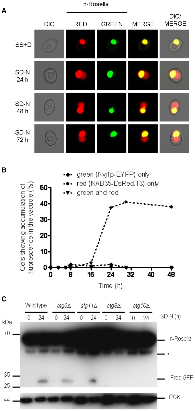
Figure 6 The deletion of the ATG6 or ATG11 genes does not abrogate the delivery of n-Rosella to the vacuole. (A) atg11Δ cells expressing n-Rosella were imaged under growing (SS+D) and nitrogen starvation (SD-N) conditions (0, 24, 48 and 72 hours after commencement of nitrogen starvation). (B) atg11Δ cells co-expressing both nuclear reporters were imaged under growing (SS+D) and nitrogen starvation (SD-N) conditions and the percentage of cells showing accumulation of fluorescence over time determined: ♦ green (Nvj1p-EYFP) only; • red (NAB35-DsRed.T3) only; ▾ green and red. (C) Wild type, atg6Δ, atg11Δ, atg8Δ and atg10Δ cells expressing n-Rosella were starved in SD(-N) medium for 0 and 24 hours, and degradation product (free GFP) levels were monitored by immunoblotting as described in Materials and Methods. Cytosolic PGK was detected as a loading control. *indicates the presumptive degradation product of n-Rosella lacking the NAB35 nuclear targeting signal.
Figure 6 The deletion of the ATG6 or ATG11 genes does not abrogate the delivery of n-Rosella to the vacuole. (A) atg11Δ cells expressing n-Rosella were imaged under growing (SS+D) and nitrogen starvation (SD-N) conditions (0, 24, 48 and 72 hours after commencement of nitrogen starvation). (B) atg11Δ cells co-expressing both nuclear reporters were imaged under growing (SS+D) and nitrogen starvation (SD-N) conditions and the percentage of cells showing accumulation of fluorescence over time determined: ♦ green (Nvj1p-EYFP) only; • red (NAB35-DsRed.T3) only; ▾ green and red. (C) Wild type, atg6Δ, atg11Δ, atg8Δ and atg10Δ cells expressing n-Rosella were starved in SD(-N) medium for 0 and 24 hours, and degradation product (free GFP) levels were monitored by immunoblotting as described in Materials and Methods. Cytosolic PGK was detected as a loading control. *indicates the presumptive degradation product of n-Rosella lacking the NAB35 nuclear targeting signal.CC BY · PubMed Central
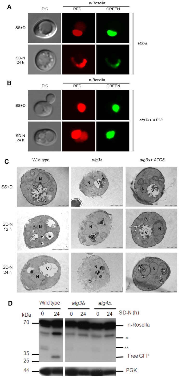
Figure 7 Deletion of the ATG3 gene abrogates the delivery of n-Rosella to the vacuole and influences nuclear morphology. (A) atg3Δ and (B) atg3Δ + ATG3 cells expressing n-Rosella were imaged under growing (SS+D) and nitrogen starvation (SD−N) conditions (24 hours after commencement of nitrogen starvation). (C) Electron microscopy images of wild type (BY4741), atg3Δ and atg3Δ + ATG3 cells expressing n-Rosella were imaged under growing (SS+D) and nitrogen starvation (SD-N) conditions. Nitrogen-starved cells were sampled for imaging after 12 and 24 hours. Examples of contacts between the nucleus and vacuoles as well as alterations in nuclear morphology are presented. N, nucleus; V, vacuole. Bar, 2 µm. (D) Wild type, atg3Δ and atg4Δ cells expressing n-Rosella were starved in SD(−N) medium for 0 and 24 hours, and the levels of free GFP degradation product monitored by immunoblotting as described in Materials and Methods. Cytosolic PGK was detected as a loading control. *indicates the presumptive degradation product of n-Rosella lacking the NAB35 nuclear targeting signal. **indicates non-specific degradation product observed only in growing cells (0 hours).
Figure 7 Deletion of the ATG3 gene abrogates the delivery of n-Rosella to the vacuole and influences nuclear morphology. (A) atg3Δ and (B) atg3Δ + ATG3 cells expressing n-Rosella were imaged under growing (SS+D) and nitrogen starvation (SD−N) conditions (24 hours after commencement of nitrogen starvation). (C) Electron microscopy images of wild type (BY4741), atg3Δ and atg3Δ + ATG3 cells expressing n-Rosella were imaged under growing (SS+D) and nitrogen starvation (SD-N) conditions. Nitrogen-starved cells were sampled for imaging after 12 and 24 hours. Examples of contacts between the nucleus and vacuoles as well as alterations in nuclear morphology are presented. N, nucleus; V, vacuole. Bar, 2 µm. (D) Wild type, atg3Δ and atg4Δ cells expressing n-Rosella were starved in SD(−N) medium for 0 and 24 hours, and the levels of free GFP degradation product monitored by immunoblotting as described in Materials and Methods. Cytosolic PGK was detected as a loading control. *indicates the presumptive degradation product of n-Rosella lacking the NAB35 nuclear targeting signal. **indicates non-specific degradation product observed only in growing cells (0 hours).CC BY · PubMed Central
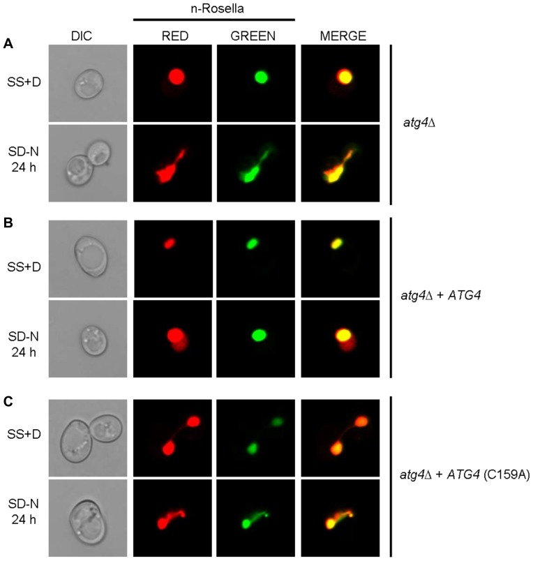
Figure 8 The absence of a functional ATG4 gene product abrogates the delivery of n-Rosella to the vacuole and influences nuclear morphology. atg4Δ (A), atg4Δ + ATG4 (B), and atg4Δ + Atg4C159A (C) cells expressing n-Rosella were imaged under growing (SS+D) and nitrogen starvation (SD−N) conditions (24 hours after commencement of nitrogen starvation).
Figure 8 The absence of a functional ATG4 gene product abrogates the delivery of n-Rosella to the vacuole and influences nuclear morphology. atg4Δ (A), atg4Δ + ATG4 (B), and atg4Δ + Atg4C159A (C) cells expressing n-Rosella were imaged under growing (SS+D) and nitrogen starvation (SD−N) conditions (24 hours after commencement of nitrogen starvation).CC BY · PubMed Central

Verification

Claims the paper makes

The claims and statistics of this paper haven’t been checked yet.

Run the check — free, no account needed. It reads the open-access text and checks the sources it cites.

A few free checks a day · papers already verified open instantly · results are public

Claims other papers make about this one

Each is checked against this paper’s own text: does it say what the citing paper says it does?

Self-check

Percentages, sums and reported differences re-computed from the paper’s own numbers.

The self-check needs the paper’s full text.

Citations

Cites

Research works printed but not linked: 48

  1. DJ Klionsky, 2007, How shall I eat thee?, Autophagy, 3, 413, 10.4161/auto.4377 · DOI
  2. A van der Vaart, 2008, A picky eater: exploring the mechanisms of selective autophagy in human pathologies., Traffic, 9, 281, 10.1111/j.1600-0854.2007.00674.x · DOI
  3. C Kraft, 2009, Selective types of autophagy in yeast., Biochim Biophys Acta, 1793, 1404, 10.1016/j.bbamcr.2009.02.006 · DOI
  4. JAKW Kiel, 2010, Autophagy in unicellular eukaryotes., Phil Trans R Soc B, 365, 819, 10.1098/rstb.2009.0237 · DOI
  5. P Roberts, 2003, Piecemeal microautophagy of nucleus in Saccharomyces cerevisiae., Mol Biol Cell, 14, 129, 10.1091/mbc.E02-08-0483 · DOI · Paper page
  6. E Kvam, 2007, Nucleus-vacuole junctions and piecemeal microautophagy of the nucleus in S. cerevisiae., Autophagy, 3, 85, 10.4161/auto.3586 · DOI
  7. R Krick, 2008, Piecemeal microautophagy of the nucleus requires the core macroautophagy genes., Mol Biol Cell, 19, 4492, 10.1091/mbc.E08-04-0363 · DOI
  8. JC Farré, 2009, Turnover of organelles by autophagy in yeast., Curr Opin Cell Biol, 21, 522, 10.1016/j.ceb.2009.04.015 · DOI
  9. JI Millen, 2009, Measuring piecemeal microautophagy of the nucleus in Saccharomyces cerevisiae., Autophagy, 5, 75, 10.4161/auto.5.1.7181 · DOI · Paper page
  10. T Sattler, 2000, Cell-free reconstitution of microautophagic vacuole invagination and vesicle formation., J Cell Biol, 151, 529, 10.1083/jcb.151.3.529 · DOI
  11. A Uttenweiler, 2007, The vacuolar transporter chaperone (VTC) complex is required for microautophagy., Mol Biol Cell, 18, 166, 10.1091/mbc.E06-08-0664 · DOI
  12. A Uttenweiler, 2008, Microautophagy in the yeast Saccharomyces cerevisiae., Methods Mol Biol, 445, 245, 10.1007/978-1-59745-157-4_16 · DOI
  13. F Dubouloz, 2005, The TOR and EGO protein complexes orchestrate microautophagy in yeast., Mol Cell, 19, 15, 10.1016/j.molcel.2005.05.020 · DOI
  14. RJ Devenish, 2008, Monitoring organelle turnover in yeast using fluorescent protein tags., Methods Enzymol, 451, 109, 10.1016/S0076-6879(08)03209-6 · DOI · Paper page
  15. X Pan, 2000, Nucleus-vacuole junctions in Saccharomyces cerevisiae are formed through the direct interaction of Vac8p and Nvj1p., Mol Biol Cell, 11, 2445, 10.1091/mbc.11.7.2445 · DOI
  16. RK Niedenthal, 1996, Green fluorescent protein as a marker for gene expression and subcellular localization in budding yeast., Yeast, 12, 773, 10.1002/(SICI)1097-0061(19960630)12:8<773::AID-YEA972>3.0.CO;2-L · DOI
  17. T Kirisako, 2000, The reversible modification regulates the membrane-binding state of Apg8/Aut7 essential for autophagy and the cytoplasm to vacuole targeting pathway., J Cell Biol, 151, 263, 10.1083/jcb.151.2.263 · DOI
  18. RS Sikorski, 1989, A system of shuttle vectors and yeast host strains designed for efficient manipulation of DNA in Saccharomyces cerevisiae., Genetics, 122, 19, 10.1093/genetics/122.1.19 · DOI
  19. C Bauer, 2001, Improved technique for electron microscope visualization of yeast membrane structures., MicroscMicroanal, 7, 530
  20. M Prescott, 1995, The functional expression of a rat cDNA encoding OSCP in the yeast Saccharomyces cerevisiae., Biochem Biophys Res Commun, 207, 943, 10.1006/bbrc.1995.1276 · DOI
  21. LS Kean, 1993, Retrograde lipid traffic in yeast: identification of two distinct pathways for internalization of fluorescent-labeled phosphatidylcholine from the plasma membrane., J Cell Biol, 123, 1403, 10.1083/jcb.123.6.1403 · DOI
  22. Y Cao, 2007, Physiological functions of Atg6/Beclin 1: a unique autophagy-related protein., Cell Res, 17, 839, 10.1038/cr.2007.78 · DOI · Paper page
  23. F Tang, 2006, Vac8p, an armadillo repeat protein, coordinates vacuole inheritance with multiple vacuolar processes., Traffic, 7, 1368, 10.1111/j.1600-0854.2006.00458.x · DOI
  24. K Suzuki, 2007, Molecular machinery of autophagosome formation in yeast, Saccharomyces cerevisiae., FEBS Lett, 581, 2156, 10.1016/j.febslet.2007.01.096 · DOI
  25. H Nakatogawa, 2009, Dynamics and diversity in autophagy mechanisms: lessons from yeast., Nat Rev Mol Cell Biol, 10, 458, 10.1038/nrm2708 · DOI
  26. SA Teter, 2001, Degradation of lipid vesicles in the yeast vacuole requires function of Cvt17, a putative lipase. J. Biol., Chem, 276, 2083
  27. UD Epple, 2003, Intravacuolar membrane lysis in Saccharomyces cerevisiae. Does vacuolar targeting of Cvt17/Aut5p affect its function?, J Biol Chem, 278, 7810, 10.1074/jbc.M209309200 · DOI
  28. Z Yang, 2006, Atg22 recycles amino acids to link the degradative and recycling functions of autophagy., Mol Biol Cell, 17, 5094, 10.1091/mbc.E06-06-0479 · DOI
  29. O Muller, 2002, Role of the Vtc proteins in V-ATPase stability and membrane trafficking., J Cell Sci, 116, 1107, 10.1242/jcs.00328 · DOI
  30. D Mijaljica, 2010, The intricacy of nuclear membrane dynamics during nucleophagy., Nucleus, 1, 213, 10.4161/nucl.11738 · DOI
  31. H Nakatogawa, 2007, Atg8, a ubiquitin-like protein required for autophagosome formation, mediates membrane tethering and hemifusion., Cell, 130, 165, 10.1016/j.cell.2007.05.021 · DOI
  32. S Subramani, 2007, A ubiquitin-like protein involved in membrane fusion., Cell, 130, 18, 10.1016/j.cell.2007.06.041 · DOI
  33. Z Yang, 2009, An overview of the molecular mechanism of autophagy., Curr Top Microbiol Immunol, 335, 1
  34. C He, 2006, Recruitment of Atg9 to the preautophagosomal structure by Atg11 is essential for selective autophagy in budding yeast., J Cell Biol, 175, 925, 10.1083/jcb.200606084 · DOI
  35. T Shintani, 2004, Autophagy in health and disease: a double-edged sword., Science, 306, 990, 10.1126/science.1099993 · DOI
  36. T Sekito, 2009, Atg17 recruits Atg9 to organize the pre-autophagosomal structure., Genes Cells, 14, 525, 10.1111/j.1365-2443.2009.01299.x · DOI
  37. JE Legakis, 2007, A cycling protein complex required for selective autophagy., Autophagy, 3, 422, 10.4161/auto.4129 · DOI
  38. WL Yen, 2007, Atg27 is required for autophagy-dependent cycling of Atg9., Mol Biol Cell, 18, 581, 10.1091/mbc.E06-07-0612 · DOI
  39. Z Xie, 2007, Autophagosome formation: core machinery and adaptations., Nat Cell Biol, 9, 1102, 10.1038/ncb1007-1102 · DOI
  40. F Reggiori, 2004, Early stages of the secretory pathway, but not endosomes, are required for Cvt vesicle and autophagosome assembly in Saccharomyces cerevisiae., Mol Biol Cell, 15, 2189, 10.1091/mbc.E03-07-0479 · DOI
  41. VM Olkkonen, 2004, Oxysterol binding proteins: in more than one place at one time?, Biochem Cell Biol, 82, 87, 10.1139/o03-088 · DOI
  42. E Kvam, 2004, Nvj1p is the outer-nuclear-membrane receptor for oxysterol-binding protein homolog Osh1p in Saccharomyces cerevisiae., J Cell Sci, 117, 4959, 10.1242/jcs.01372 · DOI
  43. E Kvam, 2005, Targeting of Tsc13p to nucleus-vacuole junctions: a role for very-long-chain fatty acids in the biogenesis of microautophagic vesicles., Mol Biol Cell, 16, 3987, 10.1091/mbc.E05-04-0290 · DOI
  44. R Krick, 2009, Piecemeal microautophagy of the nucleus: genetic and morphological traits., Autophagy, 5, 270, 10.4161/auto.5.2.7639 · DOI
  45. S Siniossoglou, 1998, A novel complex of membrane proteins required for formation of a spherical nucleus., EMBO J, 17, 6449, 10.1093/emboj/17.22.6449 · DOI
  46. H Santos-Rosa, 2005, The yeast lipin Smp2 couples phospholipid biosynthesis to nuclear membrane growth., EMBO J, 24, 1931, 10.1038/sj.emboj.7600672 · DOI
  47. S Siniossoglou, 2009, Lipins, lipids and nuclear envelope structure., Traffic, 10, 1181, 10.1111/j.1600-0854.2009.00923.x · DOI
  48. C Pereira, 2010, Mitochondrial degradation in acetic acid-induced yeast apoptosis: the role of Pep4 and the ADP/ATP carrier., Mol Microbiol, 76, 1398, 10.1111/j.1365-2958.2010.07122.x · DOI

Cited by

Sorted by citations

See all 50

Metadata

Authors
Mijaljica, D., Prescott, M., & Devenish, R. J.
Venue
PLoS ONE
Published
2012-06-29
Volume
7
Issue
6
Pages
e40013
ISSN
1932-6203
Cited by
50
References
2
Language
en

About these checks

Registry, retraction, Cites and statistics results are computed, not judged, with one exception: a printed reference that no index lists is read once by a model before it counts as a citation error. Claims are extracted by a model and kept only when their quote appears verbatim in the text; each cited source is then checked, with a model’s help, for whether it says what the claim relies on. Those verdicts can be wrong — read the claim’s quote and the cited source before relying on one.

Statistics and arithmetic are recomputed, not judged.

Confidence bands are shown only for fields calibrated on our annotated test set. None is calibrated yet, so no band is shown.

What counts as a claim ›How we measure confidence ›How we check this ›

Are you an author, or do you see a problem with this page? Request a review