

The DOI matches its Crossref record.
Rat Embryonic Mast Cells Originate in the AGM
Overview
The claims and statistics of this paper haven’t been checked yet.
Run the check — free, no account needed. It reads the open-access text and checks the sources it cites.
Integrity signals
Registry
Cites
Statistics
Figures
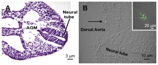
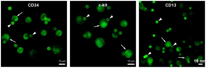
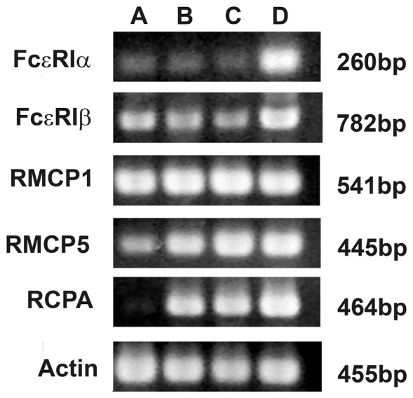
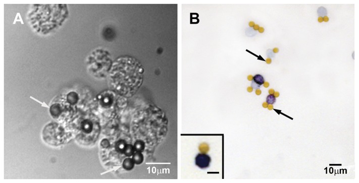
Verification
Claims the paper makes
The claims and statistics of this paper haven’t been checked yet.
Run the check — free, no account needed. It reads the open-access text and checks the sources it cites.
Claims other papers make about this one
Each is checked against this paper’s own text: does it say what the citing paper says it does?
In the citing paper
“Committed progenitors with an immature mast cell phenotype can be found in the circulation and peripheral tissues of developing mice and rats from E11.5 onwards. 54 , 56”
From Mast cell ontogeny: From fetal development to life‐long health and disease (2023) · cites this paper as [56]
In the citing paper
“In contrast, mast cells loaded with heparin‐containing granules have been reported only at E14.5‐E15.5 in the murine and rat skin. 49 , 53 , 56”
From Mast cell ontogeny: From fetal development to life‐long health and disease (2023) · cites this paper as [56]
Self-check
Percentages, sums and reported differences re-computed from the paper’s own numbers.
The self-check needs the paper’s full text.
Citations
Cites
No references could be resolved yet.
Research works printed but not linked: 45
- SC Bischoff, 2007, Role of mast cells in allergic and non-allergic immune responses: comparison of human and murine data, Nat Rev Immunol, 7, 93, 10.1038/nri2018 · DOI
- DD Metcalfe, 1997, Mast Cells, Physiol Rev, 77, 1033, 10.1152/physrev.1997.77.4.1033 · DOI
- SJ Galli, 2005, Mast cells as “tunable” effector and immunoregulatory cells: Recent advances, Annu Rev Immunol, 23, 749, 10.1146/annurev.immunol.21.120601.141025 · DOI · Paper page
- B Frossi, 2010, Exploring a regulatory role for mast cells: ‘MCregs’?, Trends Immunol, 31, 97, 10.1016/j.it.2009.12.007 · DOI
- Y Kitamura, 1979, Distribution of mast-cell precursors in hematopoietic and lymphopoietic tissues of mice, J Exp Med, 150, 482, 10.1084/jem.150.3.482 · DOI
- Y Kitamura, 1977, Development of mast cells from grafted bone marrow cells in irradiated mice, Nature, 268, 442, 10.1038/268442a0 · DOI
- M Jamur, 2010, Mast cell repopulation of the peritoneal cavity: contribution of mast cell progenitors versus bone marrow derived committed mast cell precursors, BMC Immunology, 11, 32, 10.1186/1471-2172-11-32 · DOI
- MC Jamur, 2005, Identification and characterization of undifferentiated mast cells in mouse bone marrow, Blood, 105, 4282, 10.1182/blood-2004-02-0756 · DOI · Paper page
- LK Basciano, 1986, Monoclonal antibodies that inhibit IgE binding, J Biol Chem, 261, 11823, 10.1016/S0021-9258(18)67317-X · DOI
- N Guo, 1989, Monoclonal antibody AA4, which inhibits binding of IgE to high affinity receptors on rat basophilic leukemia cells, binds to novel alpha-galactosyl derivative of ganglioside GD1b, J Biol Chem, 264, 13267, 10.1016/S0021-9258(18)51624-0 · DOI
- MC Jamur, 2001, Identification and isolation of rat bone marrow-derived mast cells using the mast cell-specific monoclonal antibody AA4, J Histochem Cytochem, 49, 219, 10.1177/002215540104900209 · DOI
- C Oliver, 1992, Binding of monoclonal antibody AA4 to gangliosides on rat basophilic leukemia cells produces changes similar to those seen with Fc epsilon receptor activation, J Cell Biol, 116, 635, 10.1083/jcb.116.3.635 · DOI
- MF Guiraldelli, 2008, The low affinity IgG receptor Fc gamma RIIB contributes to the binding of the mast cell specific antibody, mAb BGD6, Mol Immunol, 45, 2411, 10.1016/j.molimm.2007.07.041 · DOI
- Y Kitamura, 1979, Presence of mast cell precursors in fetal liver of mice, Dev Biol, 70, 510, 10.1016/0012-1606(79)90042-3 · DOI
- T Sonoda, 1983, Presence of mast cell precursors in the yolk sac of mice, Dev Biol, 97, 89, 10.1016/0012-1606(83)90066-0 · DOI · Paper page
- J Palis, 1999, Development of erythroid and myeloid progenitors in the yolk sac and embryo proper of the mouse, Development, 126, 5073, 10.1242/dev.126.22.5073 · DOI · Paper page
- A Medvinsky, 2011, Embryonic origin of the adult hematopoietic system: advances and questions, Development, 138, 1017, 10.1242/dev.040998 · DOI
- MC Jamur, 1997, Immunomagnetic isolation of rat bone marrow-derived and peritoneal mast cells, J Histochem Cytochem, 45, 1715, 10.1177/002215549704501215 · DOI
- EL Barsumian, 1981, IgE-induced histamine release from rat basophilic leukemia cell lines: isolation of releasing and nonreleasing clones, Eur J Immunol, 11, 317, 10.1002/eji.1830110410 · DOI
- A Cumano, 2001, Intraembryonic, but not yolk sac hematopoietic precursors, isolated before circulation, provide long-term multilineage reconstitution, Immunity, 15, 477, 10.1016/S1074-7613(01)00190-X · DOI
- JY Bertrand, 2005, Characterization of purified intraembryonic hematopoietic stem cells as a tool to define their site of origin, Proc Natl Acad Sci U S A, 102, 134, 10.1073/pnas.0402270102 · DOI
- J Palis, 2001, Spatial and temporal emergence of high proliferative potential hematopoietic precursors during murine embryogenesis, Proc Natl Acad Sci U S A, 98, 4528, 10.1073/pnas.071002398 · DOI
- MAS Moore, 1970, Ontogeny of the haemopoietic system: Yolk sac origin of in vivo and in vitro colony forming cells in the developing mouse embryo, Br J Haematol, 18, 279, 10.1111/j.1365-2141.1970.tb01443.x · DOI
- PM Wong, 1986, Properties of the earliest clonogenic hemopoietic precursors to appear in the developing murine yolk sac, Proc Natl Acad Sci U S A, 83, 3851, 10.1073/pnas.83.11.3851 · DOI
- A Cumano, 1996, Lymphoid potential, probed before circulation in mouse is restricted to caudal intraembryonic splanchnopleura, Cell, 86, 907, 10.1016/S0092-8674(00)80166-X · DOI
- F Dieterlen-Lievre, 1975, On the origin of haemopoietic stem cells in the avian embryo: an experimental approach, J Embryol Exp Morphol, 33, 607
- D Beaupain, 1979, Are developmental hemoglobin changes related to the origin of stem cells and site of erythropoiesis?, Blood, 53, 212, 10.1182/blood.V53.2.212.212 · DOI
- O Lassila, 1982, Erythropoiesis and lymphopoiesis in the chick yolk-sac-embryo chimeras: contribution of yolk sac and intraembryonic stem cells, Blood, 59, 377, 10.1182/blood.V59.2.377.377 · DOI
- JB Turpen, 1981, The early ontogeny of hematopoietic cells studied by grafting cytogenetically labeled tissue anlagen: Localization of a prospective stem cell compartment, Dev Biol, 85, 99, 10.1016/0012-1606(81)90239-6 · DOI
- E Dzierzak, 1998, Qualitative and quantitative aspects of haematopoietic cell development in the mammalian embryo, Immunol Today, 19, 228, 10.1016/S0167-5699(98)01258-4 · DOI
- M Tavian, 2001, The human embryo, but not its yolk sac, generates lympho-myeloid stem cells: Mapping multipotent hematopoietic cell fate in intraembryonic mesoderm, Immunity, 15, 487, 10.1016/S1074-7613(01)00193-5 · DOI
- I Godin, 1995, Emergence of multipotent hemopoietic cells in the yolk sac and paraaortic splanchnopleura in mouse embryos, beginning at 8.5 days postcoitus, Proc Natl Acad Sci U S A, 92, 773, 10.1073/pnas.92.3.773 · DOI
- A Medvinsky, 1996, Definitive hematopoiesis is autonomously initiated by the AGM region, Cell, 86, 897, 10.1016/S0092-8674(00)80165-8 · DOI
- AM Müller, 1994, Development of hematopoietic stem cell activity in the mouse embryo, Immunity, 1, 291, 10.1016/1074-7613(94)90081-7 · DOI
- A Ivanovs, 2011, Highly potent human hematopoietic stem cells first emerge in the intraembryonic aorta-gonad-mesonephros region, J Exp Med, 208, 2417, 10.1084/jem.20111688 · DOI
- J-C Boisset, 2012, On the origin of hematopoietic stem cells: Progress and controversy, Stem Cell Res, 8, 1, 10.1016/j.scr.2011.07.002 · DOI
- G Costa, 2012, Origin of blood cells and HSC production in the embryo, Trends Immunol, 33, 215, 10.1016/j.it.2012.01.012 · DOI
- I Godin, 2002, The hare and the tortoise: an embryonic haematopoietic race, Nat Rev Immunol, 2, 593, 10.1038/nri857 · DOI
- E Taylor, 2010, Hematopoietic stem cell activity in the Aorta-Gonad-Mesonephros region enhances after mid-day 11 of mouse development, Int J Dev Biol, 54, 1055, 10.1387/ijdb.103152et · DOI
- S Taoudi, 2008, Extensive Hematopoietic Stem Cell Generation in the AGM Region via Maturation of VE-Cadherin+CD45+ Pre-Definitive HSCs, Cell Stem Cell, 3, 99, 10.1016/j.stem.2008.06.004 · DOI
- MFTR de Bruijn, 2000, Definitive hematopoietic stem cells first develop within the major arterial regions of the mouse embryo, EMBO J, 19, 2465, 10.1093/emboj/19.11.2465 · DOI
- GR Johnson, 1975, Role of stem cell migration in initiation of mouse foetal liver haemopoiesis, Nature, 258, 726, 10.1038/258726a0 · DOI
- JW Combs, 1966, Maturation of mast cells, J Cell Biol, 48, 676
- LC Yong, 1997, The mast cell: origin, morphology, distribution, and function, Exp Toxicol Pathol, 49, 409, 10.1016/S0940-2993(97)80129-7 · DOI
- Q Yuan, 1998, Generation of a Novel Stem Cell Factor-Dependent Mast Cell Progenitor, J Immunol, 161, 5143, 10.4049/jimmunol.161.10.5143 · DOI
Cited by
Sorted by citations
Mast cell ontogeny: From fetal development to life‐long health and disease
Chia, S. L., Kapoor, S., Carvalho, C., Bajénoff, M., & Gentek, R. (2023) · Immunological Reviews · doi:10.1111/imr.13191 · Cited by 60
Self-citation
Metadata
- Authors
- Guiraldelli, M. F., França, C. N., de Souza, D. A., da Silva, E. Z. M., Toso, V. D., Carvalho, C. C., Jamur, M. C., & Oliver, C.
- Venue
- PLoS ONE
- Publisher
- Public Library of Science (PLoS)
- Published
- 2013-03-07
- Volume
- 8
- Issue
- 3
- Pages
- e57862
- ISSN
- 1932-6203
- PMC
- PMC3591394
- Cited by
- 5
- References
- 0
- Language
- en
About these checks
Registry, retraction, Cites and statistics results are computed, not judged, with one exception: a printed reference that no index lists is read once by a model before it counts as a citation error. Claims are extracted by a model and kept only when their quote appears verbatim in the text; each cited source is then checked, with a model’s help, for whether it says what the claim relies on. Those verdicts can be wrong — read the claim’s quote and the cited source before relying on one.
Statistics and arithmetic are recomputed, not judged.
Confidence bands are shown only for fields calibrated on our annotated test set. None is calibrated yet, so no band is shown.
What counts as a claim ›How we measure confidence ›How we check this ›
Are you an author, or do you see a problem with this page? Request a review
How to cite
Cite CoChat for the checks on this page, and the paper for its own findings.
- This page
- CoChat. (2026). Citation check: Rat Embryonic Mast Cells Originate in the AGM. https://app.cochat.ai/papers/10.1371/journal.pone.0057862
- The paper
- Guiraldelli, M. F., França, C. N., de Souza, D. A., da Silva, E. Z. M., Toso, V. D., Carvalho, C. C., Jamur, M. C., & Oliver, C. (2013). Rat Embryonic Mast Cells Originate in the AGM. PLoS ONE. https://doi.org/10.1371/journal.pone.0057862
